Comparative Study
. 2008 Aug;131(Pt 8):2028-41.
doi: 10.1093/brain/awn137. Epub 2008 Jun 28.

Primary cortical folding in the human newborn: an early marker of later functional development

Affiliations
Comparative Study

Primary cortical folding in the human newborn: an early marker of later functional development

J Dubois et al. Brain. 2008 Aug.

Abstract

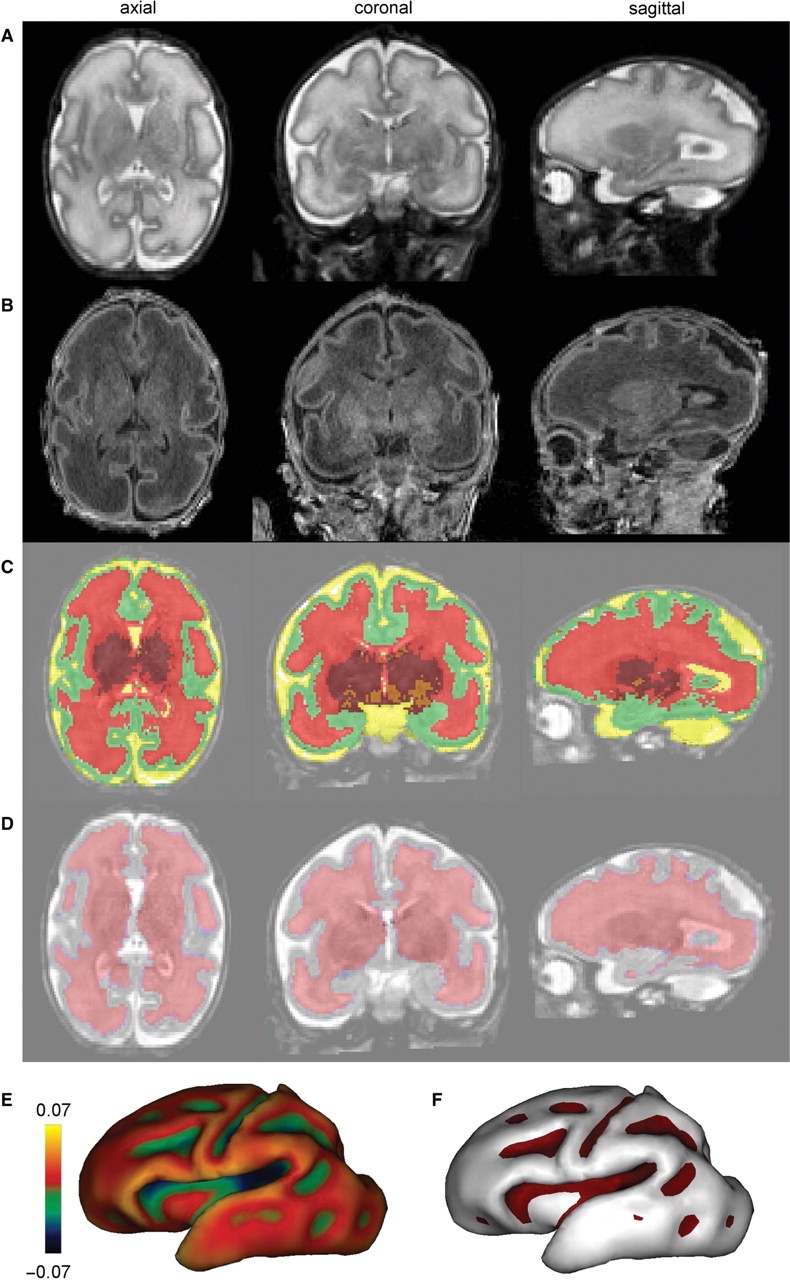
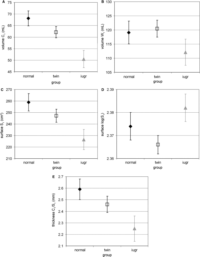
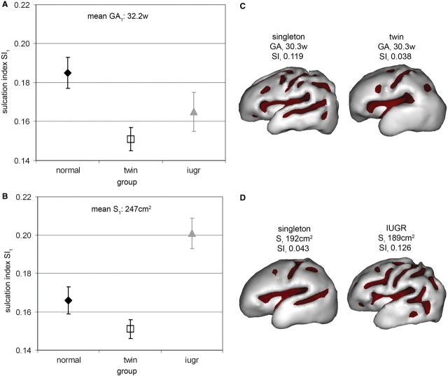
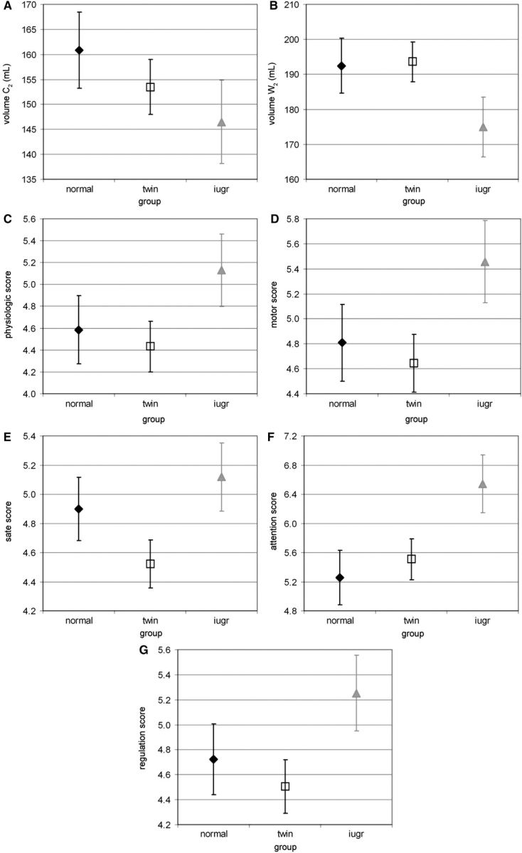
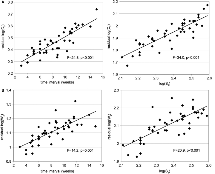
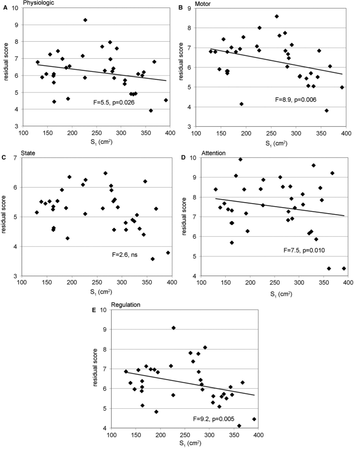
In the human brain, the morphology of cortical gyri and sulci is complex and variable among individuals, and it may reflect pathological functioning with specific abnormalities observed in certain developmental and neuropsychiatric disorders. Since cortical folding occurs early during brain development, these structural abnormalities might be present long before the appearance of functional symptoms. So far, the precise mechanisms responsible for such alteration in the convolution pattern during intra-uterine or post-natal development are still poorly understood. Here we compared anatomical and functional brain development in vivo among 45 premature newborns who experienced different intra-uterine environments: 22 normal singletons, 12 twins and 11 newborns with intrauterine growth restriction (IUGR). Using magnetic resonance imaging (MRI) and dedicated post-processing tools, we investigated early disturbances in cortical formation at birth, over the developmental period critical for the emergence of convolutions (26-36 weeks of gestational age), and defined early 'endophenotypes' of sulcal development. We demonstrated that twins have a delayed but harmonious maturation, with reduced surface and sulcation index compared to singletons, whereas the gyrification of IUGR newborns is discordant to the normal developmental trajectory, with a more pronounced reduction of surface in relation to the sulcation index compared to normal newborns. Furthermore, we showed that these structural measurements of the brain at birth are predictors of infants' outcome at term equivalent age, for MRI-based cerebral volumes and neurobehavioural development evaluated with the assessment of preterm infant's behaviour (APIB).


Figures

Fig. 1
Fig. 1
Volumetric and surfacic identification at birth: Using T2- and T1-weighted MR images (A and B), post-processing enabled the classification of cerebral tissues for volumetric measurements [(C) green: cortex, red: unmyelinated white matter, orange: myelinated white matter, maroon: basal ganglia/thalami, yellow: cerebrospinal fluid], and the segmentation of the interface between cortex and white matter for surfacic measurements (D). Based on this segmentation, the inner cortical surface was reconstructed in 3D [(E) the surface curvature is colour-coded] and the cortical sulci were identified according to negative curvatures [(F) sulci are outlined in purple], which enabled the computation of the sulcation index. Examples are presented for a newborn of 31.1-week-old GA.
Fig. 2
Fig. 2
Volumetric and surfacic measurements among groups at birth: (A) Average of the cortical (C1) and (B) white matter (W1) volumes, of the (C) inner cortical surface (S1), and of the (E) apparent cortical thickness (C1/S1) according to group (with standard error in plot bars), estimated by the models with age GA1 as co-variable (Table 2, mean GA1 = 32.2 weeks), showing lower cortical volume (A), surface (C) and apparent thickness (E) in twins and IUGR newborns compared with singletons of equivalent age. (D) Average of the logarithmic inner cortical surface, log (S1), according to group (with standard error in plot bars), estimated by the models with logarithmic cortical, log (C1), and white matter log (W1) volumes as co-variables [Table 2, mean log(C1) = 1.76, mean log(W1) = 2.06], suggesting that higher surface is related to equivalent volumes in IUGR newborns.
Fig. 3
Fig. 3
Sulcation index quantification among groups at birth. (A and B) Average of the sulcation index (SI1) according to group (with standard error in plot bars), estimated by a model with age GA1 [(A) mean GA1 = 32.2 weeks] or cortical surface S1 [(B) mean S1 = 247 cm2] as co-variable (Table 2), showing lower index in twins and IUGR newborns compared with singletons of equivalent age (A) but higher index in IUGR newborns compared with singletons and twins with similar surface (B). (C and D) Examples of inner cortical surface, with sulci outlined in colour, for a singleton and a twin of equivalent age [(C) GA1 30.3/30.3 weeks; S1 224/164 cm2; SI1 0.119/0.038], and for a singleton and an IUGR newborn with similar surface [(D) GA1 28.6/32.1 weeks; S1 192/189 cm2; SI1 0.043/0.126].
Fig. 4
Fig. 4
Volumetric and neurobehavioural measurements among groups at term equivalent age: (A and B) Average of the cortical (A: C2) and white matter (B: W2) volumes according to group (with standard error in plot bars), estimated by the models with ages GAb and GA2 as co-variables (Table 3, mean GAb = 30.5 weeks, mean GA2 = 40.7 weeks), showing a trend towards lower volumes in IUGR newborns compared with singletons of equivalent age. (CG) Average of the APIB scores [(C) autonomic or physiologic, (D) motor organizational, (E) state organizational, (F) attention–interaction, (G) self-regulation systems] according to group (with standard error in plot bars), estimated by the models with ages GAb and GA2 as co-variables (Table 3, mean GAb = 30.8 weeks, mean GA2 = 40.8 weeks), showing a trend towards higher scores in IUGR newborns compared with singletons of equivalent age.
Fig. 5
Fig. 5
Relationships between cortical surface at birth and cerebral volumes at term equivalent age: Models of cortical and white matter volumes at term [(A) log(C2), (B) log(W2)] are considered on a logarithm scale, with time interval between examinations (GA2–GA1) and inner cortical surface at birth, log(S1), as co-variables (Table 4). The plots represent the variations of ‘residual volumes’ as function of time interval, after correction for the surface effect (left column), and the variations of ‘residual volumes’ as function of surface, after correction for the time interval effect (right column). For equivalent time interval, higher volumes at term relied on higher surfaces at birth.
Fig. 6
Fig. 6
Relationships between cortical surface at birth and neurobehavioural assessment at term equivalent age: Models of APIB scores at term [(A) autonomic or physiologic, (B) motor organizational, (C) state organizational, (D) attention–interaction, (E) self-regulation systems] are considered with time interval between examinations (GA2–GA1) and inner cortical surface at birth (S1) as co-variables (Table 5). The plots represent the variations of ‘residual scores’ as function of surface, after correction for the time interval effect. The variations of ‘residual scores’ as function of time interval, after correction for the surface effect, are not presented because of their non-significance. For equivalent time interval, higher surfaces at birth implied lower APIB scores at term.

References

    1. Als H, Butler S, Kosta S, McAnulty G. The Assessment of Preterm Infants’ Behavior (APIB): furthering the understanding and measurement of neurodevelopmental competence in preterm and full-term infants. Ment Retard Dev Disabil Res Rev. 2005;11:94–102. - PMC - PubMed
    1. Als H, Lester B, Tronick E, Brazelton T. Towards a research instrument for the assessment of preterm infant's behavior (APIB) and manual for the assessment of preterm infant's behavior (APIB) In: Fitzgerald HE, Lester BM, Yogman MW, editors. Theory and research in behavioral pediatrics. New York: Plenum Publishing; 1982. pp. 35–63.
    1. Cachia A, Mangin JF, Riviere D, Kherif F, Boddaert N, Andrade A, et al. A primal sketch of the cortex mean curvature: a morphogenesis based approach to study the variability of the folding patterns. IEEE Trans Med Imaging. 2003;22:754–65. - PubMed
    1. Cachia A, Paillère-Martinot ML, Galinowski A, Januel D, de Beaurepaire R, Bellivier F, et al. Neuroimage. 2007. Cortical folding abnormalities in schizophrenia patients with resistant auditory hallucinations. DOI 10.1016/j.neuroimage.2007.08.049. - PubMed
    1. Chi JG, Dooling EC, Gilles FH. Gyral development of the human brain. Ann Neurol. 1977;1:86–93. - PubMed

Publication types