

Review
doi: 10.1038/nrmicro1928.
Architects of assembly: roles of Flaviviridae non-structural proteins in virion morphogenesis
Affiliations
- PMID: 18587411
- PMCID: PMC2764292
- DOI: 10.1038/nrmicro1928
Item in Clipboard
Review
Architects of assembly: roles of Flaviviridae non-structural proteins in virion morphogenesis
Nat Rev Microbiol.
2008 Sep.
Abstract
Viruses of the Flaviviridae family, including hepatitis C, dengue and bovine viral diarrhoea, are responsible for considerable morbidity and mortality worldwide. Recent advances in our understanding of virion assembly have uncovered commonalities among distantly related members of this family. We discuss the emerging hypothesis that physical virion components are not alone in forming the infectious particle, but that non-structural proteins are intimately involved in orchestrating morphogenesis. Pinpointing the roles of Flaviviridae proteins in virion production could reveal new avenues for antiviral therapeutics.
Figures
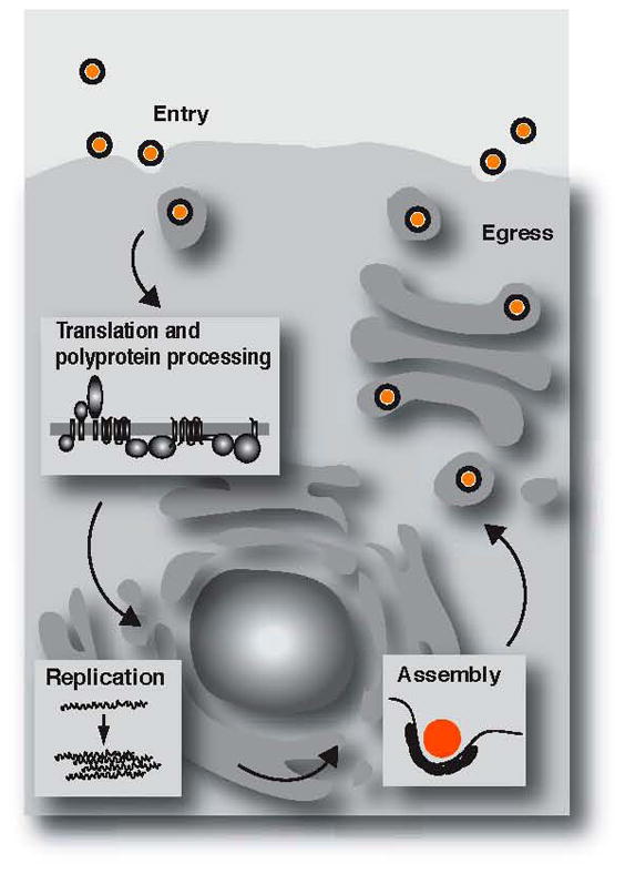
See text for details.
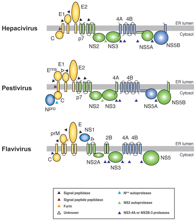
The positive-sense RNA genomes of the Flaviviridae are translated to yield a single hypothetical polyprotein, which is processed by viral and cellular enzymes. The proposed topologies of the viral proteins with respect to the endoplasmic reticulum (ER) membrane, and the enzymes involved in their liberation from the polyprotein are indicated. Structural proteins are shown in yellow, nonstructural proteins are shown in green (required for infectious virus production) or blue (have not been implicated in infectious virus production).
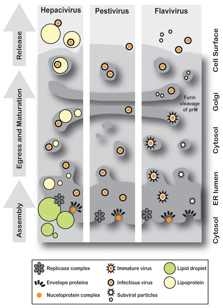
Flaviviridae particles arise through envelopment of the nucleoprotein complex by an endoplasmic reticulum (ER) membrane displaying the viral glycoproteins (E1 and E2, hepacivirus; Erns, E1, and E2, pestivirus; prM, E, flavivirus). Virions then transit from the ER lumen to the cell surface via the secretory pathway. Hepacivirus morphogenesis is believed to take place in association with membrane-apposed lipid droplets, fatty deposits at which replication complexes also accumulate. HCV particles appear to become associated with very low-density lipoproteins (VLDL) during assembly or egress,,. Pestivirus particles are also proposed to form in the ER before transport through the secretory pathway. HCV and pestivirus virions are infectious immediately or rapidly after budding,. Flavivirus particles are immature until acid-induced rearrangement of the envelope protein E, and furin-mediated cleavage of glycoprotein prM to M in a late Golgi compartment,. The formation of capsid and RNA-deficient subviral particles is a hallmark of flavivirus infections.
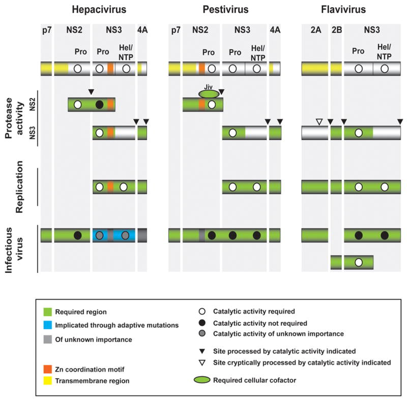
The multifunctional enzyme NS3 and its hydrophobic neighbours in the polyprotein are emerging as conserved requirements for Flaviviridae progeny virion production. The determinants of this region involved in proteolysis, RNA replication, and infectious virus production are shown. Pro, protease; Hel/NTP, helicase/nucleoside triphosphatase; yellow, hydrophobic sequences involved in membrane association; orange, zinc-coordination motifs. Green, required region; blue, implicated through adaptive mutations; dark grey, of unknown importance; white circle, enzymatic activity required; black circle, enzymatic activity not required; grey circle, enzymatic activity of unknown importance; black arrow head, site of processing by indicated catalytic activity; white arrow head, cryptic site of processing by indicated catalytic activity. NS2-mediated autoproteolysis of its carboxy terminus from NS3 is an essential step in hepacivirus and pestivirus polyprotein processing. The protease domain of NS3 is required for the catalytic activity of HCV NS2; the involvement of the zinc-binding site, but not the enzymatic activity, of NS3 suggests a structural contribution,,. Pestivirus NS2 protease activity requires only two residues of NS3,, but depends on the entire NS2 membrane-associated domain and on a cellular cofactor, Jiv,. NS3 protease activity is present in the amino-terminal domains of the hepacivirus, pestivirus, and flavivirus proteins; NS3 also possesses helicase/NTPase activity. HCV and pestivirus NS4A and flavivirus NS2B proteins are essential cofactors for NS3-mediated proteolysis. Replication requires the protease and helicase/NTPase functions of NS3. HCV and pestivirus NS2 proteins are not involved in RNA accumulation after release of NS3,,; flavivirus NS2A and NS2B are essential. Infectious virus production by HCV and pestivirus genomes requires the p7 protein,,. HCV morphogenesis also depends on a function of NS2 that is independent of its protease activity, and a role for NS3 has been suggested by emergent mutations,. Infectious pestivirus production requires the uncleaved NS2-3 precursor protein in conjunction with NS4A, again without a requirement for the known enzymatic activities of NS2 or NS3,. Flavivirus morphogenesis requires NS2A, likely in cooperation with NS3,,; in a second, proteolytic role, NS3 and its cofactor NS2B are important for capsid protein processing,,.
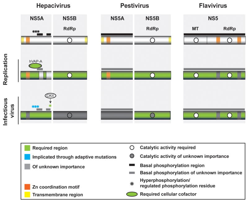
The RNA-dependent RNA polymerase and a replicase phosphoprotein have been recently implicated in Flaviviridae progeny virus production. The determinants of this region involved in RNA replication and infectious virus production are shown. RdRp, RNA-dependent RNA polymerase; MT, methyltransferase; bar, region of basal phosphorylation; star, site of regulated or hyperphosphorylation. Yellow, hydrophobic sequences involved in membrane association; orange, zinc-coordination motifs. Green, required region; blue, implicated through adaptive mutations; dark grey, of unknown importance; white circle, enzymatic activity required; grey circle, enzymatic activity of unknown importance. Replication requires the activities of NS5A and NS5B or NS5 proteins. Regions within the carboxy-terminal domains of HCV NS5A are not essential,,, but binding to a cellular factor, hVAP-A is required,. Although many HCV NS5A serines are dispensable, it is unclear if RNA replication is entirely independent of basal phosphorylation. The exact sites of phosphorylation and relevance of these modifications to pestivirus and flavivirus growth are not known. Infectious virus production may involve the hyperphosphorylated form of HCV NS5A,,,; casein kinase II (CKII)-dependent modification of a serine in the carboxy-terminal region is required. Flavivirus NS5 is required to replicate the RNA genome before its incorporation into progeny virions and changes in pestivirus NS5B have been shown to specifically abolish infectious virus production.
References
-
- Lindenbach BD, Thiel HJ, Rice CM. In: Fields Virology. Knipe DM, Howley PM, editors. Lippincott-Raven Publishers; Philadelphia: 2007. pp. 1101–1152.
-
- Gray EW, Nettleton PF. The ultrastructure of cell cultures infected with border disease and bovine virus diarrhoea viruses. J Gen Virol. 1987;68:2339–2346. - PubMed
-
- Mottola G, Cardinali G, Ceccacci A, Trozzi C, Bartholomew L, Torrisi MR, Pedrazzini E, Bonatti S, Migliaccio G. Hepatitis C virus nonstructural proteins are localized in a modified endoplasmic reticulum of cells expressing viral subgenomic replicons. Virology. 2002;293:31–43. - PubMed
Publication types
MeSH terms
Substances
Grants and funding
LinkOut - more resources
Full Text Sources
Other Literature Sources
