. 2008 Aug 15;198(4):586-93.
doi: 10.1086/590186.

Enterobacter sakazakii enhances epithelial cell injury by inducing apoptosis in a rat model of necrotizing enterocolitis

Affiliations

Enterobacter sakazakii enhances epithelial cell injury by inducing apoptosis in a rat model of necrotizing enterocolitis

Catherine J Hunter et al. J Infect Dis. .

Abstract

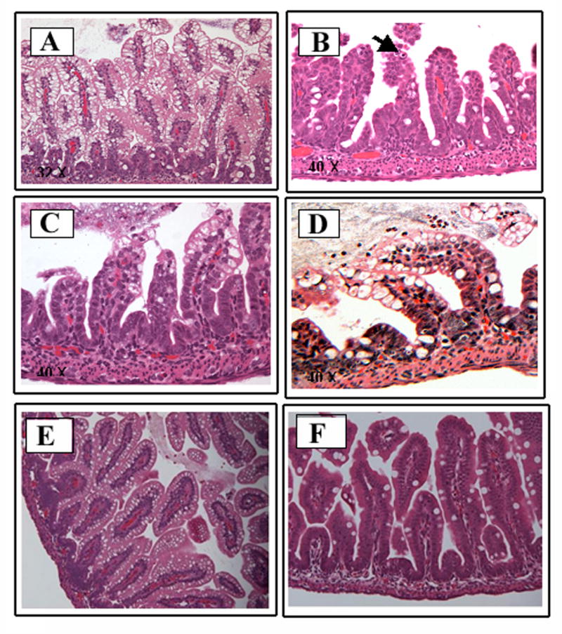
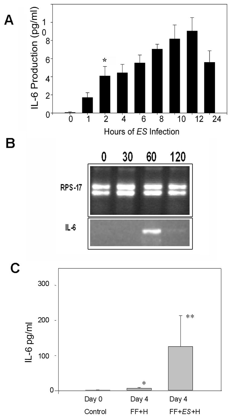
Necrotizing enterocolitis (NEC) is an inflammatory intestinal disorder that affects 2%-5% of all premature infants. Enterobacter sakazakii, a common contaminant of milk-based powdered infant formula, has been implicated as a causative agent of sepsis, meningitis, and NEC in newborn infants, with high mortality rates. However, the role played by E. sakazakii in the pathogenesis of NEC is, to date, not known. Here, we demonstrate for the first time that E. sakazakii can induce clinical and histological NEC in newborn rats. E. sakazakii was found to bind to enterocytes in rat pups at the tips of villi and to intestinal epithelial cells (IEC-6) in culture, with no significant invasion. Exposure to E. sakazakii induced apoptosis and increased the production of interleukin-6 in IEC-6 cells and in the animal model. These data suggest that E. sakazakii could be a potential pathogen that induces NEC and triggers intestinal disease by modulating enterocyte intracellular signaling pathways.


Conflict of interest statement

The authors do not have a commercial or other association that might pose a conflict of interest.

Figures

Fig. 1
Fig. 1. ES induces increased intestinal injury in the rat model of NEC
Panel A compares a FF+H+ES (left) treated rat pup with a FF+H treated rat pup (right). The FF+H+ES rat demonstrates increased abdominal girth, abdominal wall discoloration and evidence of clinical peritonitis. The FF+H rat is not distended and only a milk spot is visible through a normal abdominal wall. Panels B and C demonstrate the gross intestinal morphology after four days of FF+H+ES treatment. The intestine has patchy necrosis, with evidence of hemorrhagic intetsine and pneumatosis intestinalis. The controal rat pup has a normal appearing intestine without necrosis, and only stool visible within the lumen (Panel D).
Fig. 2
Fig. 2. Histology of intestine treated with or without ES
The parafin embedded intestine sections from each rat group are representative sections from several experiments. The sections were stained with hematoxylin and eosin at day 4. Panel A shows the preserved architecture of villi after four days of FF treatment. Panel B reveals neutrophils in the villus tip (see arrow) and alteration in villus structure in group FF+ES. Panel C demonstrates villus tip sloughing and villus blunting seen in a rat pup after four days of FF+H treatment. Panel D demonstrates the greatest degree of derrangement with luminal neutrophils, loss of normal villus architecture, and sloughing in rat pups treated with FF+H+ES. Panel E shows the preserved architecture in a rat pup subjected to FF+EC treatment whereas Panel F only demonstrates mild goblet cell hyperplasia in the setting of FF+H+EC. Histologic scoring of FF+H and FF+H+ES subjected rat pup intestine sections, which is statistically significant (p = 0.020), was shown in G. The difference between groups FF and group FF+H+ES is also statistically significant (p = 0.027).
Fig. 3
Fig. 3. Scanning electron microscopy of rat intestine with or without infection with ES
Rat pups were subjected to FF+H treatment without (A and B) or with ES (C to F) as described in Methods. Small pieces of intestine samples were prepared for SEM. Bacteria are shown by arrows. Magnification: A. ~300X; B. 3400X; C. 5000X; D. 580X E. 3200X; and F. 9600X.
Fig. 4
Fig. 4. Binding to and invasion of ES into IEC-6 cells
Confluent monolayers of IEC-6 cells were infected either with different inoculum sizes (104 to 107 cfu/well) of ES for 2 h (A) or with an inoculum size of 107 cfu/well for varying periods (B). Total cell associated (binding) or total intracellular bacteria (invasion) were determined as described in Methods. The experiments were performed at least five times and the data represent an average ± SD and expressed as relative binding or invasion taking either 6 h treatment with ES or 107 inoculum of ES as 100%. In some experiments, ES infected IEC-6 cells were stained with DAPI and observed the bacteria using transmitted light. Similarly, rat pups were infected with GFP-labeled ES to establish in vivo epithelial association of ES. Cryosections of intestines were stained with DAPI and examined with a fluorescence microscope (C).
Fig. 5
Fig. 5. Induction of apoptosis in intestinal epithelial cells by ES
IEC-6 cells were grown to 90% confluence in eight-well chamber slides and incubated with ES for varying periods. The cells were washed, fixed, and treated with TUNEL-RED reagent, and viewed under a fluorescence microscope (A). In separate experiments, infected cells were washed, released from plates with medium containing EGTA/1% BSA, stained with TUNEL-RED kit, and the number of apoptotic cells were counted (B). In addition, cryosections of intestines from rat pups subjected to FF+H or FF+H+ES treatment were also stained with DAPI and Apo Logix kit (C).
Fig. 6
Fig. 6. Expression of IL-6 in IEC-6 cells and in rat pups infected with ES
Confluent monolayers of IEC-6 cells were infected with ES for varying periods and the cytokines present in the supernatants were determined by Luminex assay. Results are representative of three independent experiments performed in triplicate (A). Total RNA was extracted from the cells and RT-PCR was performed for 30 cycles on normalized cDNA using IL-6 specific primers. RPS-17 primers were used as internal controls (B). In addition, serum samples were collected from day 4 rat pups subjected to various treatments and IL-6 production was determined by Luminex assay. The production of IL-6 from animals subjected to FF+H+ES treatment is significantly greater than the controls pups (*p<0.05 and **p<0.001).

References

    1. Mullane NR, Iversen C, Healy B, et al. Enterobacter sakazakii an emerging bacterial pathogen with implications for infant health. Minerva Pediatr. 2007;59:137–48. - PubMed
    1. Biering G, Karlsson S, Clark NC, Jonsdottir KE, Ludvigsson P, Steingrimsson O. Three cases of neonatal meningitis caused by Enterobacter sakazakii in powdered milk. J Clin Microbiol. 1989;27:2054–6. - PMC - PubMed
    1. Simmons BP, Gelfand MS, Haas M, Metts L, Ferguson J. Enterobacter sakazakii infections in neonates associated with intrinsic contamination of a powdered infant formula. Infect Control Hosp Epidemiol. 1989;10:398–401. - PubMed
    1. Kandhai MC, Reij MW, Gorris LG, Guillaume-Gentil O, van Schothorst M. Occurrence of Enterobacter sakazakii in food production environments and households. Lancet. 2004;363:39–40. - PubMed
    1. Peter CS, Feuerhahn M, Bohnhorst B, et al. Necrotising enterocolitis: is there a relationship to specific pathogens? Eur J Pediatr. 1999;158:67–70. - PubMed

Publication types

MeSH terms