. 2008 Aug 28:1227:1-11.
doi: 10.1016/j.brainres.2008.05.086. Epub 2008 Jun 13.

Brain-derived neurotrophic factor modulates expression of chemokine receptors in the brain

Affiliations

Brain-derived neurotrophic factor modulates expression of chemokine receptors in the brain

Farid Ahmed et al. Brain Res. .

Abstract

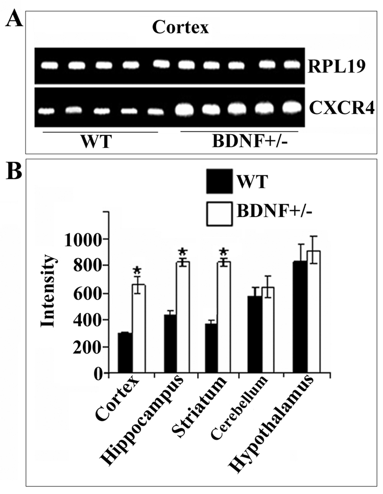
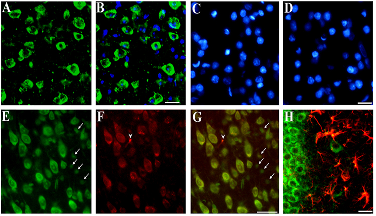
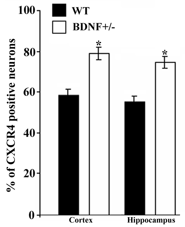
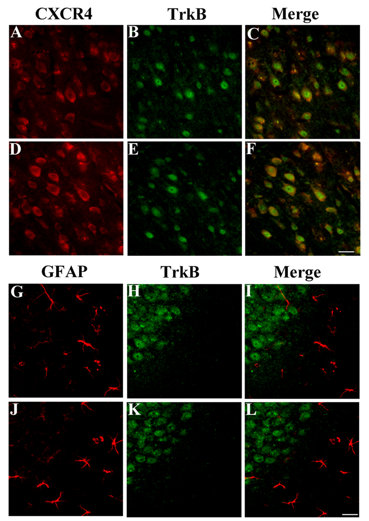
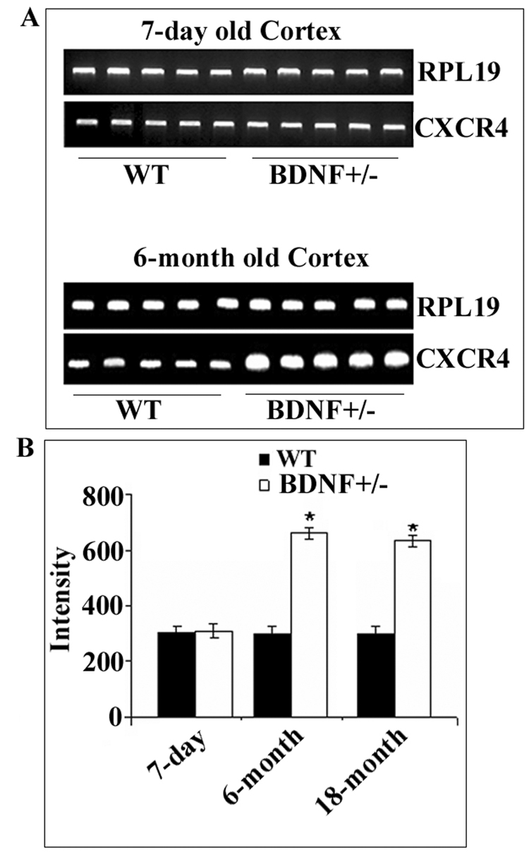
Chemokine receptors, and in particular CXCR4 and CCR5 play a key role in the neuropathogenesis of Human Immunodeficiency Virus-1 (HIV)4 associated dementia (HAD). Thus, new insight into the expression of CXCR4 in the central nervous system may help develop therapeutic compounds against HAD. Brain-derived neurotrophic factor (BDNF) is neuroprotective in vitro against two strains of the HIV envelope protein gp120 that binds to CXCR4 or CCR5. Therefore, we examined whether BDNF modulates chemokine receptor expression in vivo. The content of CXCR4 mRNA and proteins was determined in the cerebral cortex and hippocampus of 6-month-old BDNF heterozygous mice and wild type littermates by using polymerase chain reaction and immunohistochemistry, respectively. BDNF heterozygous mice exhibited an increase in CXCR4 mRNA compared to wild type. Histological analyses revealed an up-regulation of CXCR4 immunoreactivity mainly in neurons. Most of these neurons were positive for TrkB, the BDNF receptor with a tyrosine kinase activity. Increases in CXCR4 mRNA levels were observed in 18-month-old BDNF heterozygous mice but not in 7-day-old mice, suggesting that the modulatory role of BDNF occurs only in mature animals. To determine whether BDNF affects also CXCR4 internalization, SH-SY5Y neuroblastoma cells were exposed to BDNF and cell surface CXCR4 levels were measured at various times. BDNF induced CXCR4 internalization within minutes. Lastly, BDNF heterozygous mice showed higher levels of CCR5 and CXCR3 mRNA than wild type in the cerebral cortex, hippocampus and striatum. Our data indicate that BDNF may modulate the availability of chemokine receptors implicated in HIV infection.


Figures

Figure 1
Figure 1. BDNF heterozygous mice exhibit higher levels of CXCR4 mRNA than WT
RNA was extracted from the indicated brain areas of 6-month-old BDNF+/− mice or WT littermates. RT-PCR was performed using primers designed to amplify mouse CXCR4 (see Experimental Procedures). PCR reaction products were analyzed by agarose gel-electrophoresis (see Experimental Procedures). A. Representative gel showing CXCR4 and RPL19 cDNAs from the cerebral cortex. B. Semi-quantitative analysis of CXCR4 cDNA was carried out by Quantity One 1-D Analysis as described in Experimental Procedures. RPL19 was used as an internal control to normalize gel loading. Data, expressed as intensity of the cDNA band, are the mean ± SEM of five independent samples. *p<0.05 vs control.
Figure 2
Figure 2. Immunohistochemical analysis of CXCR4
Serial coronal sections (16 µm) from the frontal cortex or hippocampus were obtained from WT mice. Examples of cortical sections stained with (A) a CXCR4 antibody (green) counterstained with (B) DAPI (blue) to visualize cellular nuclei. Note that some cells are CXCR4 negative. C and D: Examples of sections in which the primary or secondary antibody, respectively, were omitted. Sections were counterstained with DAPI. E and F: Sections stained with Nissl and CXCR4 antibody, respectively (see Experimental Procedures). G: E and F were merged to show colocalization of CXCR4 and neurons (yellow). Note that in E small Nissl positive cells are CXCR4 negative (arrows). In F, one CXCR4 positive cell (arrowhead) is Nissl negative. H: Example of a hippocampal section stained with CXCR4 (green) and GFAP (red) and showing colocalization of CXCR4 and GFAP (yellow). Bars=40 µm.
Figure 3
Figure 3. BDNF+/− mice exhibit more CXCR4 immunoreactivity
Serial coronal sections obtained as described in Fig. 2 and in Experimental Procedures were used to determine the number of CXCR4 positive neurons in the frontal cortex and hippocampus. The number of CXCR4 positive neurons was determined in layer V of the cortex and CA2 region of the hippocampus in an area of 1 mm2 per section using MetaMorph® software. An average of 8000 neurons per animal per area was counted. Data, expressed as % of Nissl positive cells per section, are the mean ± SEM of 10 sections per animal (n=4 each group). *p<0.05 vs WT.
Figure 4
Figure 4. CXCR4 co-localizes with TrkB in neurons
Cortical sections from WT (A–C) and BDNF+/− mice (D–F) were stained with CXCR4 antibody (red) and an antibody that recognizes the extracellular domain of TrkB (αtrkB23–36, green). In C and F, please note that a number of CXCR4 positive cells are also TrkB positive. Sections from the hippocampus of WT (G–I) and BDNF+/− mice (J–L) were stained for GFAP (red, G and J) and TrkB (green, H and K). No astrocytes were TrkB positive. Bar=50 µm.
Figure 5
Figure 5. Age-dependent increase of CXCR4 in BDNF+/− mice
Cortical CXCR4 mRNA levels were measured in 7-day-old pups, 6 and 18-month-old BDNF+/− mice and WT littermates. RNA extraction and RT-PCR was carried out as described in Experimental Procedures. A. Representative gels showing CXCR4 and RPL19 cDNAs from the cortex of 7-day old and 6-month old mice. B. Semi-quantitative analysis of CXCR4 mRNA was carried out by Quantity One 1-D analysis. Data, expressed as Intensity of cDNA bands, are the mean ± SEM of five independent samples. *p<.005 vs WT.
Figure 6
Figure 6. BDNF increases internalization of CXCR4
SY5YTB8 cells (Kim et al., 1999) were incubated with medium control (0.1% BSA) or medium containing BDNF (10 ng/ml) for 5 or 30 min or 3 hr. Cells were then washed and treated with 2 mM NHS-LC-biotin for 30 min at 4°C to biotinylate cell surface proteins. Biotinylated proteins were precipitated with immobilized streptavidin. Precipitates were then electrophoresed on SDS-polyacrylamide gels and subjected to CXCR4 immunoblotting. A. Example of immunoblot. B. Immunoreactive bands were scanned and quantified by Quantity One 1-D Analysis Software. Data, expressed as intensity of immunoreactive bands, are the mean ± SEM of three independent experiments. *p<0.05 vs control.
Figure 7
Figure 7. BDNF decreases CXCR4 mRNA levels
SY5YTB8 cells were exposed to medium control (0.1% BSA) or containing BDNF (10 ng/ml) for 4 and 8 hr and CXCR4 mRNA levels were measured by RT-PCR using primers designed to amplify human CXCR4 (see Table 1). Bands corresponding to CXCR4 cDNA were quantified using Quantity One 1-D Analysis Software. Data, expressed as intensity of bands, are the mean ± SEM of three independent experiments done (n=9). *p<0.05 vs control.
Figure 8
Figure 8. Modulation of chemokine receptors by BDNF is area-specific
Total RNA was extracted from the indicated brain areas of 6-month-old BDNF+/− mice or WT littermates. RT-PCR was performed using primers designed to amplify mouse CCR5 and CXCR3. A. Representative gels showing CCR5, CXCR3 and RPL19 cDNAs from the indicated brain areas. B. Semi-quantitative analysis of chemokine receptor cDNAs was carried out by Quantity One 1-D analysis. Data, expressed as intensity of cDNA bands, are the mean ± SEM of five independent samples. *p<0.05; **p<0.001 vs WT.

References

    1. Bachis A, Major EO, Mocchetti I. Brain-derived neurotrophic factor inhibits human immunodeficiency virus-1/gp120-mediated cerebellar granule cell death by preventing gp120 internalization. J Neurosci. 2003;23:5715–5722. - PMC - PubMed
    1. Bachis A, Mocchetti I. Brain-derived neurotrophic factor is neuroprotective against human immunodeficiency virus-1 envelope proteins. Ann NY Acad Sci. 2005;1053:247–257. - PubMed
    1. Bachis A, Aden SA, Nosheny RL, Andrews PM, Mocchetti I. Axonal transport of Human Immunodeficiency Virus Type 1 envelope glycoprotein 120 is found in association with neuronal apoptosis. J. Neurosci. 2006;26:6771–6780. - PMC - PubMed
    1. Bagri A, Gurney T, He X, Zou YR, Littman DR, Tessier-Lavigne M, Pleasure SJ. The chemokine SDF1 regulates migration of dentate granule cells. Development. 2002;129:4249–4260. - PubMed
    1. Banisadr G, Fontanges P, Haour F, Kitabgi P, Rostene W, Parsadaniantz SM. Neuroanatomical distribution of CXCR4 in adult rat brain and its localization in cholinergic and dopaminergic neurons. Eur J Neurosci. 2002;16:1661–1671. - PubMed

Publication types

MeSH terms