. 2008 Sep 5;381(3):748-62.
doi: 10.1016/j.jmb.2008.06.026. Epub 2008 Jun 17.

Different requirements of the kinase and UHM domains of KIS for its nuclear localization and binding to splicing factors

Affiliations

Different requirements of the kinase and UHM domains of KIS for its nuclear localization and binding to splicing factors

Valérie Manceau et al. J Mol Biol. .

Abstract

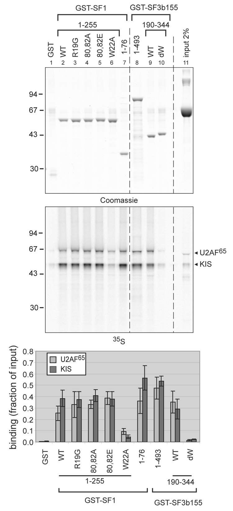
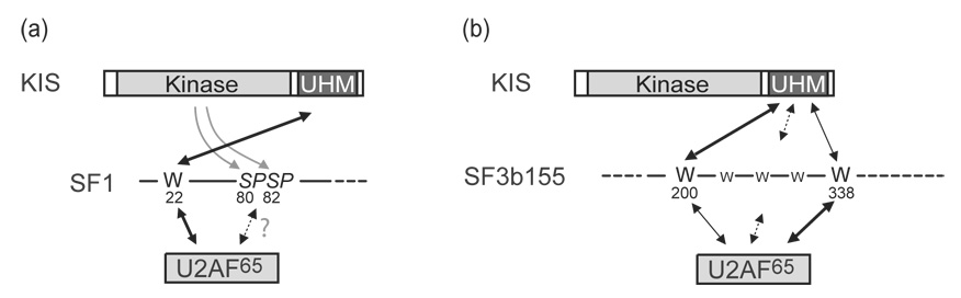
The protein kinase KIS is made by the juxtaposition of a unique kinase domain and a C-terminal domain with a U2AF homology motif (UHM), a sequence motif for protein interaction initially identified in the heterodimeric pre-mRNA splicing factor U2AF. This domain of KIS is closely related to the C-terminal UHM domain of the U2AF large subunit, U2AF(65). KIS phosphorylates the splicing factor SF1, which in turn enhances SF1 binding to U2AF(65) and the 3' splice site, an event known to take place at an early step of spliceosome assembly. Here, the analysis of the subcellular localization of mutated forms of KIS indicates that the kinase domain of KIS is the necessary domain for its nuclear localization. As in the case of U2AF(65), the UHM-containing C-terminal domain of KIS is required for binding to the splicing factors SF1 and SF3b155. The efficiency of KIS binding to SF1 and SF3b155 is similar to that of U2AF(65) in pull-down assays. These results further support the functional link of KIS with splicing factors. Interestingly, when compared to other UHM-containing proteins, KIS presents a different specificity for the UHM docking sites that are present in the N-terminal region of SF3b155, thus providing a new insight into the variety of interactions mediated by UHM domains.


Figures

Figure 1
Figure 1. Domains organization of the proteins used in this study
(a) KIS is formed by the juxtaposition of a serine/threonine kinase domain and a C-terminal noncanonical RNA Recognition Motif of the UHM type (U2AF Homology Motif). This C-terminal domain is highly homologous to the C-terminal UHM domain of U2AF65. (b) In addition to its C-terminal UHM domain necessary for interaction with SF1,, and SF3b155 ,,, U2AF65 possesses two classical RRM domains involved in RNA binding, while its N-terminal RS domain also contacts the branch site during spliceosome assembly. Heterodimerisation with U2AF35 involves an ULM (UHM-Ligand Motif) indicated by a W for the crucial tryptophan residue that is required for UHM binding. (c) SF1 is expressed as multiple spliced forms differing by their C-terminal proline rich domains. The most abundant SF1HL1 isoform of HeLa cells is presented. The C-terminal proline rich region is proposed to mediate protein interactions in particular with WW motifs containing proteins as FBP11 and CA150. The N-terminal ULM domain (W) is required for binding to U2AF65. An extended KH-QUA2 domain mediates binding of SF1 to RNA–,. In between the RNA and U2AF65 binding regions two serine residues within a SPSP conserved motif are mostly in a phosphorylated state in cells and substrate for KIS in vitro and in vivo (ref and our unpublished results). (d) SF3b155 is part of the proteins that form the complex SF3b an integral part of U2snRNP and of the U11/U12 di-snRNP. The C-terminus of SF3b155 contains 22 tandem helical repeats (HEAT repeats) and was shown by electron cryomicroscopy to form a structural element of the outer shell of the complex SF3b enclosing p14 another component of SF3b. Nevertheless, a minimal peptide comprising amino-acids [396–424] of SF3b155 binds p14 ,,,. The N-terminal domain of SF3b155 contains seven potential ULMs in the [190–344] region necessary for U2AF65 binding. In addition numerous TP dipeptides in this region constitute potential phosphorylation sites in agreement with SF3b155 being phosphorylated in vivo and in vitro by CyclinE-cdk2. Interaction of the nuclear phosphatase 1 inhibitor NIPP1 is dependent on phosphorylation of the N-terminus of SF3b155.
Figure 2
Figure 2. Subcellular localization of KIS and mutated forms in CHO cells
(a–h) Different HA-tagged forms of KIS were transiently expressed in Chinese Hamster Ovary (CHO) cells and their subcellular localization was analysed by indirect immunofluorescence microscopy using an anti HA antibody. Typical views with an X60 objective and corresponding to similar exposure times are presented (bar = 10 µm). The primary structure of each mutant is depicted at the bottom of each picture. In contrast to most constructs which presented a variety of situations with either only nuclear or nuclear and cytoplasmic localizations, the two mutants with major deletions of the kinase domain (KIS[212–419] and KIS[293–419]) never presented a nuclear enrichment (e,f). (i) The mean values and standard deviation of the quantification of three experiments are presented. In transfected cells, nuclear enrichment was considered when a clear contrasted brighter staining of the nucleus compared to the cytoplasm was observed.
Figure 3
Figure 3. Interaction of KIS with SF3b155
(a) KIS interacts with SF3b155 in the two-hybrid assay. Different interactions were tested in the two-hybrid system in yeast. Cells expressing the bait plasmids were mated to cells expressing the prey plasmids and expression of the reporter genes HIS3 that allows growth on medium lacking histidine and of the reporter gene lacZ revealed by the activity of beta-galactosidase are presented. KIS (fragment [120–419]), U2AF65 and U2AF35 interacted with SF3b155 (fragment [171–775]29) but not with a fragment of tsg101 used as a negative control. (b) Co-precipitation of KIS with SF3b155. GST-pull-down assays were performed to test the binding of KIS to SF3b155. Extracts of HEK293 cells that had been transfected with a KIS expressing vector were mixed with GST or with GST fused to a N-terminal part of SF3b155 (SF3b155n: amino-acids [1–493]) and pull-down were performed with glutathione beads. 20% of the extract used as “input” and the proteins retained on the beads were analysed by SDS PAGE and immunoblotting with an anti-KIS polyclonal antibody (left panel). A similar experiment was conducted using recombinant KIS partially purified from bacteria (right panel). A truncated form of KIS most probably originating from internal cleavage by thrombin is also detected (asterix). The picture shows typical results obtained in duplicate in two experiments. (c) Preferential binding of KIS to SF1 and SF3b155. KIS was in vitro translated using [35S]-methionine in a reticulocyte lysate, then diluted in interaction buffer containing 1 µg/µL BSA and mixed with about 20 pmoles of each GST-fusion protein to test their interaction by co-precipitation on glutathione beads. The proteins bound to the beads (lanes 2–7) and 2% of the amount of the diluted solution of KIS used for each interaction (input 2%, lane 8) were separated by SDS PAGE. The gel was stained with Coomassie blue (top panel) revealing the GST-fusion proteins (lanes 2–7) and BSA as the major protein in the KIS dilution (input 2%, lane 8). Once dried, the gel was analysed using a Phosphorimager to quantify the binding of KIS to each of the GST-fusion protein as described in the Material and Methods section (bottom panel). SF1f: fragment [1–255] of human SF1; CTD: C-terminus of polymerase II; p27: p27kip1. The mean values of three experiments with standard deviation are presented showing a clear preferential co-precipitation of KIS with SF1f and SF3b155n over the other GST-fusion proteins. (d) Competition of SF1 and SF3b155 for binding to KIS. SF1f (produced by cleavage of GST-SF1f by precision protease (Amersham) and ion exchange purification) was used as a competitor in pull-down assays of KIS with GST-SF1f or GST-SF3b155n performed as in (c). A ten fold excess of SF1f greatly reduced the co-precipitation of in vitro translated KIS with both GST-fusion proteins. The mean values and standard deviation of duplicates are presented.
Figure 4
Figure 4. KIS phosphorylates SF1f much more efficiently than SF3b155 in vitro
In vitro kinase assays were performed using either recombinant GST-KIS or a soluble cell extract of mouse embryonic fibroblasts as a source of kinases as indicated. About 20 pmoles of each purified protein SF1f, GST-SF3b155n and GST-p27kip1 were used as substrates. Phosphorylation reactions were performed with 100 µM [γ-32P]ATP for 30 minutes and products were separated on SDS PAGE that was Coomassie stained (top) and analysed with a phosphorimager for quantification of the radioactivity (bottom). The amount of phosphate (pmoles) that was incorporated in each substrate is indicated beneath the lanes.
Figure 5
Figure 5. The KIS UHM domain is necessary for interaction with SF1 and SF3b155
(a) Full length KIS and U2AF65 and mutant forms of KIS as schematically presented were in vitro translated in the presence of [35S]-methionine and tested for their interaction with GST-SF1f (SF1 fragment [1–255]) and GST-SF3b155n (fragment [1–493]) in GST pull-down assays. The autoradiogram of the SDS PAGE gel of a typical experiment is presented. 2% of the inputs were loaded on the same gel (lanes 1 to 6) to allow the quantification of the retention of the labelled proteins on the beads as described for Fig. 3c. (b) Representation of the means of retention rate and standard deviation for four experiments.
Figure 6
Figure 6. Effect of mutations of SF1 and SF3b155 on binding to KIS and U2AF65
Different mutant forms of GST-SF1 and GST-SF3b155 as indicated above the lanes, were compared in pull-down assays with a mixture of [35S]-labelled in vitro translated U2AF65 and KIS diluted in interaction buffer containing 1 µg/µL BSA as described for Fig. 3c. After SDS PAGE of the binding reactions, the gel was stained with Coomassie blue and digitalized with an infrared scanner (top panel). Retention of KIS and U2AF65 was quantified with a phosphorimager (middle panel) as explained in the Material and Methods section. The mean values of three experiments with standard deviation are presented (bottom panel), showing that mutations of tryptophan residues within the ULM peptides of the SF1f and SF3b155r proteins reduced KIS and U2AF65 co-precipitation.
Figure 7
Figure 7. Different properties of SF3b155 ULMs in mediating interactions with U2AF65 and KIS
(a) The binding of in vitro translated [35S]-labelled U2AF65 and KIS to the wild type SF3b155r (residues [190–344], lane 1), SF3b155r with the seven tryptophan mutated to alanine (lane 2) and the mutants in which each of the seven tryptophan residues were independently reintroduced (lanes 3–9) were tested. The experiments were performed as for Fig. 3c. (b) The efficiency of binding was calculated as the ratio of binding to the wild type SF3b155r. The mean values of three experiments (each interaction reaction being performed in duplicate) are presented with standard deviation. One-way ANOVA statistical analysis was conducted with a Bonferroni post-hoc test yielding a p<0.001 for a significant difference of binding of U2AF65 on SF3b155-W338 compared to SF3b155-W200, and a significant difference of binding of KIS to SF3b155-W200 compared to SF3b155-W338. (c) Alignment of the seven potential ULMs of SF3b155r with that of SF1. The key tryptophan residues are in boldface. Basic residues N-terminal to the key tryptophan are boxed in grey. Lysine 15 of SF1 that might contribute to the particular binding of SF1 to KIS is underlined (see Discussion). The best binding sites for KIS and U2AF65, ULM-W200 and ULM-W338 present a higher homology with that of SF1.
Figure 8
Figure 8
Schematic representation for the comparison of interactions of KIS and U2AF65 with the N-terminal regions of SF1 and SF3b155. Contacts additional to those represented might contribute to these interactions. (a) Thick arrows indicate the major contribution of W22 of SF1 for the binding of U2AF65 and KIS. Additionally KIS phosphorylates serines 80 and 82 which increases the binding of SF1 to U2AF65 potentially by direct contact (broken line arrow). (b) Thick arrows indicate the predominant contribution of W200 and W338 for the binding of KIS and U2AF65 to the N-terminal region of SF3b155. Additional interactions depending on other tryptophan residues are indicated by broken lines arrows.

References

    1. Hunter T. Signaling--2000 and beyond. Cell. 2000;100:113–127. - PubMed
    1. Maris C, Dominguez C, Allain FH. The RNA recognition motif, a plastic RNA-binding platform to regulate post-transcriptional gene expression. Febs J. 2005;272:2118–2131. - PubMed
    1. Maucuer A, Camonis JH, Sobel A. Stathmin interaction with a putative kinase and coiled-coil-forming protein domains. Proc Natl Acad Sci U S A. 1995;92:3100–3104. - PMC - PubMed
    1. Maucuer A, Ozon S, Manceau V, Gavet O, Lawler S, Curmi P, Sobel A. KIS is a protein kinase with an RNA recognition motif. J Biol Chem. 1997;272:23151–23156. - PubMed
    1. Bieche I, Manceau V, Curmi PA, Laurendeau I, Lachkar S, Leroy K, Vidaud D, Sobel A, Maucuer A. Quantitative RT-PCR reveals a ubiquitous but preferentially neural expression of the KIS gene in rat and human. Brain Res Mol Brain Res. 2003;114:55–64. - PubMed

Publication types

MeSH terms