. 2008 Aug 26;155(3):888-901.
doi: 10.1016/j.neuroscience.2008.04.081. Epub 2008 Jun 6.

Novel role of adrenergic neurons in the brain stem in mediating the hypothalamic-pituitary axis hyperactivity caused by prenatal alcohol exposure

Affiliations

Novel role of adrenergic neurons in the brain stem in mediating the hypothalamic-pituitary axis hyperactivity caused by prenatal alcohol exposure

I Y Choi et al. Neuroscience. .

Abstract

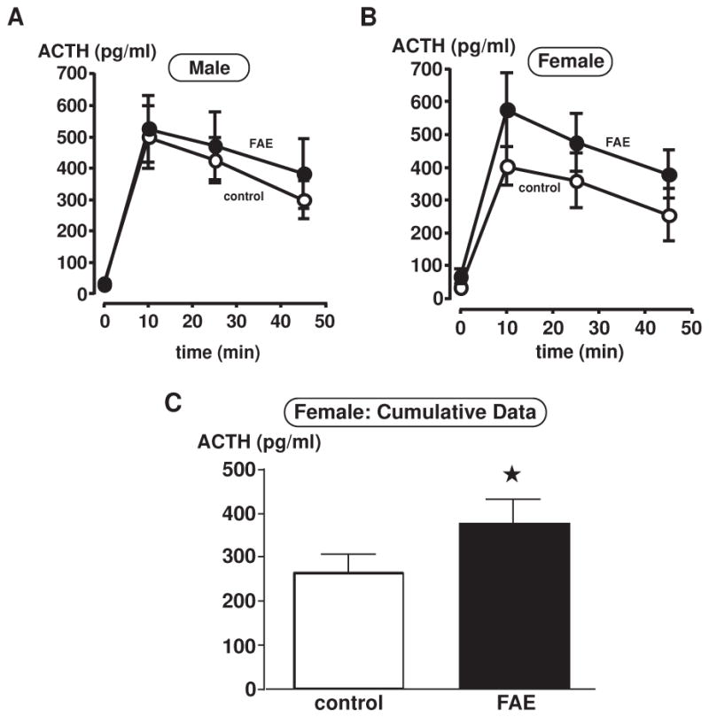
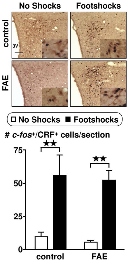
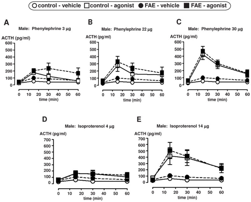
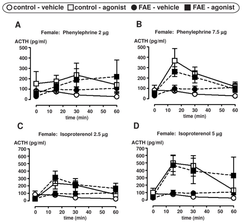
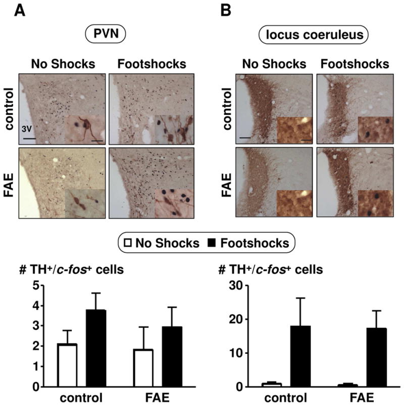
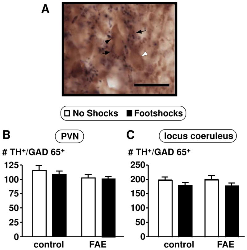
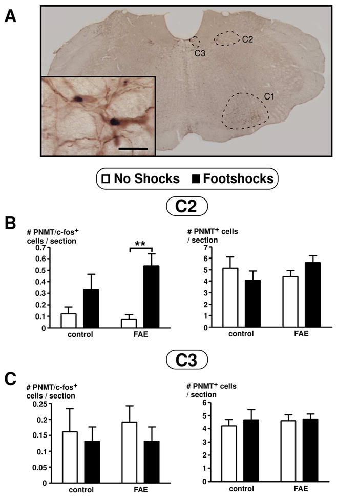
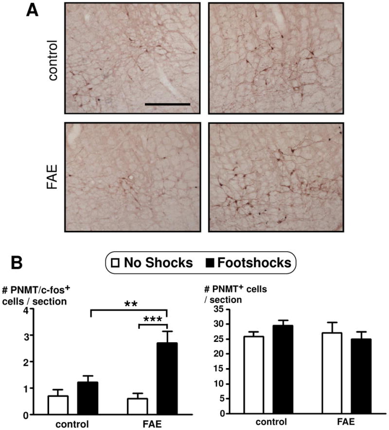
Exposure to alcohol during embryonic development leads to changes in the hypothalamic-pituitary-adrenal (HPA) axis such that adult offspring release more adrenocorticotrophic hormone (ACTH) than controls when exposed to stress. In the present work, we tested the hypothesis that changes in the activity of the catecholaminergic system modulate, at least in part, this upregulation of the HPA axis. Pregnant Sprague-Dawley rats were exposed to alcohol 6 h daily during gestation days 7-18 using the vapor chamber model, which generated mean blood alcohol levels of 188.6+/-10 mg/dl. All experiments were performed on 2 to 3-month-old offspring. We first measured the ACTH response to i.c.v. injection of adrenergic receptor agonists. In rats exposed to footshocks, we then investigated the activity of corticotrophin-releasing factor (CRF) as well as indexes of catecholamine ir, namely tyrosine hydroxylase (TH) immunopositive neurons in the paraventricular nucleus (PVN), TH immunopositive neurons in the locus coeruleus, and phenylethanolamine N-methyltransferase (PNMT) immunopositive neurons in the brain stem. While adult females exposed to alcohol during fetal development (FAE) displayed the expected enhanced ACTH response to stress, there were no significant differences in response to adrenergic receptor agonists or in shock-induced CRF/TH ir and neuronal activity, as determined by c-fos colocalization. In contrast, FAE female offspring exposed to footshocks showed a significant increase in the activity of adrenergic neurons in the C1 region of the brain stem, a population of cells that project to the PVN. Collectively, these results suggest that while FAE-induced hyperactivity of the HPA axis is not accompanied by significant changes in PVN CRF or TH-ir neurons, it is characterized by an upregulation of C1 adrenergic neurons of the brain stem. This novel finding should lead to the functional characterization of this brain region in the FAE model.


Figures

Figure 1
Figure 1
Plasma ACTH measurements in control and FAE male (A) and female (B–C) rats with inescapable footshock stress. Plasma was taken at 0 min (prior to footshocks), 10, 25 and 45 min upon footshock initiation. ACTH levels are presented in pg/ml. Each data point displays mean ± SEM (n = 6–7/group). (C) Bar graph displays cumulative data, where ACTH levels are collapsed over time. There is only a significant main effect of FAE in female rats (*, p = 0.037).
Figure 2
Figure 2
Colocalization of c-fos in CRF immunoreactive cells in the PVN of female rats. Pictures display PVN at 10X objective (scale bar = 100 μm) and insets at 60X oil objective (scale bar = 20 μm). Double immunohistochemistry techniques labelled c-fos as black and CRF as brown. Cell counts were obtained throughout the rostral-caudal extent of the PVN. Results are displayed as the number of CRF and c-fos positive cells per section. Rats were either undisturbed (no shocks) or exposed to inescapable footshock stress (footshocks) for one hour prior to transcardial perfusion. There was a significant main effect of footshock stress (p < 0.0001, n = 12). Bonferroni post-hoc test reveals a significance between no shocks and footshocks within each group (control and FAE) **, p < 0.01.
Figure 3
Figure 3
Plasma ACTH measurements in male control and FAE rats after i.c.v. infusion of an adrenergic receptor agonist. ACTH levels are presented in pg/ml at four time points (0, 15, 30, and 60 min) relative to the agonist infusion, with 0 min time point conducted just prior to agonist infusion. Graphs display mean ± SEM (n = 4–9/group, n = 4 only applies to the FAE rats infused with 22 μg of phenylephrine). Alpha 1 agonist (phenylephrine HCl) infusion results are displayed in figures A–C. Beta agonist (isoproterenol HCl) infusion results are displayed in figures D and E.
Figure 4
Figure 4
Plasma ACTH measurements in female control and FAE rats after i.c.v. infusion of an adrenergic receptor agonist. ACTH levels are presented in pg/ml at four time points (0, 15, 30, and 60 min) relative to the agonist infusion, with 0 min time point conducted just prior to agonist infusion. Graphs display ± SEM (n = 6–8/group). Alpha 1 agonist (phenylephrine HCl) infusion results are displayed in figures A and B. Beta agonist (isoproterenol HCl) infusion results are displayed in figures C and D.
Figure 5
Figure 5
Colocalization of c-fos in TH immunoreactive cells in the PVN (A) and locus coeruleus (B) of female rats. Pictures were taken with a 10X objective (scale bar = 100μm) and insets with a 60X oil objective (scale bar = 20 μm). Double immunohistochemical techniques stained c-fos in black and TH in brown. Cell counts were obtained throughout the rostral-caudal extent of the PVN and locus coeruleus. Results are displayed as the number of TH and c-fos positive cells per section. No shocks and footshocks conditions are described in Figure 2. There was only a significant main effect of footshock stress in the locus coeruleus (p = 0.003, n = 12).
Figure 6
Figure 6
Number of TH positive neurons with GAD65 positive terminals contacting the cell body in female rats. (A) Picture was taken with a 60X oil objective (scale bar = 20 μm). Image shows a representation of the double immunohistochemical technique which stained GAD65 in black and TH in brown. Black arrowhead indicates GAD65 positive stain, white arrowhead indicates TH positive stain, and black arrows indicate double labelled cells. Cell counts were obtained for TH and GAD65 throughout the rostral-caudal extent of the PVN (B) and locus coeruleus (C). Results are displayed as the number of TH and GAD65 positive cells per section. No shocks and footshocks conditions are described in Figure 2. There were no significant differences across any groups (p = 0.075, n = 12–13).
Figure 7
Figure 7
PNMT and c-fos colocalization visualized using double immunohistochemical technique in the brain stem of female rats (A). The three adrenergic regions (C1, C2, and C3) are outlined as indicated. Inset picture display colocalization of PNMT in brown and c-fos in black. Picture was taken with a 2X dry objective and inset picture using a 60X oil objective (inset scale bar = 20 μm). (B–C) Number of PNMT and colocalization of c-fos in PNMT immunoreactive cells in the C2 (B) and C3 (C) regions of the brain stem of female rats. Results are displayed as the number of PNMT and c-fos positive cells per section (on left panel) or number of PNMT positive cells per section (on right panel), as indicated. Cell counts were obtained throughout the rostral-caudal extent of the C2 and C3 areas of the brain stem. There was only a significant main effect of stress in the C2 area (p = 0.001, n = 12). Bonferroni post-hoc test revealed significance as indicated by asterisks (**, p < 0.01). There were no significant effects observed in the C3 area.
Figure 8
Figure 8
Number of PNMT and colocalization of c-fos in PNMT immunoreactive cells in the C1 region of the brain stem of female rats. (A) Double immunohistochemical techniques stained c-fos in black and PNMT in brown. Picture was taken with a 10X dry objective and display panels showing comparison of the C1 are of the brain stem for each of the four conditions, as identified (scale bar = 200 μm). (B) Results are displayed as the number of PNMT and c-fos positive cells per section (on left panel) or number of PNMT positive cells per section (on right panel), as indicated. Cell counts were obtained throughout the rostral-caudal extent of the C1 area of the brain stem. No shocks and footshocks condition are described in Figure 2. There was a significant main effect of stress (p < 0.001, n = 12) and a significant main effect of FAE (p = 0.029). There was also a significant interaction of FAE pre-treatment and stress (p = 0.014). Bonferroni post-hoc test revealed significance as indicated by asterisks (**, p < 0.01; ***, p < 0.0005).

References

    1. Aird F, Halasz I, Redei E. Ontogeny of hypothalamic corticotropin-releasing factor and anterior pituitary pro-opiomelanocortin expression in male and female offspring of alcohol-exposed and adrenalectomized dams. Alcohol Clin Exp Res. 1997;21:1560–1566. - PubMed
    1. Allan AM, Wu H, Paxton LL, Savage DD. Prenatal ethanol exposure alters the modulation of the gamma-aminobutyric acidA1 receptor-gated chloride ion channel in adult rat offspring. J Pharmacol Exp Ther. 1998;284:250–257. - PubMed
    1. Amelita M, Estacio C, Tukamura H, Reyes B, Uenoyama Y, I’Anson H, Maeda K. Involvement of brainstem catecholaminergic inputs to the hypothalamic paraventricular nucleus in estrogen receptor a expression in thie nucleus during different stress conditions in female rats. Endocrinology. 2004;145:4917–4926. - PubMed
    1. Angelogianni P, Gianoulakis C. Prenatal exposure to ethanol alters the ontogeny of the β-endorphin response to stress. Alcoholism: Clin Exper Res. 1989;13:564–571. - PubMed
    1. Augustine G. Neural Signaling: Molecular signaling within neurons. In: Purves D, Augustine GJ, Fitzpatrick D, Halle W, LaMantia A-S, McNamara JO, White L, editors. Neuroscience. Sunderland: Sinauer Associates Inc; 2008. pp. 153–176.

Publication types

MeSH terms