

Peripheral pulmonary nodules: relationship between multi-slice spiral CT perfusion imaging and tumor angiogenesis and VEGF expression
- PMID: 18590539
- PMCID: PMC2474637
- DOI: 10.1186/1471-2407-8-186
Peripheral pulmonary nodules: relationship between multi-slice spiral CT perfusion imaging and tumor angiogenesis and VEGF expression
Abstract
Background: The aim of this study is to investigate the relationship between 16-slice spiral CT perfusion imaging and tumor angiogenesis and VEGF (vascular endothelial growth factor) expression in patients with benign and malignant pulmonary nodules, and differential diagnosis between benign and malignant pulmonary nodules.
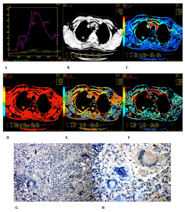
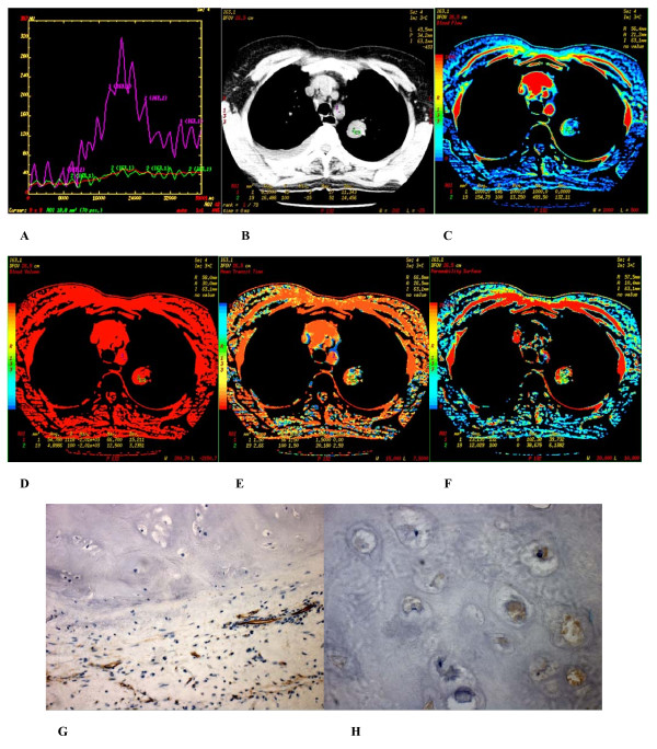
Methods: Sixty-four patients with benign and malignant pulmonary nodules underwent 16-slice spiral CT perfusion imaging. The CT perfusion imaging was analyzed for TDC (time density curve), perfusion parametric maps, and the respective perfusion parameters. Immunohistochemical findings of MVD (microvessel density) measurement and VEGF expression was evaluated.
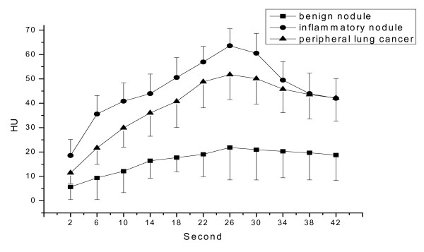
Results: The shape of the TDC of peripheral lung cancer was similar to those of inflammatory nodule. PH (peak height), PHpm/PHa (peak height ratio of pulmonary nodule to aorta), BF (blood flow), BV (blood volume) value of peripheral lung cancer and inflammatory nodule were not statistically significant (all P > 0.05). Both showed significantly higher PH, PHpm/PHa, BF, BV value than those of benign nodule (all P < 0.05). Peripheral lung cancer showed significantly higher PS (permeability surface) value than that of inflammatory nodule and benign nodule (all P < 0.05). BV, BF, PS, MTT, PH, PHpm/PHa, and MVD among three groups of peripheral lung cancers were not significantly (all P > 0.05). In the case of adenocarcinoma, BV, BF, PS, PHpm/PHa, and MVD between poorly and well differentiation and between poorly and moderately differentiation were statistically significant (all P < 0.05). The peripheral lung cancers with VEGF positive expression showed significantly higher PH, PHpm/PHa, BF, BV, PS, and MVD value than those of the peripheral lung cancer with VEGF negative expression, and than those of benign nodule with VEGF positive expression (all P < 0.05). When investigating VEGF negative expression, it is found that PH, PHpm/PHa, and MVD of inflammatory nodule were significantly higher than those of peripheral lung cancer, PS of inflammatory nodule were significantly lower than that of peripheral lung cancer (all P < 0.05). PH, PHpm/PHa, BF, and BV of benign nodule were significantly lower than those of inflammatory nodule (all P < 0.05), rather than PS and MTT (mean transit time) (all P > 0.05). PH, PHpm/PHa, BV, and PS of benign nodule were significantly lower than those of peripheral lung cancer (all P < 0.05). In the case of VEGF positive expression, MVD was positively correlated with PH, PHpm/PHa, BF, BV, and PS of peripheral lung cancer and PS of benign nodule (all P < 0.05).
Conclusion: Multi-slice spiral CT perfusion imaging closely correlated with tumor angiogenesis and reflected MVD measurement and VEGF expression. It provided not only a non-invasive method of quantitative assessment for blood flow patterns of peripheral pulmonary nodules but also an applicable diagnostic method for peripheral pulmonary nodules.
Figures
References
-
- Vaupel P, Kallinowski F, Okunieff P. Blood flow, oxygen and nutrient supply, and metabolic microenvironment of human tumors: a review. Cancer Res. 1989;49:6449–6465. - PubMed
-
- Schaefer JF, Vollmar J, Schick F, Vonthein R, Seemann MD, Aebert H, Dierkesmann R, Friedel G, Claussen CD. Solitary pulmonary nodules: dynamic contrast-enhanced MR imaging-perfusion differences in malignant and benign lesions. Radiology. 2004;232:544–553. doi: 10.1148/radiol.2322030515. - DOI - PubMed
-
- Swensen SJ, Brown LR, Colby TV, Weaver AL, Midthun DE. Lung nodule enhancement at CT: prospective findings. Radiology. 1996;201:447–455. - PubMed
Publication types
MeSH terms
Substances
LinkOut - more resources
Full Text Sources
Medical
Miscellaneous
