. 2008 Sep 1;321(1):101-10.
doi: 10.1016/j.ydbio.2008.06.004. Epub 2008 Jun 13.

barx1 is necessary for ectomesenchyme proliferation and osteochondroprogenitor condensation in the zebrafish pharyngeal arches

Affiliations

barx1 is necessary for ectomesenchyme proliferation and osteochondroprogenitor condensation in the zebrafish pharyngeal arches

Steven M Sperber et al. Dev Biol. .

Abstract

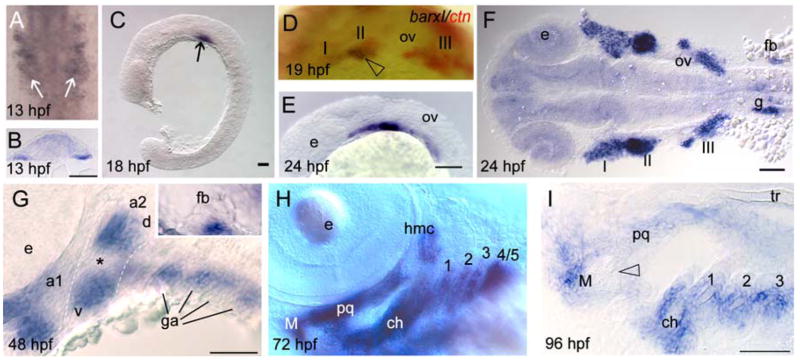
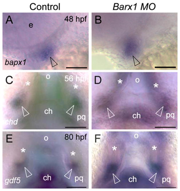
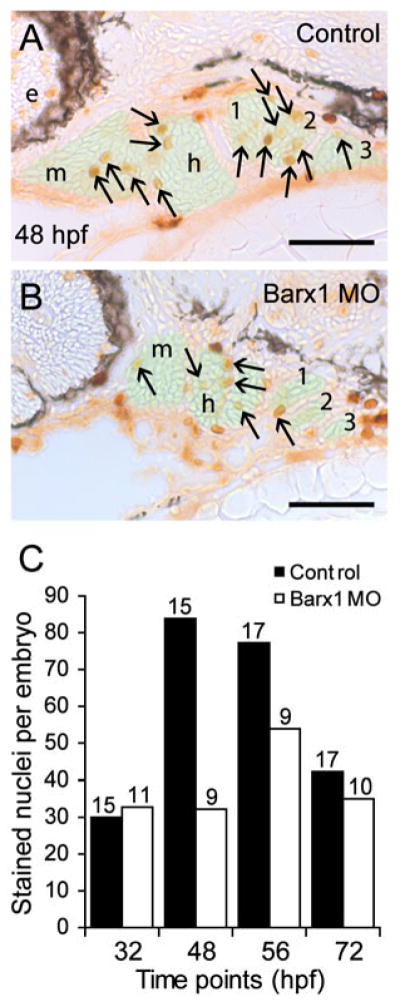
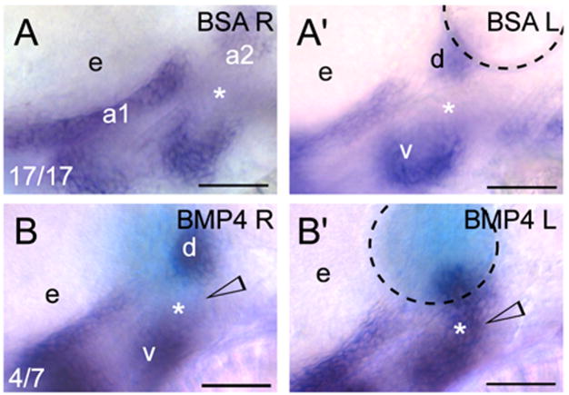
Barx1 modulates cellular adhesion molecule expression and participates in specification of tooth-types, but little is understood of its role in patterning the pharyngeal arches. We examined barx1 expression during zebrafish craniofacial development and performed a functional analysis using antisense morpholino oligonucleotides. Barx1 is expressed in the rhombencephalic neural crest, the pharyngeal arches, the pectoral fin buds and the gut in contrast to its paralogue barx2, which is most prominently expressed in the arch epithelium. Additionally, barx1 transient expression was observed in the posterior lateral line ganglia and developing trunk/tail. We show that Barx1 is necessary for proliferation of the arch osteochondrogenic progenitors, and that morphants exhibit diminished and dysmorphic arch cartilage elements due to reductions in chondrocyte differentiation and condensation. Attenuation of Barx1 results in lost arch expression of osteochondrogenic markers col2a1, runx2a and chondromodulin, as well as odontogenic marker dlx2b. Further, loss of barx1 positively influenced gdf5 and chordin, markers of jaw joint patterning. FGF signaling is required for maintaining barx1 expression, and that ectopic BMP4 induces expression of barx1 in the intermediate region of the second pharyngeal arch. Together, these results indicate an essential role for barx1 at early stages of chondrogenesis within the developing zebrafish viscerocranium.


Figures

Figure 1
Figure 1
Molecular analysis of zebrafish barx1. (A) The coding region of barx1 consists of four exons (boxes, numbered with Roman numerals) containing the homeobox (HB, blue) and the Bar Basic Region (BBR, green); UTR’s are indicated by outlined boxes. Target sites of the barx1 ATG morpholino (Bx MO) and the Splice acceptor site morpholino (Sa MO) are indicated. (B) Phylogenetic tree comparing zebrafish (z) Barx1 with the human (h), mouse (m), and chicken (c) Barx amino acid sequences, as examined using Clustal W. GenBank Accession Nos.: (h1) NM021570, (m1) NM007526, (c1) NM204193, (z1) NM001024949, (h2) NM003658, (m2) NM013800, (c2) NM204896, (z2) XM001342008 respectively. (C) Syntenic relationship between zebrafish chromosome 11 fragment contig: CR548622.8 (Ensembl release 49, March 2008) and the human chromosome 9q22.32 (loci distances indicated in Mb). The barx1 and phf2, PHD finger protein 2, are linked in zebrafish and humans.
Figure 2
Figure 2
Barx1 expression. Embryos at (A, B) 13 hpf, (C)18 hpf, (D) 19 hpf, (E, F) 24 hpf, (G) 48 hpf, (H) 72 hpf, and (I) 96 hpf, were examined by in situ hybridization. (A) barx1 expression in the migrating neural crest in bilateral domains adjacent to the hindbrain (arrows). Anterior is at the top. (B) coronal section through the hindbrain. (C) lateral view of barx1 expression (arrow). (D) double in situ of barx1 (dark blue, arrowhead) and crestin (red) co-expression in the neural crest streams indicated by Roman numerals. (E, F) expression in the cranial neural crest streams. (G, G inset) barx1 expression in the pharyngeal arches and fin bud; a1, first arch; a2, second arch; d, dorsal expression domain; ga, gill arches; v, ventral expression domain; asterisk indicates the a2 intermediate region. (H) lateral oblique view of barx1 expression in the chondrocytes. (I) parasagittal section, open arrowhead indicates jaw joint. ch, ceratohyal; ceratobranchials are numbered; e, eye; fb, fin bud; g, gut primordium; hmc, hyomandibular condensation; M, Meckel’s cartilage; ov, otic vesicle; pq, palatoquadrate; tr, trabecula. Scale bar: (B and I) 50 μm; (C, E, F, and G) 100 μm.
Figure 3
Figure 3
Attenuation of barx1 expression perturbs viscerocranial morphology and cartilage patterning. Larvae injected with (A) control MO (4 ng), and (B and C) Bx MO (4 ng). Alcian Blue staining of control MO (D) and Bx MO injected (E and F) 120 hpf larvae. Dissection of the first and second arches of control (G) and mildly affected larvae (H). (I and J) parasagittal sections of 72 hpf embryos. (J) Bx MO (4 ng) injected compared with (I) control MO injected (4 ng). Anterior is to the left; (A–C, G–J) lateral views, (D–F) ventral view. (A–C) Arrows indicate mouth, and arrowheads the pharyngeal arches; (D–F) open arrowheads indicate the ceratohyal cartilage; (G, H) asterisk, perturbed fusion; black arrowhead, joint fusion between Meckel’s cartilage (M) and the palatoquadrate (pq); cb, ceratobranchials; ch, ceratohyal; ep, ethmoid plate; hm, hyomandibular; ov, otic vesicle; sy, symplectic element. Scale bars: (A–C) 250 μm; (D–H) 100 μm; (J) 50 μm. (K, L) Effectiveness and specificity of the barx1 (Bx) and splice acceptor (Sa) morpholinos. (K) Western analysis of in vivo attenuation of barx1:GFP translation in 24 hpf zebrafish embryos. (L) total RNA extracts (18S and 28S bands shown) used for RT-PCR of barx1. β-actin used as a control. (M) Percentage of affected embryos comparing control MO, Bx MO and Sa MO, and rescue by co-injection of barx1 mRNA with a 5-bp mismatch to the ATG target site. Affected embryos are defined as those exhibiting micrognathia as a result of a reduction or loss of arch cartilage elements, as seen by Alcian Blue staining at 120 hpf. (1) Control MO 4 ng; (2) Bx MO 4 ng; (3) Sa MO 4 ng; (4) Bx MO 2 ng + Sa MO 2 ng. Rescue: (5) gfp mRNA 25 pg; (6) barx1 mRNA 25 pg; (7) Bx MO + Sa MO 2 ng each + barx1 mRNA 25 pg. Number of embryos indicated on top of each bar.
Figure 4
Figure 4
Initial patterning of the arches is maintained in barx1 morphants. (A, C, E and G) control MO injected embryos, (B, D, F and H) embryos co-injected with Bx MO + Sa MO, 2 ng each. Neural crest expression of inca (A, B) and dlx2a (C and D) at 24 hpf. (E, F) neurod expression at 24 hpf in neural ganglia precursors; (G and H) hand2 expression in ventral endoderm at 32 hpf. Neural crest streams marked in Roman numerals, gill arches indicated by numbers; e, eye; m, mandibular arch; h, hyoid arch; ov, otic vesicle; ad/av/f, anterodorsal/anteroventral lateral line/facial placode/ganglia; o, octavel/statoacoustic ganglia precursors; p, posterior lateral line placode/ganglion. Scale bar: 100 μm.
Figure 5
Figure 5
Barx1 is necessary for expression of chondrogenic markers within the pharyngeal arches. (A, C, E, G, I and K) control embryos; (B, D, F, H, J and L) barx1 morphant embryos (see Fig. 4). Whole-mount in situ hybridization for (A, B) goosecoid (gsc), (C, D) sox9a, (E, F) runx2b, (G, H) type II collagen (col2a1), (I, J) chondromodulin (chm), and (K, L) dlx2b. Anterior is to the left; open arrowheads indicate sites of altered marker expression in the morphants; bcc, basicranial commissures; cb, ceratobranchials; ch, ceratohyal; cl, cleithrum; d, diencephalon; ep, ethmoid plate; fb, fin bud; hsc, hyosymplectic condensation; ov, otic vesicle; pq, palatoquadrate; t, telencephalon. Scale bar: 100 μm.
Figure 6
Figure 6
Loss of barx1 enhances chordin and gdf5 expression in the jaw joint Whole-mount in situ hybridization for (A, B) bapx1, (C, D) chordin (chd), and (E, F) gdf5. (A, C, E) control uninjected embryos; (B, D, F) barx1 morphants (see Fig. 4). Embryonic ages as indicated. (A, B) lateral view, anterior to the left. (C–F) ventral view, anterior at the top; asterisk indicates Meckel’s cartilage, open arrowheads indicate jaw joint expression domains; e, eye; ch, ceratohyal; o orifice; pq, palatoquadrate. Scale bar: (A–F) 50 μm.
Figure 7
Figure 7
Barx1 positively influences cellular proliferation within the pharyngeal arches. (A, B) Sagittal serial sections (10 μm) of the pharyngeal arch region stained with Phosphohistone-H3 antibody. (A) Control embryo at 48 hpf. (B) barx1 morphant. The neural crest derived mesenchyme is overlaid in green; arrows indicate stained nuclei; e, eye; m, mandibular arch; h, hyoid arch; gill arches are numbered. Anterior is to the left. Scale bar: 50 μm. (C) Averaged cell proliferation in control uninjected embryos (black bars) and barx1 morphants (see Fig. 4; white bars) was determined within the mesenchyme as shown in (A,B) at the indicated four time points. Approximately fifty serial sections per embryo were examined. Number of embryos examined is indicated above each bar. Scale bar: (A, B) 50 μm.
Figure 8
Figure 8
Barx1 expression in fgf mutant embryos. (A, B) Wild-type sibling and lia/fgf3 mutant. Arrows indicate sites of barx1 expression in the gill arches. (C, D and E) Wild-type sibling and left and right views of an ace/fgf8 mutant embryo. Left/right asymmetric expression indicated in the mutant (arrowhead); asterisk indicates reduced expression in the third gill arch. (F, G) Overstained control (DMSO) and embryo treated with the FGF receptor inhibitor SU5402 from 24 hpf to 48 hpf. Lateral views with anterior to the left, panel (D) was flipped for comparison; d, dorsal and v, ventral expression domains of the second arch; e, eye; gill arches are numbered; g, gut primordium; h, hyoid arch; m, mandibular arch; ov, otic vesicle. Scale bar: (A–G) 50 μm.
Figure 9
Figure 9
Exogenous BMP signaling influences barx1 expression within the zebrafish pharyngeal arches. (A, B) Whole-mount in situ hybridization of barx1 in the left (bead implanted) and right (control) side of single embryos. Right-side images were flipped for easier comparison. (A, A′) Control BSA white bead, (B, B′) BMP4 blue bead. Anterior is to the left; d and v, dorsal and ventral expression domains in the second arch; e, eye. Arrowhead indicates sites of barx1 misexpression; asterisk indicates intermediate region, normally devoid of barx1 expression at this stage. Number of affected embryos over the total embryos with successful bead implantation is indicated in the panels (A, B). Black dashed line outlines the bead. Scale bar: 50 μm.

Similar articles

Cited by

References

    1. Abu-Issa R, Smyth G, Smoak I, Yamamura K-i, Meyers EN. Fgf8 is required for pharyngeal arch and cardiovascular development in the mouse. Development. 2002;129:4613–4625. - PubMed
    1. Akimenko MA, Ekker M, Wegner J, Lin W, Westerfield M. Combinatorial expression of three zebrafish genes related to distal-less: part of a homeobox gene code for the head. J Neurosci. 1994;14:3475–86. - PMC - PubMed
    1. Albertson RC, Yelick PC. Roles for fgf8 signaling in left-right patterning of the visceral organs and craniofacial skeleton. Dev Biol. 2005;283:310–21. - PubMed
    1. Angelo S, Lohr J, Lee KH, Ticho BS, Breitbart RE, Hill S, Yost HJ, Srivastava D. Conservation of sequence and expression of Xenopus and zebrafish dHAND during cardiac, branchial arch and lateral mesoderm development. Mech Dev. 2000;95:231–7. - PubMed
    1. Barlow AJ, Bogardi JP, Ladher R, Francis-West PH. Expression of chick Barx-1 and its differential regulation by FGF-8 and BMP signaling in the maxillary primordia. Dev Dyn. 1999;214:291–302. - PubMed

Publication types