Comparative Study
. 2008 Oct;213(2):257-67.
doi: 10.1016/j.expneurol.2008.05.025. Epub 2008 Jun 12.

Activation of p38 MAP kinase is involved in central neuropathic pain following spinal cord injury

Affiliations
Comparative Study

Activation of p38 MAP kinase is involved in central neuropathic pain following spinal cord injury

Eric D Crown et al. Exp Neurol. 2008 Oct.

Abstract

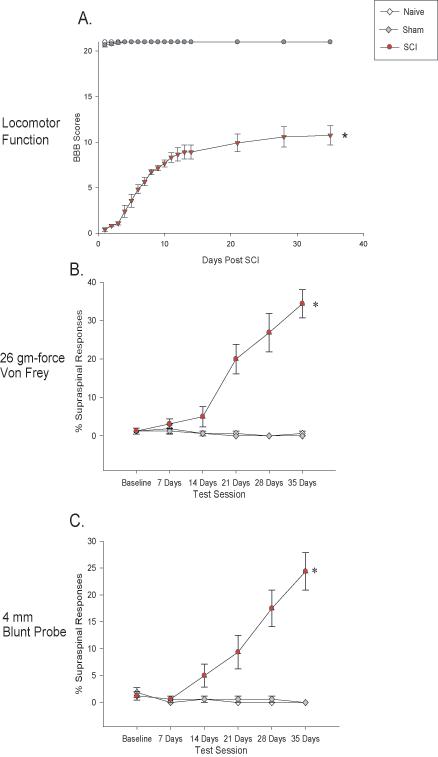
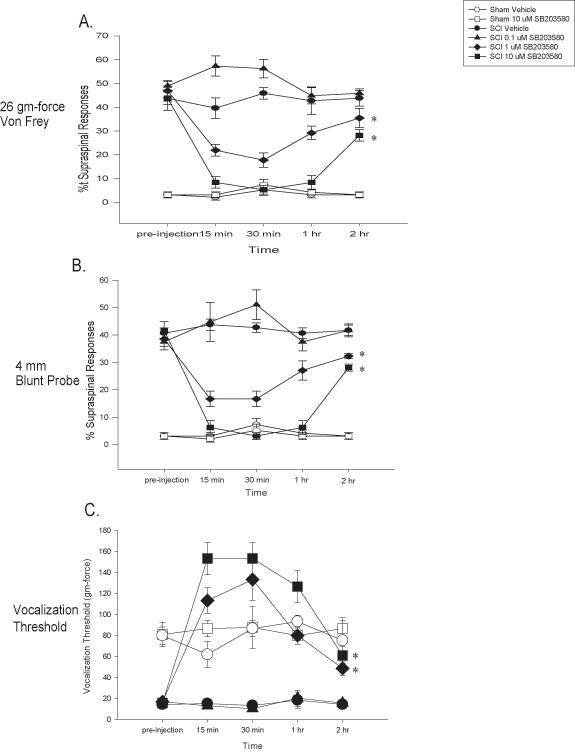
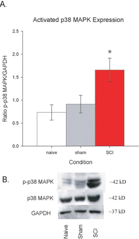
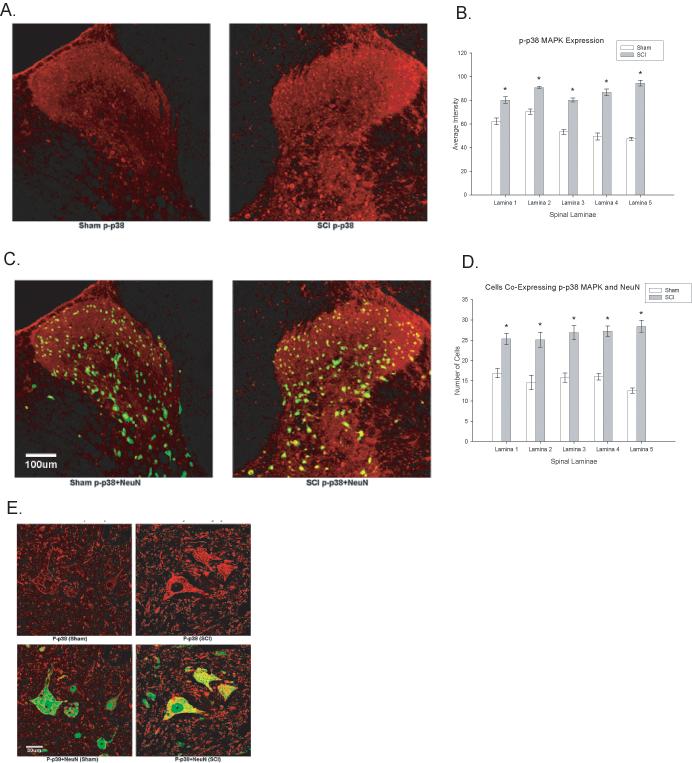
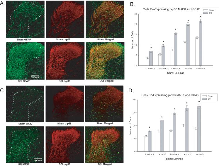
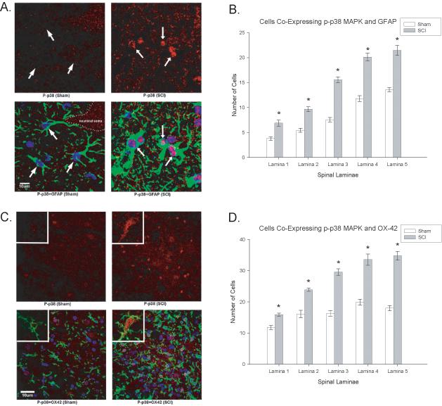
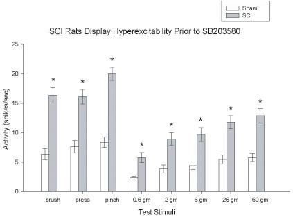
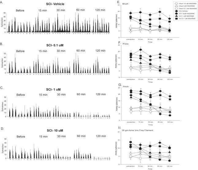
Recent work regarding chronic central neuropathic pain (CNP) following spinal cord injury (SCI) suggests that activation of key signaling molecules such as members of the mitogen activated protein kinase (MAPK) family play a role in the expression of at-level mechanical allodynia. Previously, we have shown that the development of at-level CNP following moderate spinal cord injury is correlated with increased expression of the activated (and thus phosphorylated) forms of the MAPKs extracellular signal related kinase and p38 MAPK. The current study extends this work by directly examining the role of p38 MAPK in the maintenance of at-level CNP following spinal cord injury. Using a combination of behavioral, immunocytochemical, and electrophysiological measures we demonstrate that increased activation of p38 MAPK occurs in the spinal cord just rostral to the site of injury in rats that develop at-level mechanical allodynia after moderate SCI. Immunocytochemical analyses indicate that the increases in p38 MAPK activation occurred in astrocytes, microglia, and dorsal horn neurons in the spinal cord rostral to the site of injury. Inhibiting the enzymatic activity of p38 MAPK dose dependently reverses the behavioral expression of at-level mechanical allodynia and also decreases the hyperexcitability seen in thoracic dorsal horn neurons after moderate SCI. Taken together, these novel data are the first to demonstrate causality that increased activation of p38 MAPK in multiple cell types play an important role in the maintenance of at-level CNP following spinal cord injury.


Figures

Figure 1
Figure 1
Recovery of locomotor function and development of at-level mechanical allodynia following contusive SCI. The recovery of locomotor function (as assessed using the BBB Score) following 150 kdyne, 1 s dwell time impact using the Infinite Horizons device. SCI rats displayed significant impairment in locomotor function relative to naïve and sham rats (panel A, p < 0.0001). The percentage of supraspinal nociceptive responses (y axis) made by naïve (Naïve), sham (Sham), and spinal injured rats (SCI) tested weekly for 35 days post injury is graphed. The SCI rats showed a significant increase in supraspinal responses to both von Frey stimulation (panel B, *p < 0.0001) and stimulation with a 4 mm blunt probe (panel C, *p < 0.0001).
Figure 2
Figure 2
Inhibition of p38 MAPK activation reverses at-level mechanical allodynia after SCI. The inhibitor of p38 MAPK activation, SB203580, dose dependently reversed at-level mechanical allodynia when subjects are tested at 35 days post injury. The percentage of supraspinal nociceptive responses (y axis) made by sham (Sham), and spinal injured rats (SCI) tested is graphed. Prior to SB203580 injection or vehicle, SCI rats displayed at-level mechanical allodynia (as evidenced by an increased percentage of supraspinal nociceptive responses). SB203580 dose dependently reversed this at-level mechanical allodynia to both von Frey (panel A, * p < 0.0001) and blunt probe (panel B, * p < 0.0001) stimulation. Panel C demonstrates that SB203580 dose dependently reversed decreases in vocalization thresholds to mechanical stimuli at the level of the spinal cord injury (* p < 0.0001). In all cases, SB203580 had no effect on sham rats. All data are represented as mean ± SEM.
Figure 3
Figure 3
Activated p38 MAPK is upregulated in the spinal cord by 35 days post injury. A. Western blot data from naïve, sham, and SCI rats (normalized to GAPDH expression) indicated that SCI rats had significantly greater levels of activated p38 MAPK than the other groups (*p < 0.0001). B. Representative Western blots for p-p38 MAPK (top panel) and total p38 MAPK (middle panel), with GAPDH (bottom panel) included as the loading control. All data are represented as mean ± SEM.
Figure 4
Figure 4
Spinal cord injury increases activated p38 MAPK expression within the spinal cord. A. Example of phosphorylated p38 MAPK (p-p38 MAPK) expression in sham (left panel) and SCI (right panel) rats. B. Quantification of the significant increase in p-p38 MAPK expression in laminae I-V following SCI (p < 0.05). C. Colocalization of NeuN with p-p38 MAPK in the spinal cord dorsal horn of sham (left panel) and SCI (right panel) rats. D. Quantification of the significant increase in p-p38 MAPK expression in the neurons following SCI (p < 0.05). E. High magnification images of NeuN positive cells in sham (left panels) and SCI (right panels) rats to illustrate differences in p-p38 MAPK expression. All data are represented as mean ± SEM.
Figure 5
Figure 5
Activated p38 MAPK expression is seen in astrocytes and microglia of spinal cord injured rats that develop at-level neuropathic pain. Spinal cord injury increased expression of GFAP in astrocytes (Figure 5A, left panels) and significantly increased the expression of OX-42 in microglia (Figure 5C, left panels). Histogram density measures of immunofluorescent staining of GFAP (Figure 5B) and OX-42 Figure 5D) indicated significantly increased expression in laminae of the dorsal horn just rostral to the spinal injury. Colocalization with activated p38 MAPK was significantly upregulated and colocalized with astrocytes (Figure 5A, right panels) and microglia (Figure 5B, right panels) in the dorsal horn of injured (right panels) rats relative to sham (left panels) rats (*p < 0.0001). The colocalization is evident at the cellular level in Figure 6.
Figure 6
Figure 6
SCI demonstrates higher magnification of increased cellular localization of activated p38 (p-p38, red) in both astrocytes (Figure 6A, GFAP positive, green) and microglia (Figure 6B, OX-42 positive, green). Note that the p-p38 is increased in the nuclear compartment of both cell types. Quantification of the significant increases in p-p38 MAPK in astrocytes (GFAP, 6B) and microglia (OX-42, 6D) in the dorsal horn laminae of SCI rats relative to sham rats (*p<0.001). Quantification of the significant increases in p-p38 MAPK colocalization with GFAP (6B) and OX-42 (6D) positive cells (astrocytes and microglia, respectively) in the dorsal horn laminae of SCI rats relative to sham rats (*p < 0.001). All data are represented as mean ± SEM.
Figure 7
Figure 7
SCI produces dorsal horn neuronal hyperexcitability at the site of injury. SCI significantly increased evoked neuronal responses to brush, press, pinch and graded von Frey stimuli relative to sham rats (*p < 0.001) as demonstrated by the histograms of dorsal horn neuronal responses in spikes/sec. All data are represented as mean ± SEM.
Figure 8
Figure 8
Inhibition of p38 MAPK activation significantly decreases background activity and evoked neuronal responses to brush, press, pinch, and graded von Frey stimulation at the level of SCI. A-D Spike frequency histograms of neuronal activity to brush, press, pinch and von Frey stimuli prior to SB203580 administration, and at 15, 30, 60, and 120 minutes after vehicle (A), 0.1 uM (B), 1 uM (C), or 10 uM (D) SB203580 administration. Mean activity (± SEM) for background and responses to brush (E), press (F), and pinch (G) for sham (open symbols) and SCI (filled symbols) rats given vehicle or SB203580 over the 120 minute recording period. SB203580 not only significantly depressed background activity but also dose dependently produced significant decreases in neuronal responding to brush, press, and pinch (*p < 0.001). H. Mean activity (± SEM) for responding to von Frey stimuli applied to the skin immediately rostral to the site of SCI for the 120 minutes following drug administration. SB203580 dose dependently depressed neuronal responding to the 2 gm, 6 gm, 26 gm, and 60 gm von Frey stimuli (*p < 0.001). For ease of presentation, only data for the 26 gm von Frey filament (the same filament used in the behavioral studies) is shown. All data are represented as mean ± SEM.

References

    1. Attal N, Guirimand F, Brasseur L, Gaude V, Chauvin M, Bouhassira D. Effects of IV morphine in central pain: a randomized placebo-controlled study. Neurol. 2002;58:554–563. - PubMed
    1. Basso DM, Beattie MS, Bresnahan JC. A sensitive and reliable locomotor rating scale for open field testing in rats. J Neurotrauma. 1995;12:1–21. - PubMed
    1. Bigbee AJ, Hoang TX, Havton LA. At-level neuropathic pain is induced by lumbosacral ventral root avulsion injury and ameliorated by root reimplantation into the spinal cord. Exp Neurol. 2007;204:273–282. - PMC - PubMed
    1. Boyle DL, Jones TL, Hammaker D, Svensson CI, Rosengren S, Albani S, Sorkin L, Firestein GS. Regulation of peripheral inflammation by spinal p38 MAP kinase in rats. PLoS Med. 2006;3:e338. - PMC - PubMed
    1. Bu X, Huang P, Qi Z, Zhang N, Han S, Fang L, Li J. Cell type-specific activation of p38 MAPK in the brain regions of hypoxic preconditioned mice. Neurochem Int. 2007;51:459–466. - PubMed

Publication types

MeSH terms