. 2008 Sep 12;381(4):928-40.
doi: 10.1016/j.jmb.2008.06.038. Epub 2008 Jun 20.

The SOCS box domain of SOCS3: structure and interaction with the elonginBC-cullin5 ubiquitin ligase

Affiliations

The SOCS box domain of SOCS3: structure and interaction with the elonginBC-cullin5 ubiquitin ligase

Jeffrey J Babon et al. J Mol Biol. .

Abstract

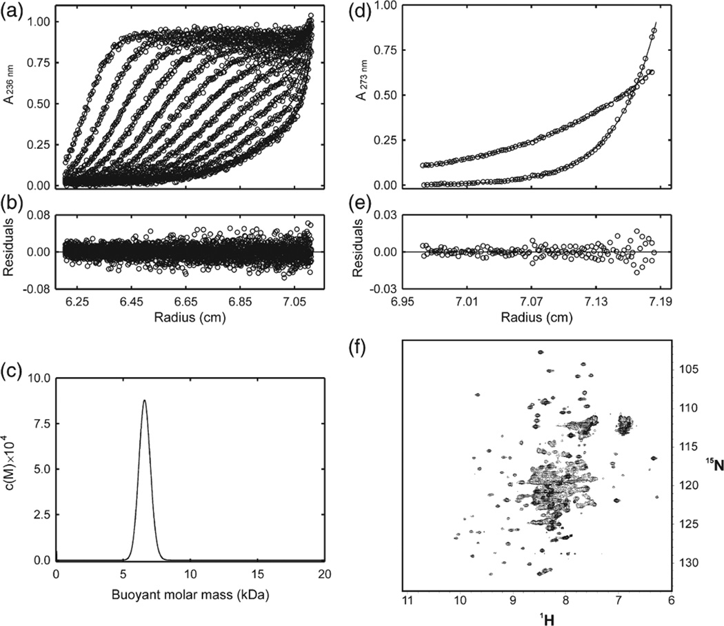
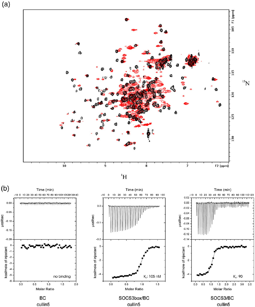
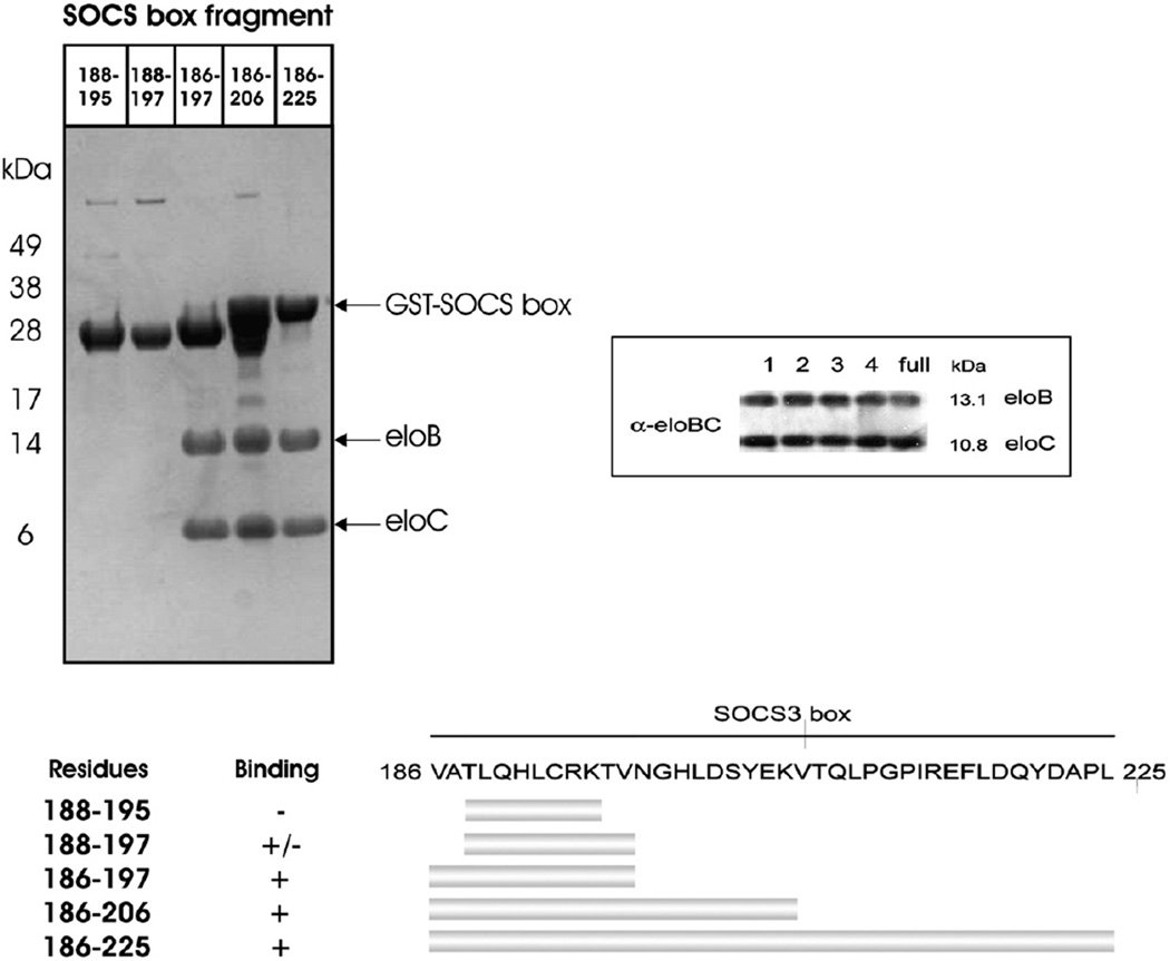
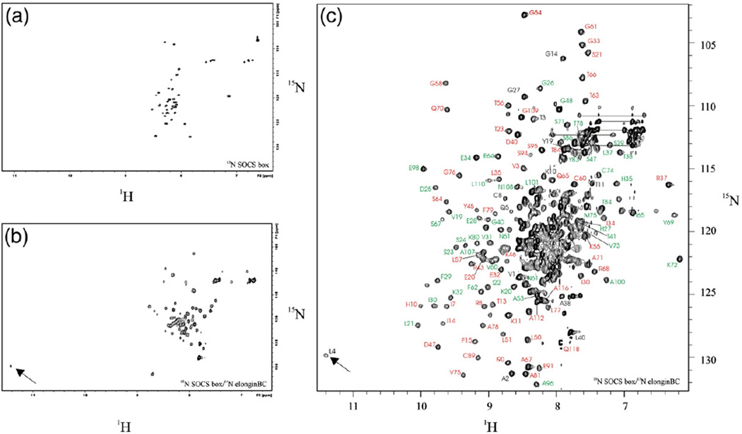
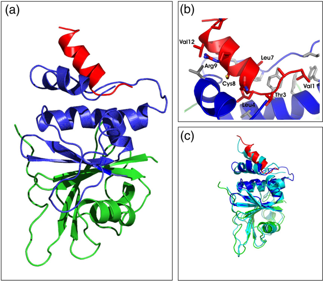
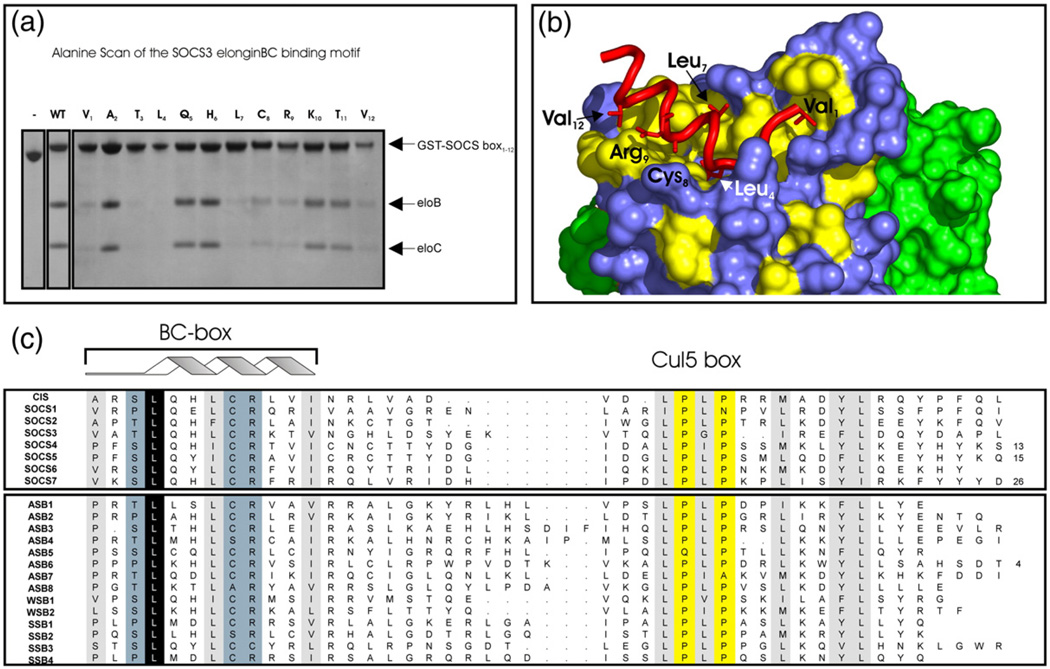
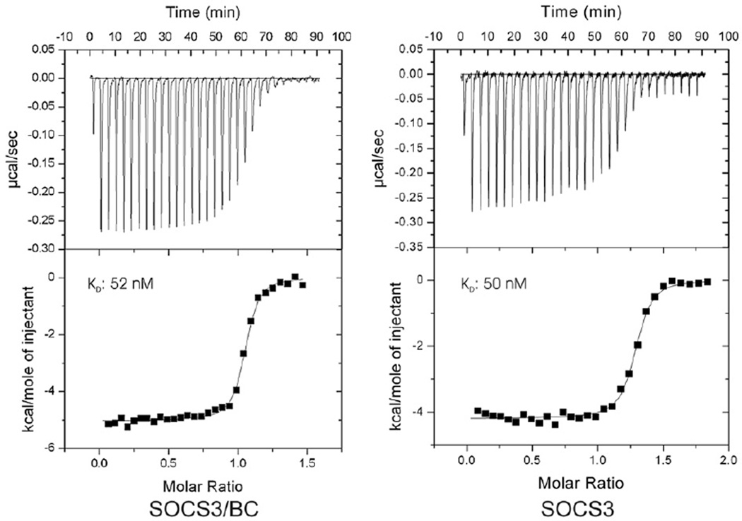
Suppressor of cytokine signalling 3 (SOCS3) is responsible for regulating the cellular response to a variety of cytokines, including interleukin 6 and leukaemia inhibitory factor. Identification of the SOCS box domain led to the hypothesis that SOCS3 can associate with functional E3 ubiquitin ligases and thereby induce the degradation of bound signalling proteins. This model relies upon an interaction between the SOCS box, elonginBC and a cullin protein that forms the E3 ligase scaffold. We have investigated this interaction in vitro using purified components and show that SOCS3 binds to elonginBC and cullin5 with high affinity. The SOCS3-elonginBC interaction was further characterised by determining the solution structure of the SOCS box-elonginBC ternary complex and by deletion and alanine scanning mutagenesis of the SOCS box. These studies revealed that conformational flexibility is a key feature of the SOCS-elonginBC interaction. In particular, the SOCS box is disordered in isolation and only becomes structured upon elonginBC association. The interaction depends upon the first 12 residues of the SOCS box domain and particularly on a deeply buried, conserved leucine. The SOCS box, when bound to elonginBC, binds tightly to cullin5 with 100 nM affinity. Domains upstream of the SOCS box are not required for elonginBC or cullin5 association, indicating that the SOCS box acts as an independent binding domain capable of recruiting elonginBC and cullin5 to promote E3 ligase formation.


Figures

Fig. 1
Fig. 1
NMR and analytical ultracentrifugation analysis of elonginBC. (a) Radial profiles of 0.92 mg/mL elonginBC monitored at 236 nm during centrifugation at 40,000 rpm (○). The sedimentation velocity data were analyzed in terms of a continuous buoyant molar mass distribution (−) using the continuous mass distribution model [c(M)] resident in the program SEDFIT. Ninety scans were used in the analysis; however, for clarity, only every seventh is presented. (b) Plot of residuals for the analysis of the experimental data in (a). (c) c(M) distribution describing the experimental data in (b). The distribution exhibits a single, well-defined maximum corresponding to a buoyant (reduced) molar mass of 6.5 kDa. This corresponds to a physical mass of 24.5 kDa under the conditions used. The predicted mass of a 1:1 complex is 25.6 kDa. (d) Radial profiles of 0.92 mg/mL elonginBC monitored at 273 nm after centrifugation for 24 h at 20,000 and at 35,000 rpm. The sedimentation equilibrium data were globally analyzed in terms of a single ideal solute (−) using the species analysis model resident in the program SEDPHAT. The buoyant molar mass recovered from the analysis was 6.7 kDa. This corresponds to a physical mass of 25.1 kDa under the conditions used. (e) Plot of residuals for the analysis of the experimental data in (d). (f) 1H–15N HSQC analysis of elonginBC. Recombinant elonginBC, 0.25 mM in 20 mM potassium phosphate, 2.5 mM DTT, pH 6.7, was recorded at 310 K on a Bruker Avance 500 spectrometer. Note the concentration of broad overlapped peaks in the centre of the spectrum indicating conformational exchange.
Fig. 2
Fig. 2
SOCS3 and elonginBC forms a stable ternary complex that binds tightly to cullin5. Overlay of the 15N HSQC spectrum of elonginBC alone shown in red with that of elonginBC (15N labelled) in complex with the SOCS3 box (unlabelled) shown in black. The broad peaks in the centre of the elonginBC-alone spectrum are shifted and well resolved in the ternary complex, indicating little, if any, conformational exchange once the SOCS box is bound. (b) ITC analysis of the SOCS3 SOCS box–elonginBC–cullin5 interaction. GST–cullin5 (80 µM, NTD) was titrated into 10 µM elonginBC, SOCS3box/elonginBC or SOCS3/elonginBC in 30×10-µL injections using a VP-ITC unit (Microcal). Both protein and peptide were prepared in 20 mM potassium phosphate and 100 mM NaCl supplemented with 2 mM 2-mercaptoethanol. Only the SOCS3box–elonginBC and SOCS3–elonginBC ternary complexes bound to cullin5 with measurable affinity. These titration curves fitted well to a single-site model with a Kd of 105±10 nM, ΔH −3700±500 cal/mol, N=0.96 and Kd of 90±10 nM, ΔH −4100±200 cal/mol, N=1.05, respectively (average of duplicate experiments).
Fig. 3
Fig. 3
Determination of the minimal elonginBC binding epitope on the SOCS3 SOCS box. The SOCS box fragments were produced as GST fusions to aid stability during expression and co-expressed with elonginBC. Glutathione Sepharose was used to pull down GST-labelled proteins present in the cell lysate (upper left panel). As shown, a fragment comprising residues 1–12 was the smallest segment of the SOCS box capable of co-purifying with elonginBC with similar affinity to the entire SOCS box (1–40). The successful co-expression of elonginBC in all cultures was shown by Western blot (upper right panel) and a schematic view of the mutants used and the results are also shown (bottom panel).
Fig. 4
Fig. 4
The SOCS boxis unstructured inisolation and becomes partially structured upon elonginBC association. 1H–15N HSQC spectra of the SOCS box, in isolation and bound to elonginBC. (a) Recombinant 15N-labelled SOCS3 SOCS box was prepared by thrombin digestion of a GST–SOCS box fusion protein. 15N HSQC spectrum of 0.1 mM protein in 20 mM potassium phosphate and 2.5 mM DTT, pH 6.7, was recorded at 310 K and 500 MHz. (b) The sample from (a) was mixed with a threefold excess of unlabelled SOCS box–elonginBC ternary complex and HSQC analysis was performed as before. The 15N-labelled SOCS box was able to compete with the unlabelled domain in the ternary complex and thus bind elonginBC. The downfield amide peak at 11.3 ppm arises from Leu4 of the SOCS box (arrow). The range of peak intensities seen is a further indication of conformational exchange. (c) Backbone amide assignment of the SOCS box–elonginBC ternary complex. HSQC analysis of a fully 15N labelled ternary complex was performed using conditions identical to that of (a) and (b). SOCS box assignments are shown in black, elonginC assignments in green and elonginB assignments in red. Some assignments are omitted for clarity.
Fig. 5
Fig. 5
Tertiary structure of an SOCS3 SOCS box–elonginBC complex. (a) Ribbon diagram of the SOCS3 SOCS box (red) in complex with elonginC (blue) and elonginB (green). The interaction of the SOCS box occurs exclusively with elonginC and is mediated mostly by hydrophobic interactions. ElonginBC forms a tightly associated complex with the core of the association being a continuous β-sheet formed by residues from both proteins. A number of side-chain hydrophobic interactions further stabilise the complex. (b) Close view of the SOCS box elonginC interface with hydrophobic side chains from the SOCS box (red) labelled. The hydrophobic residues from elonginC (blue) that form the interface are shown in grey stick representation. (c) Ribbon diagram of the SOCS3 SOCS box in complex with elonginBC [same color scheme as in (a)] shown overlaid on the SOCS2/elonginBC structure (cyan).
Fig. 6
Fig. 6
Key residues required for elonginBC binding. An Ala scan of the SOCS3 SOCS box to determine residues required for elonginBC binding was performed. Co-expression of 12 SOCS box domain constructs, each containing a single Ala mutation, with elonginBC was performed in E. coli, and glutathione Sepharose was used to pull down GST-labelled proteins present in the cell lysate. (a) The following mutations completely interfered with elonginBC binding: Val1, Thr3, Leu4, Leu7, Cys8, Arg9 and Val12. Of these, only the L4A mutation completely abolished binding. (c) Residues identified by Ala scan are highlighted on a surface representation of elonginBC where hydrophobic residues on the surface of elonginC are shown in yellow. The BC box of SOCS3 (red) is shown in cartoon representation with important side chains displayed in “stick” representation. ElonginC is shown in blue and elonginB in green.
Fig. 7
Fig. 7
Isothermal titration analyses of the SOCS3–gp130 interaction in the presence and absence of elonginBC. ITC analyses show that a ternary SOCS–elonginBC complex is able to bind a tyrosine-phosphorylated peptide from the gp130 receptor with similar affinity to SOCS3 alone. Phosphopeptide (160 µM) was titrated into 20 µM SOCS3/elonginBC (left) or 20 µM SOCS3 (right) in 30×10-µL injections using a VP-ITC unit (Microcal). Both protein and peptide were prepared in Tris-buffered saline supplemented with 2 mM 2-mercaptoethanol. The titration curves fitted well to a single-site model with a Kd of 50±4 nM, ΔH −5030±30 kcal/mol, N 1.03 (SOCS3/BC) and Kd of 52±7 nM, ΔH −4200±30 kcal/mol, N 1.26. SOCS3, both as an isolated protein and in complex with elonginBC, lacked the PEST motif.

References

    1. Starr R, Willson TA, Viney EM, Murray LJ, Rayner JR, Jenkins BJ, et al. A family of cytokine-inducible inhibitors of signalling. Nature. 1997;387:917–921. - PubMed
    1. Leonard WJ, O’Shea JJ. Jaks and STATs: biological implications. Annu. Rev. Immunol. 1998;16:293–322. - PubMed
    1. Kimura A, Kinjyo I, Matsumura Y, Mori H, Mashima R, Harada M, et al. SOCS3 is a physiological negative regulator for granulopoiesis and granulocyte colony-stimulating factor receptor signaling. J. Biol. Chem. 2004;279:6905–6910. - PubMed
    1. Croker BA, Metcalf D, Robb L, Wei W, Mifsud S, DiRago L, et al. SOCS3 is a critical physiological negative regulator of G-CSF signaling and emergency granulopoiesis. Immunity. 2004;20:153–165. - PubMed
    1. Roberts AW, Robb L, Rakar S, Hartley L, Cluse L, Nicola NA, et al. Placental defects and embryonic lethality in mice lacking suppressor of cytokine signaling 3. Proc. Natl Acad. Sci. USA. 2001;98:9324–9329. - PMC - PubMed

Publication types

MeSH terms

Associated data

LinkOut - more resources