. 2008 Sep 12;381(4):956-74.
doi: 10.1016/j.jmb.2008.06.041. Epub 2008 Jun 20.

Involvement of the second extracellular loop and transmembrane residues of CCR5 in inhibitor binding and HIV-1 fusion: insights into the mechanism of allosteric inhibition

Affiliations

Involvement of the second extracellular loop and transmembrane residues of CCR5 in inhibitor binding and HIV-1 fusion: insights into the mechanism of allosteric inhibition

Kenji Maeda et al. J Mol Biol. .

Abstract

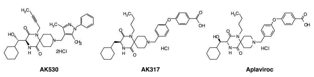
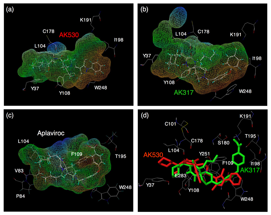
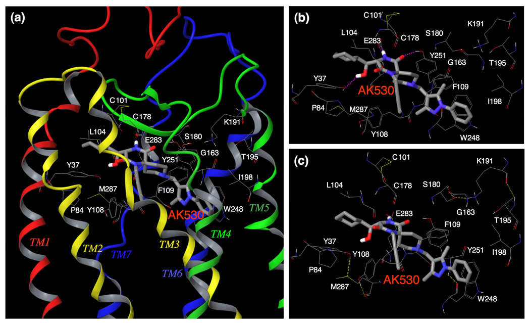
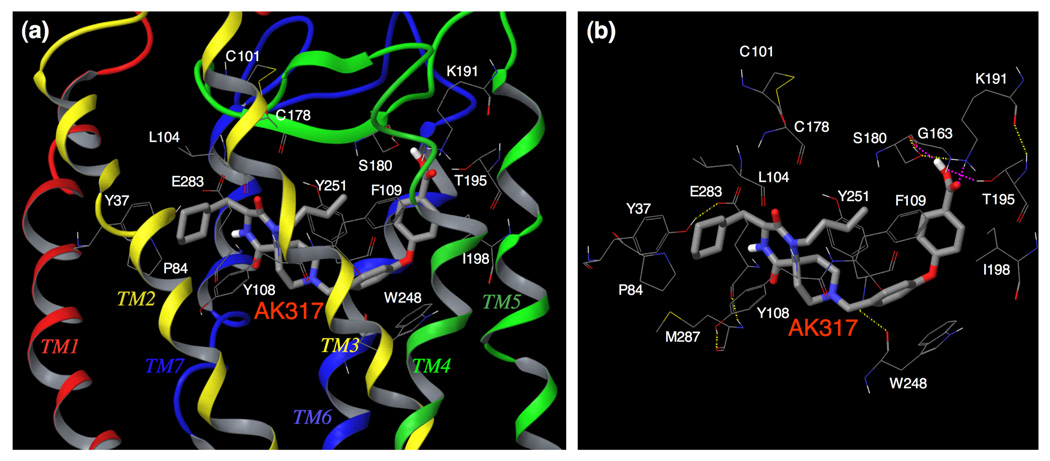
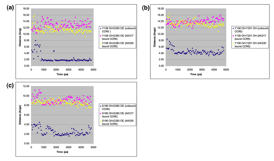
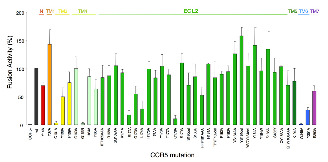
C-C chemokine receptor 5 (CCR5), a member of G-protein-coupled receptors, serves as a coreceptor for human immunodeficiency virus type 1 (HIV-1). In the present study, we examined the interactions between CCR5 and novel CCR5 inhibitors containing the spirodiketopiperazine scaffolds AK530 and AK317, both of which were lodged in the hydrophobic cavity located between the upper transmembrane domain and the second extracellular loop (ECL2) of CCR5. Although substantial differences existed between the two inhibitors--AK530 had 10-fold-greater CCR5-binding affinity (K(d)=1.4 nM) than AK317 (16.7 nM)-their antiviral potencies were virtually identical (IC(50)=2.1 nM and 1.5 nM, respectively). Molecular dynamics simulations for unbound CCR5 showed hydrogen bond interactions among transmembrane residues Y108, E283, and Y251, which were crucial for HIV-1-gp120/sCD4 complex binding and HIV-1 fusion. Indeed, AK530 and AK317, when bound to CCR5, disrupted these interhelix hydrogen bond interactions, a salient molecular mechanism enabling allosteric inhibition. Mutagenesis and structural analysis showed that ECL2 consists of a part of the hydrophobic cavity for both inhibitors, although AK317 is more tightly engaged with ECL2 than AK530, explaining their similar anti-HIV-1 potencies despite the difference in K(d) values. We also found that amino acid residues in the beta-hairpin structural motif of ECL2 are critical for HIV-1-elicited fusion and binding of the spirodiketopiperazine-based inhibitors to CCR5. The direct ECL2-engaging property of the inhibitors likely produces an ECL2 conformation, which HIV-1 gp120 cannot bind to, but also prohibits HIV-1 from utilizing the "inhibitor-bound" CCR5 for cellular entry--a mechanism of HIV-1's resistance to CCR5 inhibitors. The data should not only help delineate the dynamics of CCR5 following inhibitor binding but also aid in designing CCR5 inhibitors that are more potent against HIV-1 and prevent or delay the emergence of resistant HIV-1 variants.


Figures

Fig. 1
Fig. 1
Structures of small molecule CCR5 inhibitors, AK530, AK317, and aplaviroc (APL).
Fig. 2
Fig. 2. The trans-membrane helices and extra-cellular loop regions of CCR5
(a) A side-view of the TM domains. TM2, TM3, and TM4 are above the plane of the paper, and TM6 and TM7 are below the plane. The following assignments have been made for TM helices: TM1, residues 27 to 57; TM2, residues 64 to 93; TM3, residues 97 to 130; TM4, residues 142 to 165; TM5, residues 191 to 219; TM6, residues 232 to 259; TM7, residues 279 to 300. (b) A top-view of the extra-cellular loop regions. The following assignments have been made for loops: N-terminus, 1 to 26; ECL1, residues 94 to 96; ECL2, residues 166 to 190; ECL3, residues 260 to 278.
Fig. 3
Fig. 3
Transmembrane residues Y108(TM3) / E283(TM7) / Y251(TM6) / M287(TM7) / S180(ECL2) of unliganded CCR5, forming a hydrogen bond network. Y37 has hydrogen bond interaction with H175. This shows that transmembrane residues, implicated in CCR5 inhibitor binding, have direct interactions with ECL2 residues in the unliganded receptor. It is assumed that these intramolecular interactions are responsible for maintaining a conformation of ECL2 that is favorable for binding with HIV-1 gp120/CD4 complex. AK530 interacts with Y37, Y108, E283, and Y251 in the binding cavity and disrupts the intramolecular interactions of Y108 and Y251 with E283, and those of E283 with the ECL2 (Fig. 5). It is assumed that these structural changes, after inhibitor binding, alter the conformation of the ECL2, resulting in loss of association with gp120. The analysis strongly suggests that the loss of hydrogen bonds between helices may cause allosteric conformational changes in ECL2 leading to the inhibition of HIV-1 gp120 binding to CCR5.
Fig. 4
Fig. 4
Lipophilic potential mapped on the binding cavity of CCR5 inhibitors. The lipophilic potential mapped onto the binding cavity when AK530, AK317, and APL bind to CCR5 are shown in (a), (b), and (c), respectively. The predominantly lipophilic region of the cavity is shown in brown (bottom region of the cavity which is towards the cytoplasmic region of CCR5). The blue regions are predominantly hydrophilic (present towards the extra-cellular region), and the green regions are moderately lipophilic. The figure was generated using MOLCAD. The shape of the binding cavity is slightly different for AK530 and AK317 as the receptor conformations are slightly different when these molecules bind to CCR5. The unoccupied volumes of the cavities suggest optimization ideas for improving the potency of these molecules. (d) The binding modes of AK530 and AK317 superimposed. AK530 is shown in red and AK317 in green. Note that the binding orientation in the vicinity of TM helices 5 and 6 differ. AK317 binds towards and around ECL2 residues, whereas AK530 bends towards the intracellular domain. AK530 has a high binding affinity probably because it binds “deeper” into the cavity. On the other hand, by being able to interact with ECL2 residues, AK317 maintains comparable anti-HIV-1 potency with AK530 even though its binding affinity is about ten fold lower.
Fig. 5
Fig. 5. Amino acid residues forming the binding cavity within CCR5 for AK530
(a) The amino acid residues forming the binding cavity of CCR5 and the binding mode of AK530 is shown. The TM regions and the ECL2 enclose the cavity. Every TM helix has at least one residue that contributes towards forming the binding pocket for AK530. (b) AK530 is predicted to have hydrogen bond interactions with Y37(TM1), Y251(TM6), and E283(TM7), and has favorable hydrophobic interactions with several binding site residues including P84, L104, F109, and I198. The benzene ring of AK530 forms a π-π interaction with W248. (c) The hydrogen bond networks, involving multiple transmembrane domains, define the shape of CCR5 cavity for AK530. There is one network of hydrogen bonds involving Y37 (TM1), M287 (TM7), and Y108 (TM3). Another network involves S180 (ECL2), G163 (TM4), K191 (TM5), and T195 (TM5). The hydrogen bond networks are different for the unbound and inhibitor bound CCR5.
Fig. 6
Fig. 6. Amino acid residues forming the binding cavity within CCR5 for AK317
(a) The binding mode of AK317 within CCR5 is shown. (b) The intramolecular hydrogen bond interactions of CCR5 defining the binding cavity, and the binding interactions of AK317 with CCR5 are shown. AK317 has hydrogen bond interactions with S180, K191, and T195 (shown in pink dotted line). Other residues in the binding cavity are predicted to have hydrophobic interactions with AK317. As in the case of AK530, there are several intra-molecular hydrogen bond networks (shown in yellow dotted line) that define the shape of the CCR5 binding cavity for AK317. There is a network involving, S180 (ECL2), G163 (TM4), K191 (TM5), and T195 (TM5), and another involving Y37 (TM1), E283 (TM7), M287 (TM7), and Y108 (TM3). The conformation involving amino acid residues in the latter network differs from that of the case of AK530 binding. It appears that CCR5 undergoes different conformational changes to accommodate different inhibitors.
Fig. 7
Fig. 7
Key interatomic distances from molecular dynamics simulation of AK317-bound, AK530-bound, and inhibitor-unbound CCR5. Molecular dynamics simulation for 4800 ps for AK317- and AK530-bound CCR5 was conducted and critical interatomic distances between key amino acids were determined. A hydrogen bond is present if the interatomic distance is less than 3Å. (a) Distance between Y108 (hydroxyl hydrogen, PDB atom type OH) and E283 (carboxylate oxygen, PDB atom type OE). In the inhibitor-unbound conformation, there is a strong hydrogen bond interaction between Y108 in TM3 and E283 in TM7. Y108 and E283 have to move away from each other to form the binding cavity for the inhibitor to bind, and there is no hydrogen bond between these residues after AK317 and AK530 bind. (b) Distance between Y108 and Y251 hydroxyl oxygen): The tyrosines have moved away from each other after inhibitor binding. (c) Distance between E283 (carboxylate oxygen, PDB atom type OE) and S180 (hydroxyl hydrogen, PDB atom type OH). In the unbound conformation, E283 in TM7 has hydrogen bond interactions with S180 in ECL2. This hydrogen bond is disrupted for AK317 and AK530 bound CCR5.
Fig. 8
Fig. 8
Inhibition of the binding of anti-CCR5 mAbs by AK530, AK317, and APL. Inhibition by three CCR5 inhibitors of the binding of anti-CCR5 mAbs [45531 (a), 45523 (b), 2D7 (c), and 45549 (d)], which recognize the extracellular domain(s) of CCR5, is illustrated. CCR5-overexpressing CHO cells were incubated with each of FITC-conjugated anti-CCR5 mAbs in the presence of various concentrations of a CCR5 inhibitor and the fluorescence intensity on the cells was determined. Each value was compared to that obtained without an inhibitor and is shown as % control.
Fig. 9
Fig. 9
Effects of an amino acid substitution(s) and deletion(s) on HIV-1 gp120-elicited cell-cell fusion. CD4+ MAGI cells which expressed sufficient numbers of wild-type or mutated CCR5 were cultured with HIV-1 env+, tat+ 293T cells for 6 hours, and the fusion efficiency was determined with the luciferase activity (luminescence levels) using the reporter gene activation assay. The magnitude of luminescence levels with mutant CCR5 is expressed as % fusion (% control compared to the luminescence level with wild-type CCR5). Note that three single amino acid substitutions (E172A, L174A, and C178A), which resulted in a substantial reduction in the fusion level (> 70%), are located in ECL2 that has an antiparallel β-hairpin structure (Fig. 2 and Fig. 3), while C101A and G163R are in TM3 and TM4, respectively, and W248A and Y251A are in TM6.

Similar articles

Cited by

References

    1. Raport CJ, Gosling J, Schweickart VL, Gray PW, Charo IF. Molecular cloning and functional characterization of a novel human CC chemokine receptor (CCR5) for RANTES, MIP-1beta, and MIP-1alpha. J Biol Chem. 1996;271:17161–17166. - PubMed
    1. Alkhatib G, Combadiere C, Broder CC, Feng Y, Kennedy PE, Murphy PM, Berger EA. CC CKR5: a RANTES, MIP-1alpha, MIP-1beta receptor as a fusion cofactor for macrophage-tropic HIV-1. Science. 1996;272:1955–1958. - PubMed
    1. Wu L, Gerard NP, Wyatt R, Choe H, Parolin C, Ruffing N, Borsetti A, Cardoso AA, Desjardin E, Newman W, Gerard C, Sodroski J. CD4-induced interaction of primary HIV-1 gp120 glycoproteins with the chemokine receptor CCR-5. Nature. 1996;384:179–183. - PubMed
    1. Trkola A, Dragic T, Arthos J, Binley JM, Olson WC, Allaway GP, Cheng-Mayer C, Robinson J, Maddon PJ, Moore JP. CD4-dependent, antibody-sensitive interactions between HIV-1 and its co- receptor CCR-5. Nature. 1996;384:184–187. - PubMed
    1. Deng H, Liu R, Ellmeier W, Choe S, Unutmaz D, Burkhart M, Di Marzio P, Marmon S, Sutton RE, Hill CM, Davis CB, Peiper SC, Schall TJ, Littman DR, Landau NR. Identification of a major co-receptor for primary isolates of HIV-1. Nature. 1996;381:661–666. - PubMed

Publication types

MeSH terms

LinkOut - more resources