. 2008 Sep-Oct;125(9-10):865-73.
doi: 10.1016/j.mod.2008.06.005. Epub 2008 Jun 12.

Isolation of new polar granule components in Drosophila reveals P body and ER associated proteins

Affiliations

Isolation of new polar granule components in Drosophila reveals P body and ER associated proteins

Travis Thomson et al. Mech Dev. 2008 Sep-Oct.

Abstract

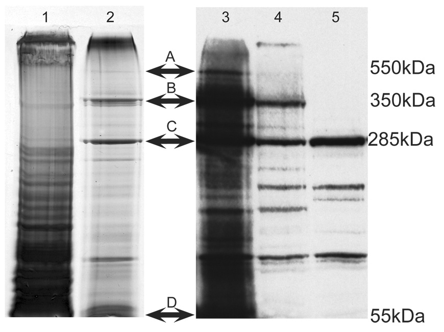
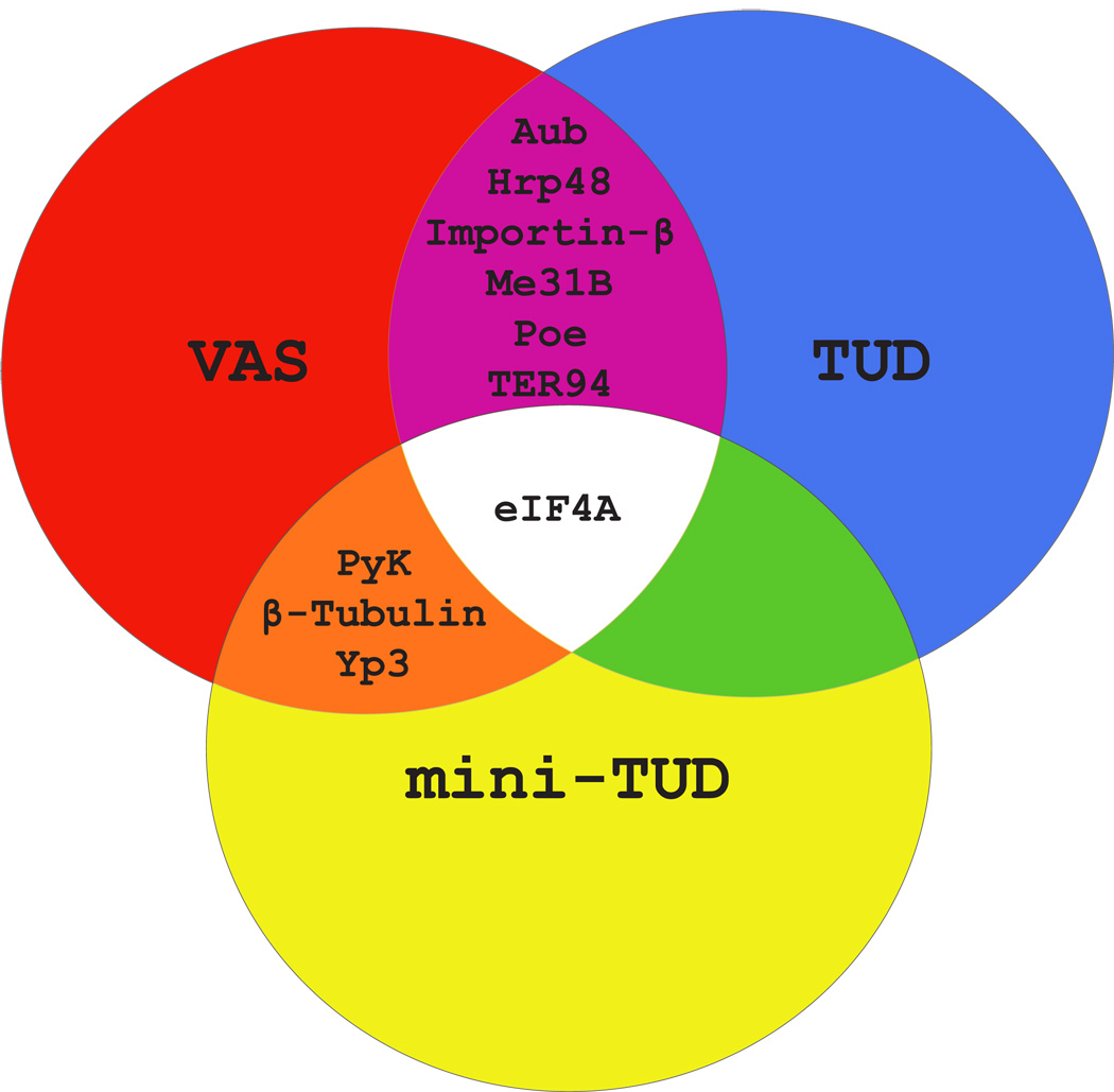
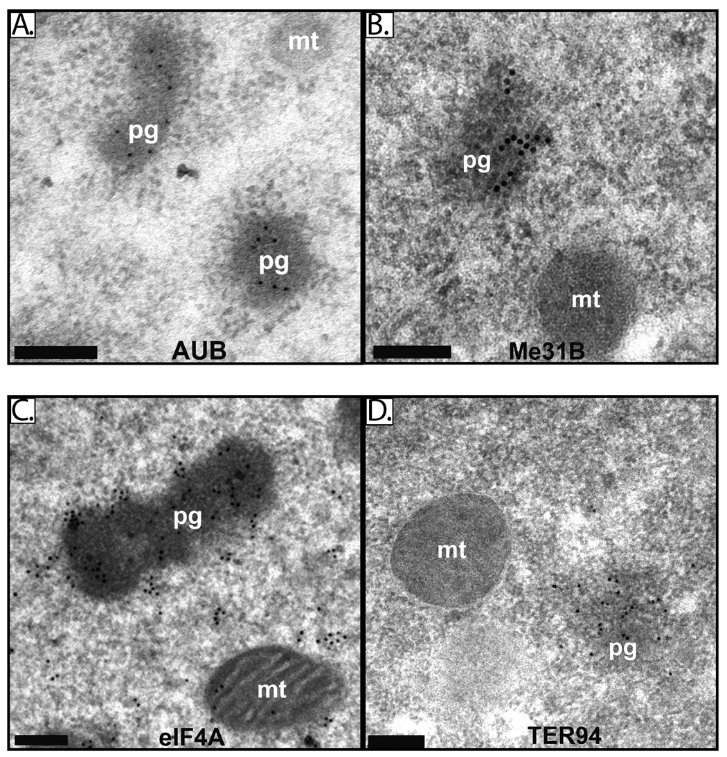
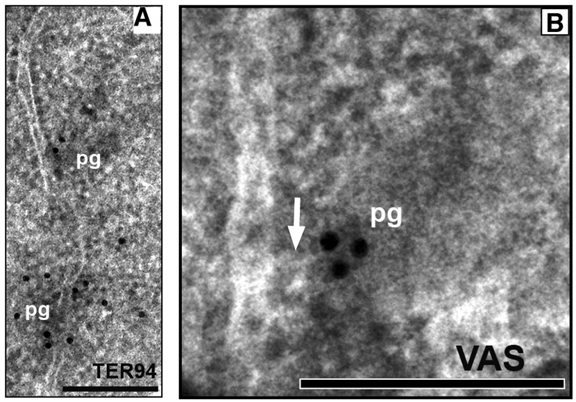
Germ plasm, a specialized cytoplasm present at the posterior of the early Drosophila embryo, is necessary and sufficient for germ cell formation. Germ plasm is rich in mitochondria and contains electron dense structures called polar granules. To identify novel polar granule components we isolated proteins that associate in early embryos with Vasa (VAS) and Tudor (TUD), two known polar granule associated molecules. We identified Maternal expression at 31B (ME31B), eIF4A, Aubergine (AUB) and Transitional Endoplasmic Reticulum 94 (TER94) as components of both VAS and TUD complexes and confirmed their localization to polar granules by immuno-electron microscopy. ME31B, eIF4A and AUB are also present in processing (P) bodies, suggesting that polar granules, which are necessary for germ line formation, might be related to P bodies. Our recovery of ER associated proteins TER94 and ME31B confirms that polar granules are closely linked to the translational machinery and to mRNP assembly.


Figures

Figure 1
Figure 1. Isolation of TUD complexes
Figure 1. Isolation of TUD complexes. Lanes 1 and 2 are silver-stained 4.5% SDS-PAGE of immunoprecipitates from BMH-crosslinked embryos using pre-immune serum (lane 1) or anti-TUD (lane 2). Bands A and B were reproducible in TUD immunoprecipitates, and these bands were excised and microsequenced. In some isolations uncrosslinked or "free" TUD was present (C). Lanes 3–5 are immunoblots using the anti-TUD antiserum. Lanes 3 and 4 were loaded with crosslinked extracts subjected to TUD immunoprecipitation, lane 3 was loaded with less total protein than lane 4. Lane 5 was loaded with extract that was immunoprecipitated but not BMH-crosslinked. Immunoglobulin-G (IgG) heavy chain is present at the bottom of the gel (D).
Figure 2
Figure 2. TUD and VAS protein complexes contain some common components
A Venn diagram proteins present in more than one of the three types of complex (TUD, mini-TUDΔ3 and VAS).
Figure 3
Figure 3. aub, me31B, eIF4A and TER94 mutations interact genetically with vas and tud
Embryos (stage 10) were collected from females of the indicated genotype, stained with anti-VAS, and pole cells were counted. n represents the total number of embryos scored per genotype, and the bar graphs show the average number of pole cells and standard error. Females heterozygous for aub, me31B, eIF4A, or TER94 and either vasPH165 or tudtux46 produce embryos with significantly fewer pole cells than wild type or females heterozygous for only a single mutation. The differences between singly heterozygous females and transheterozygous combinations with vasPH165 or tudtux46 are statistically significant as determined by Student t-test (all p values < 0.001).
Figure 4
Figure 4. AUB, Me31B, eIF4A and TER94 are components of polar granules
Electron micrographs of immunostained embryos (stages 1–2) illustrate enrichment of AUB (A), Me31B (B) eIF4A (C) and TER94 (D) on polar granules. Polar granules are denoted as “pg” and mitochondria as “mt”. Black bar = 0.2µM.
Figure 5
Figure 5. Polar granules contact ER
A. Electron micrograph of germ plasm of an early embryo (stage 1–2) showing contact between ER and two electron dense polar granules immunostained for TER94. B. Electron micrograph of germ plasm of an early embryo (stage 1–2) immunostained for VAS showing contact between ER and polar granule. The ER appears to protrude toward the polar granule (arrow). Polar granules are denoted as “pg”. Black bar = 0.2µM.

References

    1. Amikura R, Hanyu K, Kashikawa M, Kobayashi S. Tudor protein is essential for the localization of mitochondrial RNAs in polar granules of Drosophila embryos. Mech Dev. 2001;107:97–104. - PubMed
    1. Anne J, Ollo R, Ephrussi A, Mechler BM. Arginine methyltransferase Capsuleen is essential for methylation of spliceosomal Sm proteins and germ cell formation in Drosophila. Development. 2007;134:137–146. - PubMed
    1. Arkov AL, Wang JY, Ramos A, Lehmann R. The role of Tudor domains in germline development and polar granule architecture. Development. 2006;133:4053–4062. - PubMed
    1. Bardsley A, McDonald K, Boswell RE. Distribution of tudor protein in the Drosophila embryo suggests separation of functions based on site of localization. Development. 1993;119:207–219. - PubMed
    1. Boswell RE, Mahowald AP. tudor, a gene required for assembly of the germ plasm in Drosophila melanogaster. Cell. 1985;43:97–104. - PubMed

Publication types

MeSH terms