

Effects of age at onset of deafness and electrical stimulation on the developing cochlear nucleus in cats
- PMID: 18590947
- PMCID: PMC2575007
- DOI: 10.1016/j.heares.2008.05.007
Effects of age at onset of deafness and electrical stimulation on the developing cochlear nucleus in cats
Abstract
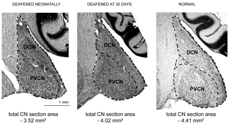
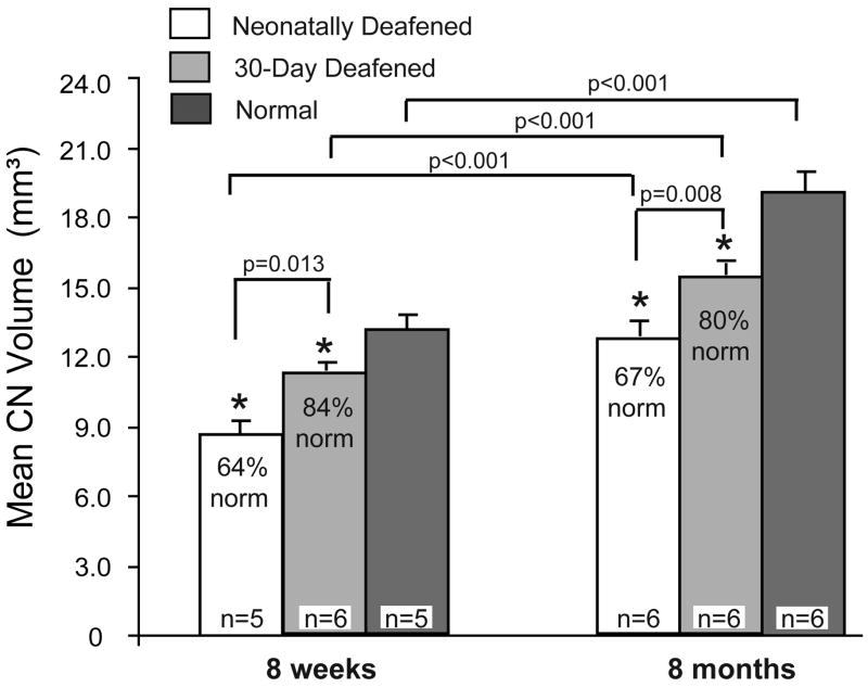
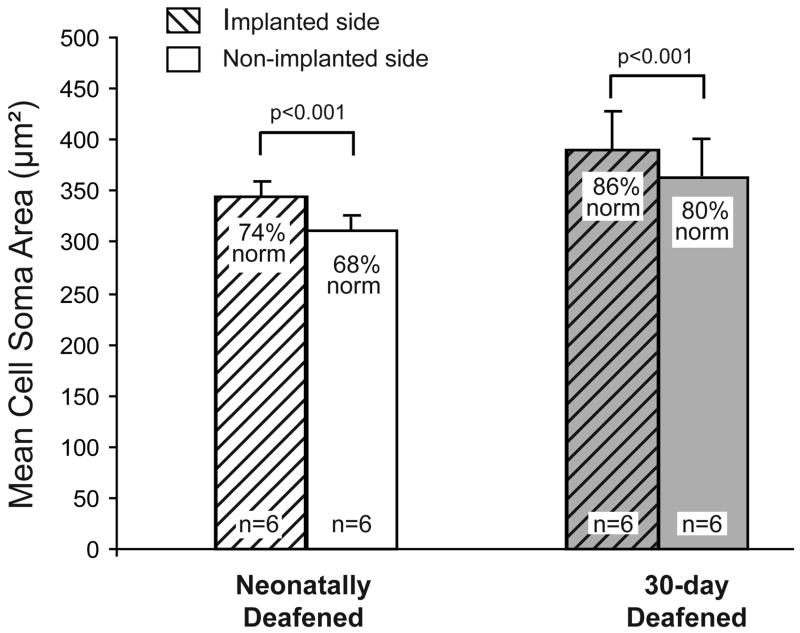
This study examined the effects of deafness and intracochlear electrical stimulation on the anatomy of the cochlear nucleus (CN) after a brief period of normal auditory development early in life. Kittens were deafened by systemic ototoxic drug injections either as neonates or starting at postnatal day 30. Total CN volume, individual CN subdivision volumes, and cross-sectional areas of spherical cell somata in the anteroventral CN (AVCN) were compared in neonatally deafened and 30-day deafened groups at 8 weeks of age and in young adults after approximately 6 months of electrical stimulation initiated at 8 weeks of age. Both neonatal and early acquired hearing loss resulted in a reduction in CN volume as compared to normal hearing cats. Comparison of 8- and 32-week old groups indicated that the CN continued to grow in both deafened groups despite the absence of auditory input. Preserving normal auditory input for 30 days resulted in a significant increase in both total CN volume and cross-sectional areas of spherical cell somata, as compared to neonatally deafened animals. Restoring auditory input in these developing animals by unilateral intracochlear electrical stimulation did not elicit any difference in CN volume between the two sides, but resulted in 7% larger spherical cell size on the stimulated side. Overall, the brief period of normal auditory development and subsequent electrical stimulation maintained CN volume at 80% of normal and spherical cell size at 86% of normal ipsilateral to the implant as compared to 67% and 74%, respectively, in the neonatally deafened group.
Figures

Similar articles
-
Effects of brain-derived neurotrophic factor (BDNF) on the cochlear nucleus in cats deafened as neonates.Hear Res. 2016 Dec;342:134-143. doi: 10.1016/j.heares.2016.10.011. Epub 2016 Oct 20. Hear Res. 2016. PMID: 27773647 Free PMC article.
-
Factors influencing neurotrophic effects of electrical stimulation in the deafened developing auditory system.Hear Res. 2008 Aug;242(1-2):86-99. doi: 10.1016/j.heares.2008.06.002. Epub 2008 Jun 7. Hear Res. 2008. PMID: 18573324 Free PMC article.
-
Changes in the cat cochlear nucleus following neonatal deafening and chronic intracochlear electrical stimulation.Hear Res. 1994 Apr;74(1-2):29-37. doi: 10.1016/0378-5955(94)90173-2. Hear Res. 1994. PMID: 8040097
-
Deafness-induced changes in the auditory pathway: implications for cochlear implants.Audiol Neurootol. 2001 Nov-Dec;6(6):305-18. doi: 10.1159/000046843. Audiol Neurootol. 2001. PMID: 11847461 Review.
-
Effect of chronic electrical stimulation on cochlear nucleus neuron size in normal hearing kittens.Acta Otolaryngol. 1993 Jul;113(4):489-97. doi: 10.3109/00016489309135851. Acta Otolaryngol. 1993. PMID: 8379304 Review.
Cited by
-
Brain Morphological Modifications in Congenital and Acquired Auditory Deprivation: A Systematic Review and Coordinate-Based Meta-Analysis.Front Neurosci. 2022 Mar 28;16:850245. doi: 10.3389/fnins.2022.850245. eCollection 2022. Front Neurosci. 2022. PMID: 35418829 Free PMC article.
-
Vestibulo-ocular reflex responses to a multichannel vestibular prosthesis incorporating a 3D coordinate transformation for correction of misalignment.J Assoc Res Otolaryngol. 2010 Sep;11(3):367-81. doi: 10.1007/s10162-010-0208-5. Epub 2010 Feb 23. J Assoc Res Otolaryngol. 2010. PMID: 20177732 Free PMC article.
-
Volumes of cochlear nucleus regions in rodents.Hear Res. 2016 Sep;339:161-74. doi: 10.1016/j.heares.2016.07.003. Epub 2016 Jul 18. Hear Res. 2016. PMID: 27435005 Free PMC article.
-
Age-Related Hearing Loss: Sensory and Neural Etiology and Their Interdependence.Front Aging Neurosci. 2022 Feb 17;14:814528. doi: 10.3389/fnagi.2022.814528. eCollection 2022. Front Aging Neurosci. 2022. PMID: 35250542 Free PMC article. Review.
-
Bilateral effects of unilateral cochlear implantation in congenitally deaf cats.J Comp Neurol. 2010 Jun 15;518(12):2382-404. doi: 10.1002/cne.22339. J Comp Neurol. 2010. PMID: 20437534 Free PMC article.
References
-
- Anson BJ, Donaldson JA. Surgical anatomy of the temporal bone. W.B. Saunders Company; 1981. The ear: developmental anatomy; pp. 23–57.
-
- Birnholz JC, Benacerraf BR. The development of human fetal hearing. Science. 1983;222:516–518. - PubMed
-
- Born DE, Rubel EW. Afferent influences on brain stem auditory nuclei in the chicken: neuron number and size following cochlea removal. J Comp Neurol. 1985;231:435–445. - PubMed
-
- Busby PA, Clark GM. Gap detection by early-deafened cochlear-implant subjects. J Acoust Soc Am. 1999;105(3):1841–52. - PubMed
-
- Chao TK, Burgess BJ, Eddington DK, Nadol JB., Jr Morphometric changes in the cochlear nucleus in patients who had undergone cochlear implantation for bilateral profound deafness. Hear Res. 2002;174:196–205. - PubMed
Publication types
MeSH terms
Substances
Grants and funding
LinkOut - more resources
Full Text Sources
Medical
Miscellaneous
