Review
. 2008 Oct;93(10):890-8.
doi: 10.1136/adc.2008.142836. Epub 2008 Jun 30.

Cerebrovascular disease and stroke

Affiliations
Review

Cerebrovascular disease and stroke

J Pappachan et al. Arch Dis Child. 2008 Oct.

Abstract

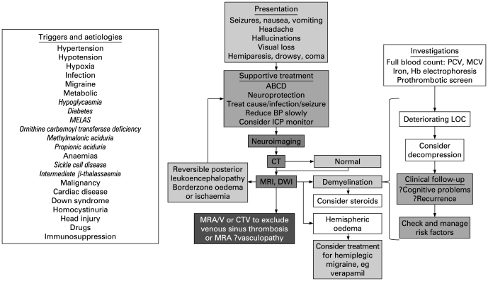
Stroke and cerebrovascular disorders are important causes of morbidity and mortality in children; they are already amongst the top 10 causes of childhood death and are probably increasing in prevalence. Acute treatment of stroke syndromes in adults is now evidence based. However, paediatric stroke syndromes are far less common and the differential diagnosis is very wide, but the individual health resource implications are much greater because of the life-long treatment costs in survivors. Recognition and consultation with a paediatric neurologist should be rapid so that children can benefit from regional services with emergency neurological, neuroradiological and neurosurgical intervention and paediatric intensive care. This review focuses on the epidemiology, presentation, differential diagnosis, generic/specific emergency management and prognosis of acute stroke in children. Its aim is to educate and guide management by general paediatricians and to emphasise the importance of local guidelines for the initial investigation and treatment and appropriate transfer of these children.


Conflict of interest statement

Competing interests: None.

Figures

Figure 1
Figure 1
Neuroimaging in children with stroke. Row 1: CT scans; row 2: T2-weighted MRI scans; row 3: vascular imaging (A–D: magnetic resonance angiography (MRA); E: MR venography (MRV); F: watershed infarction); row 4: additional imaging which may be useful in difficult cases (A–D: conventional angiography; E: venous thrombosis on MRI; F: potentially reversible signal abnormality on diffusion weighted imaging). Column 1: haemorrhage; column 2: extracranial dissection; column 3: transient cerebral arteriopathy; column 4: moyamoya; column 5: venous sinus thrombosis; column 6: posterior circulation stroke and stroke mimics – posterior leukoencephalopathy, “covert” watershed ischaemia, hemiplegic migraine. A: Haemorrhage; A1: spontaneous intracerebral haemorrhage with midline shift; A2: MRI showing haemorrhage from mycotic aneurysm in a patient with subacute bacterial endocarditis; A3: mycotic aneurysm on MRA; A4: conventional arteriography showing arteriovenous malformation. B: Dissection; B1: infarct in a child who had suffered a minor head injury 24 h before; B2: large cerebral infarct after head injury; B3: fat-saturated T1 MRI of the neck showing haemorrhage in the vessel wall; B4: conventional arteriography showing tapering “rat’s tail” appearance characteristic of extracranial dissection. C: Transient cerebral arteriopathy; C1: infarct in a child with stuttering stroke onset; C2: infarct in a child with recent varicella; C3: short segment of middle cerebral artery stenosis (MRA from child in C2); C4: conventional arteriography from child in C1 showing longer segment of middle cerebral artery stenosis (the infarct had extended in size on the post-arteriography CT scan). D: Moyamoya; D1: bilateral frontal infarction in a child with livedo reticularis; D2: bilateral frontal infarction in a child with sickle cell anaemia; D3: bilateral middle cerebral artery stenosis with collateral formation obvious on MRA; D4: conventional arteriography showing attenuation of major intracranial vessels and collaterals. E: Venous sinus thrombosis; E1: bilateral thalamic signal change in severe iron deficiency anaemia; E2: occipital signal change in nephrotic syndrome; E3: sagittal sinus thrombosis in systemic lupus erythaematosus presenting with psychiatric symptoms; E4: transverse sinus thrombosis on plain MRI in child in E1. F: Posterior circulation stroke and stroke mimics; F1: cerebellar infarction in a boy with vertebral dissection; F2: bilateral occipital signal change suggestive of posterior leukoencephalopathy; F3: bilateral watershed infarction after facial infection in sickle cell anaemia (MRA and MRV were normal); F4: diffusion-weighted imaging shows potentially reversible pathology in a patient with hemiplegic migraine and normal T2-weighted MRI.
Figure 2
Figure 2
Flow diagram for the diagnosis and management of haemorrhagic stroke. ABCD, airway, breathing, circulation, disability; AVM, arteriovenous malformation; BP, blood pressure; CT, computed tomography; CTV, CT venography; ICH, intracerebral haemorrhage; ICP, intracranial pressure; IIH, idiopathic intracranial hypertension; LOC, level of consciousness; LP, lumbar puncture; MCV, mean cell volume; MRA, magnetic resonance angiography; MRV, magnetic resonance venography; PCV, packed cell volume; SAH, subarachnoid haemorrhage; US, ultrasound.
Figure 3
Figure 3
Flow diagram for the diagnosis and management of haemorrhagic stroke. ABCD, airway, breathing, circulation, disability; MCA, middle cerebral artery; MRA, magnetic resonance angiography; MRV, magnetic resonance venography; PHACES, posterior fossa malformations, haemangiomas, arterial anomalies, cardiac defects, eye abnormalities, and sternal or ventral defects; RPLS, reversible posterior leukoencephalopathy syndrome; SCD, sickle cell disease; VST, venous sinus thrombosis.
Figure 4
Figure 4
Flow diagram for the diagnosis and management of venous sinus thrombosis. IIH, idiopathic intracranial hypertension. ABCD, airway, breathing, circulation, disability; CTV, computed tomography venography; LOC, level of consciousness; MRV, magnetic resonance venography.
Figure 5
Figure 5
Flow diagram for the management of stroke mimics. ABCD, airway, breathing, circulation, disability; BP, blood pressure; CTV, CT venography; DWI, diffusion weighted imaging; LOC, level of consciousness; MRA, magnetic resonance angiography; MRV, magnetic resonance venography.

References

    1. Zahuranec DB, Brown DL, Lisabeth LD, et al. Is it time for a large, collaborative study of pediatric stroke? Stroke 2005;36:1825–9 - PubMed
    1. Telfer P, Coen P, Chakravorty S, et al. Clinical outcomes in children with sickle cell disease living in England: a neonatal cohort in East London. Haematologica 2007;92:905–12 - PubMed
    1. Mazumdar M, Heeney MM, Sox CM, et al. Preventing stroke among children with sickle cell anemia: an analysis of strategies that involve transcranial Doppler testing and chronic transfusion. Pediatrics 2007;120:e1107–16 - PubMed
    1. Laugesaar R, Kolk A, Tomberg T, et al. Acutely and retrospectively diagnosed perinatal stroke: a population-based study. Stroke 2007;38:2234–40 - PubMed
    1. Braun KP, Kappelle LJ, Kirkham FJ, et al. Diagnostic pitfalls in paediatric ischaemic stroke. Dev Med Child Neurol 2006;48:985–90 - PubMed

Publication types