Comparative Study
. 2008 Sep;76(9):4359-67.
doi: 10.1128/IAI.00669-08. Epub 2008 Jun 30.

A Candida albicans mannoprotein deprived of its mannan moiety is efficiently taken up and processed by human dendritic cells and induces T-cell activation without stimulating proinflammatory cytokine production

Affiliations
Comparative Study

A Candida albicans mannoprotein deprived of its mannan moiety is efficiently taken up and processed by human dendritic cells and induces T-cell activation without stimulating proinflammatory cytokine production

Donatella Pietrella et al. Infect Immun. 2008 Sep.

Abstract

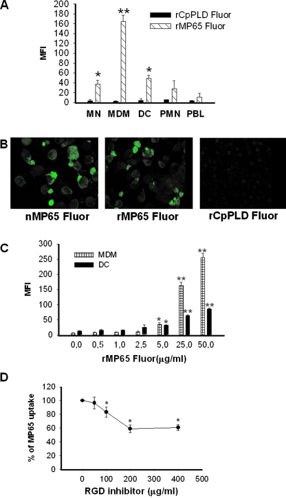
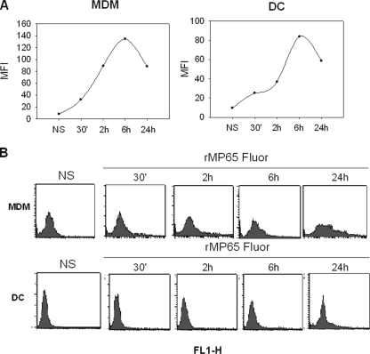
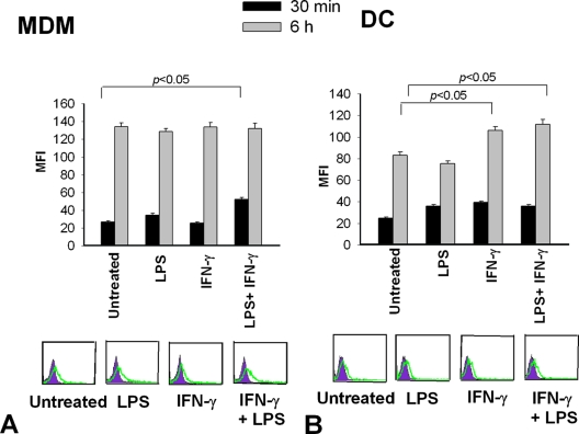
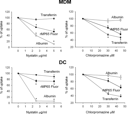
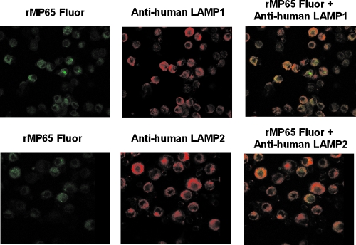
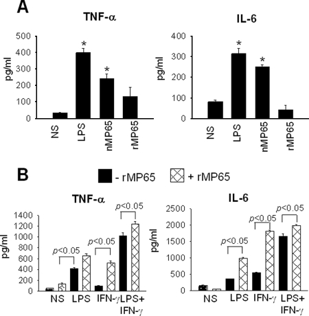
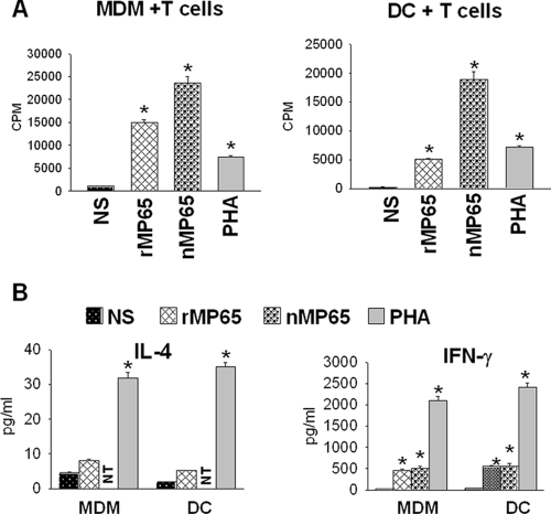
Mannoproteins are cell wall components of pathogenic fungi and play major virulence and immunogenic roles with both their mannan and protein moieties. The 65-kDa mannoprotein (MP65) of Candida albicans is a beta-glucanase adhesin recognized as a major target of the human immune response against this fungus, and its recombinant product (rMP65; devoid of the mannan moiety) is presently under consideration as a vaccine candidate. Here we investigated cellular and molecular aspects of the interaction of rMP65 with human antigen-presenting cells. We also assessed the ability of rMP65 to initiate a T-cell response. Both the native mannosylated MP65 (nMP65) and the recombinant product were efficiently bound and taken up by macrophages and dendritic cells. However, contrarily to nMP65, rMP65 did not induce tumor necrosis factor alpha and interleukin-6 release from these cells. On the other hand, rMP65 was rapidly endocytosed by both macrophages and dendritic cells, in a process involving both clathrin-dependent and clathrin-independent mechanisms. Moreover, the RGD sequence inhibited rMP65 uptake to some extent. After internalization, rMP65 partially colocalized with lysosomal membrane-associated glycoproteins 1 and 2. This possibly resulted in efficient protein degradation and presentation to CD4(+) T cells, which proliferated and produced gamma interferon. Collectively, these results demonstrate that the absence of the mannan moiety does not deprive MP65 of the capacity to initiate the pattern of cellular and molecular events leading to antigen presentation and T-cell activation, which are essential features for further consideration of MP65 as a potential vaccine candidate.


Figures

FIG. 1.
FIG. 1.
Uptake of rMP65 fluor by different cell types. (A) MN, MDM, DC, PMN, and PBL treated for 30 min at 37°C with rMP65 fluor (5 μg/ml) or rCpPLD fluor (5 μg/ml) were analyzed by flow cytometry to assess rMP65 and rCpPLD uptake. The rMP65 fluor uptake by different cell types is expressed as the mean fluorescence intensity (MFI). Data are expressed as means ± SEM for three independent experiments. *, P < 0.05; **, P < 0.001 (rMP65 fluor-treated cells versus rCpPLD fluor-treated cells). (B) rMP65 fluor, nMP65 fluor, and rCpPLD fluor uptake by DC, tested using confocal microscopy. DC were treated with 25 μg/ml of rMP65 fluor, rCpPLD fluor, or nMP65 fluor for 2 h at 37°C. The cells were then fixed and analyzed with a confocal microscope. (C) MDM or DC were stimulated for 30 min at 37°C with different concentrations of rMP65 fluor (0.5, 1, 2.5, 5, 25, and 50 μg/ml). The rMP65 fluor uptake by different cell types is expressed as the MFI. Data are expressed as means ± SEM for three independent experiments. *, P < 0.05; **, P < 0.001 (rMP65-treated cells versus untreated cells). (D) MDM were pretreated with various concentrations of RGDS or control peptide for 15 min, and then rMP65 fluor (25 μg/ml) was added. Data are expressed as means ± SEM for three independent experiments. *, P < 0.05 (RGDS-treated cells versus untreated cells).
FIG. 2.
FIG. 2.
Kinetics of rMP65 fluor uptake by MDM and DC. MDM or DC left untreated (NS) or treated with 5 μg/ml of rMP65 fluor were incubated at 37°C for different times (30 min and 2, 6, and 24 h). (A) rMP65 fluor uptake by different cell types is expressed as the MFI. (B) Fluorescence-activated cell sorting histograms represent the background cell fluorescence (NS) and cell fluorescence after different times of incubation. Data shown are from one representative experiment out of four performed with similar results.
FIG. 3.
FIG. 3.
rMP65 fluor uptake by activated MDM and DC. MDM (A) or DC (B) were left untreated or activated with LPS (1 μg/ml) or IFN-γ (100 ng/ml), alone or in combination, for 30 min at 37°C. The cells were incubated with 5 μg/ml of rMP65 fluor for 30 min or 6 h at 37°C. The rMP65 fluor uptake by different cell types is expressed as the MFI. Data are expressed as means ± SEM for four independent experiments. Statistical analysis was performed with ANOVA and corrected by the Bonferroni test for multiple comparisons. In the lower part of the figure, filled histograms represent the autofluorescence of cells, while the green lines represent fluorescence of rMP65 fluor uptake by cells for a 30-min incubation. Results of cytofluorimetric analysis presented are from one representative experiment.
FIG. 4.
FIG. 4.
Inhibition of clathrin-independent and clathrin-dependent uptake of rMP65 fluor. MDM or DC were left untreated or treated with nystatin (2.5 or 5 μg/ml) for 30 min at 4°C or with chlorpromazine (30 or 45 μM) for 30 min at 37°C. The cells were then stimulated for 2 h with 25 μg/ml of rMP65 fluor, transferrin fluor, and albumin fluor. Results are expressed as percentages of rMP65 fluor, albumin fluor, and transferrin fluor uptake. Data are expressed as means ± SEM for three independent experiments. *, P < 0.05 (nystatin- or chlorpromazine-treated cells versus untreated cells).
FIG. 5.
FIG. 5.
Colocalization of rMP65 with LAMP-1 and LAMP-2. DC were treated with rMP65 fluor (25 μg/ml) for 2 h at 37°C. The cells were then fixed, permeabilized, and incubated for 1 h with primary antibodies (mouse anti-human LAMP-1 and mouse anti-human LAMP-2). After incubation, the cells were treated for 30 min with secondary antibodies (anti-mouse immunoglobulin G conjugated with Cy). Cells were fixed and analyzed with a confocal microscope. The data reported are from one experiment out of three performed with similar results.
FIG. 6.
FIG. 6.
TNF-α and IL-6 production from activated MDM after stimulation with rMP65. (A) MDM were left untreated (NS) or treated with rMP65, nMP65 (5 μg/ml), or LPS (1 μg/ml) for 18 h at 37°C. After incubation, supernatants were recovered and tested for the presence of TNF-α and IL-6. Data are expressed as means ± SEM for four independent experiments. *, P < 0.05 (rMP65-treated cells versus untreated cells). (B) MDM were left untreated (NS) or treated with LPS (1 μg/ml) or IFN-γ (100 ng/ml), alone or in combination, for 30 min at 37°C. After activation, cells were left untreated (−rMP65) or treated with 5 μg/ml rMP65 (+rMP65) for 18 h at 37°C. Data are expressed as means ± SEM for four independent experiments. Statistical analysis was performed with ANOVA and corrected by the Bonferroni test for multiple comparisons. *, P < 0.05 (rMP65-treated cells versus untreated cells).
FIG. 7.
FIG. 7.
Proliferation of T cells and IL-4 and IFN-γ secretion in supernatant fluids from MDM or DC treated with rMP65 and cocultured with autologous T cells. MDM or DC left untreated (NS) or treated with nMP65 (5 μg/ml) or rMP65 (5 μg/ml) were added to autologous T cells. PHA (10 μg/ml) was used as a positive control. (A) Proliferation was measured after 4 days of incubation. (B) The supernatant fluids were recovered after 5 days of incubation and tested for IL-4 and IFN-γ by ELISA. NT, not tested. Data are expressed as means ± SEM for four independent experiments. *, P < 0.05 (rMP65-, nMP65-, or PHA-treated cells versus untreated cells).

References

    1. Banchereau, J., and R. M. Steinman. 1998. Dendritic cells and the control of immunity. Nature 392245-252. - PubMed
    1. Calderone, R., S. Suzuki, R. Cannon, T. Cho, D. Boyd, J. Calera, H. Chibana, D. Herman, A. Holmes, H. W. Jeng, H. Kaminishi, T. Matsumoto, T. Mikami, J. M. O'Sullivan, M. Sudoh, M. Suzuki, Y. Nakashima, T. Tanaka, G. R. Tompkins, and T. Watanabe. 2000. Candida albicans: adherence, signaling and virulence. Med. Mycol. 38(Suppl. 1)125-137. - PubMed
    1. Cao, L., K. M. Chan, D. Chen, N. Vanittanakom, C. Lee, C. M. Chan, T. Sirisanthana, D. N. Tsang, and K. Y. Yuen. 1999. Detection of cell wall mannoprotein Mp1p in culture supernatants of Penicillium marneffei and in sera of penicilliosis patients. J. Clin. Microbiol. 37981-986. - PMC - PubMed
    1. Cassone, A. 2008. Fungal vaccines: real progress from real challenges. Lancet Infect. Dis. 8114-124. - PubMed
    1. Ciervo, A., F. Mancini, and A. Cassone. 2007. Transcription, expression, localization and immunoreactivity of Chlamydophila pneumoniae phospholipase D protein. Microb. Pathol. 4396-105. - PubMed

Publication types