. 2008 Sep;76(9):4269-81.
doi: 10.1128/IAI.01735-07. Epub 2008 Jun 30.

A Mycobacterium tuberculosis Rpf double-knockout strain exhibits profound defects in reactivation from chronic tuberculosis and innate immunity phenotypes

Affiliations

A Mycobacterium tuberculosis Rpf double-knockout strain exhibits profound defects in reactivation from chronic tuberculosis and innate immunity phenotypes

Eleanor Russell-Goldman et al. Infect Immun. 2008 Sep.

Abstract

Resuscitation-promoting factors (Rpfs), apparent peptidoglycan hydrolases, have been implicated in the reactivation of dormant bacteria. We previously demonstrated that deletion of rpfB impaired reactivation of Mycobacterium tuberculosis in a mouse model. Because M. tuberculosis encodes five Rpf paralogues, redundant functions among the family members might obscure rpf single-knockout phenotypes. A series of rpf double knockouts were therefore generated. One double mutant, DeltarpfAB, exhibited several striking phenotypes. Consistent with the proposed cell wall-modifying function of Rpfs, DeltarpfAB exhibited an altered colony morphology. Although DeltarpfAB grew comparably to the parental strain in axenic culture, in vivo it exhibited deficiency in reactivation induced in C57BL/6 mice by the administration of nitric oxide synthase inhibitor (aminoguanidine) or by CD4(+) T-cell depletion. Notably, the reactivation deficiency of DeltarpfAB was more severe than that of DeltarpfB in aminoguanidine-treated mice. A similar deficiency was observed in DeltarpfAB reactivation from a drug-induced apparently sterile state in infected NOS2(-/-) mice upon cessation of antimycobacterial therapy. Secondly, DeltarpfAB showed a persistence defect not seen with the DeltarpfB or DeltarpfA single mutants. Interestingly, DeltarpfAB exhibited impaired growth in primary mouse macrophages and induced higher levels of the proinflammatory cytokines tumor necrosis factor alpha and interleukin 6. Simultaneous reintroduction of rpfA and rpfB into the double-knockout strain complemented the colony morphology and macrophage cytokine secretion phenotypes. Phenotypes related to cell wall composition and macrophage responses suggest that M. tuberculosis Rpfs may influence the outcome of reactivation, in part, by modulating innate immune responses to the bacterium.


Figures

FIG. 1.
FIG. 1.
Southern blots demonstrating the unmarking of the rpfA and rpfD deletion mutants. (A) Genomic DNAs prepared from putative rpf double-knockout strains, from the original single ΔrpfA strain, or from Erdman wt were digested with BstEII and probed with a 32P-labeled probe consisting of the upstream flanking region of the rpfA gene. The expected band sizes for the Erdman wt, ΔrpfA, and ΔrpfA::res strains are indicated to the right of the blot. (B) Genomic DNAs prepared from putative rpf double-knockout strains, from the original single ΔrpfD strain, or from Erdman wt were digested with SmaI and probed with a 32P-labeled probe consisting of the upstream flanking region of the rpfD gene. The expected band sizes for the Erdman wt, ΔrpfD, and ΔrpfD::res strains are indicated to the right of the blot. Panels A and B show that, for all of the putative double-deletion mutants, the original “parental” strain did possess an unmarked (or “resolved” [res]) rpfA gene (A) or rpfD gene (B). The positions of size markers (kb) are shown on the left of each panel.
FIG. 2.
FIG. 2.
Southern blots demonstrating that the second rpf deletions were introduced into the unmarked ΔrpfA::res and ΔrpfD::res strains. Genomic DNAs were digested with SmaI (A and C), BamHI (B), or NcoI (D) and probed with the upstream (B and C) or downstream (A and D) flanking regions of the genes targeted for deletion. The expected band sizes for Erdman wt and the various rpf deletion mutants are indicated on the right of each blot. Correct rpf double-deletion mutant clones include ΔrpfBD-2 and -3 (A), ΔrpfAB-1, -2, and -3 (A), ΔrpfCD-2 and -3 (B), ΔrpfAC-2 (B), ΔrpfAD-2 (C), ΔrpfDE-2 and -3 (D), and ΔrpfAE-1 and -2 (D). The positions of size markers (kb) are shown on the left of each panel.
FIG. 3.
FIG. 3.
ΔrpfAB exhibits altered colony morphology. Colonies of ΔrpfAB (B) are smoother and more regular than those of the Erdman wt strain (A); the ΔrpfAB colonies also have a more prominent translucent “halo.”
FIG. 4.
FIG. 4.
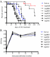
ΔrpfAB and ΔrpfBD strains are reactivation deficient. (A) Mortality. C57BL/6 mice were infected with ∼100 CFU of M. tuberculosis Erd wt or the indicated rpf double-deletion mutant: ΔrpfAB, ΔrpfBD, ΔrpfDE, ΔrpfCD, ΔrpfAD, ΔrpfAE, or ΔrpfAC. Reactivation was induced at 18 weeks postinfection by AG administration, with survival monitored over time (n = 8 to 12 mice per group). (B) Lung titers. C57BL/6 mice were infected by aerosol with the indicated strains, and mice were sacrificed at various time points to determine the numbers of CFU in the lung (n = 3 mice per group). The error bars indicate standard errors.
FIG. 5.
FIG. 5.
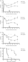
ΔrpfAB is impaired in both persistence and AG-induced reactivation. (A to C) Organ bacterial burdens. C57BL/6 mice were infected by aerosol with ∼400 CFU of the Erd wt or ΔrpfAB strain, and bacterial counts in the lung (A), spleen (B), and liver (C) were monitored over time (n = 3 or 4 mice per time point, except for the final time point, where n = 2). The error bars indicate standard errors. (D) Mortality. To induce reactivation, mice were given AG at 13 weeks postinfection as described in Materials and Methods, and survival was monitored. n = 13 for the Erd strain and 8 for the ΔrpfAB strain. *, P value for the Erd strain versus the ΔrpfAB strain, <0.05.
FIG. 6.
FIG. 6.
The ΔrpfAB strain exhibits delayed reactivation in alternative models, induced by CD4 depletion in immunocompetent mice (A and B) or a modified Cornell model in NOS2−/− mice (C and D). (A) Pulmonary bacterial burden. C57BL/6 mice were infected by aerosol with ∼400 to 600 CFU of the Erd wt or the ΔrpfAB strain, and the bacterial burden in the lung was monitored over time (n = 3 mice per time point). The error bars indicate standard errors. *, P values for the Erd strain versus the ΔrpfAB strain, <0.05. (B) Mortality. Erdman-infected or ΔrpfAB-infected mice were administered GK1.5 α-CD4 antibody or rat IgG as a control beginning during the chronic persistent phase of infection (6 months postinfection). n = 9 to 11 mice per group. (C) Pulmonary bacterial burden. NOS2−/− mice were infected by aerosol with ∼250 to 400 CFU of the Erd wt or ΔrpfAB strain, and the bacterial burden in the lung was monitored over time (n = 2 mice per time point). The period of INH/PZA administration, beginning at 16 days postinfection and continuing for 3 months, is indicated. (D) Mortality. Erdman-infected or ΔrpfAB-infected NOS2−/− mice were observed for mortality. The survival curves are significantly different, as indicated. Although the results of a single experiment are shown, the experiment has since been repeated with similar results.
FIG. 7.
FIG. 7.
The ΔrpfAB strain is attenuated for growth within macrophages. (A and B) Resting macrophages. Bone marrow-derived macrophages (BMDM) from C57BL/6 mice were infected with Erdman wt, ΔrpfAB clone 1 (ΔrpfAB-1), or ΔrpfAB clone 2 (ΔrpfAB-2) at an MOI of 1 (A) or 5 (B), and bacterial numbers were monitored at the indicated time points. (C) Activated macrophages. C57BL/6 BMDM, which had been activated with IFN-γ and LPS, were infected with Erdman wt, ΔrpfAB clone 1, or ΔrpfAB clone 2, and bacterial numbers were assessed as described above. For panels A, B, and C: ψ, P values for the Erd strain versus the ΔrpfAB-1 strain, < 0.05; *, P values for the Erd strain versus the ΔrpfAB-2 strain, <0.05. The error bars indicate standard errors. (D) Axenic culture. Growth of the Erd wt and the ΔrpfAB strain was monitored in 7H9 medium.
FIG. 8.
FIG. 8.
The ΔrpfAB strain induces increased macrophage production of TNF-α and IL-6. C57BL/6 BMDM were infected with Erdman wt (light-gray bars) or ΔrpfAB clone 2 (dark-gray bars) at an MOI of 1 (A and C) or 5 (B and D) or left uninfected (white bars). Supernatants collected at 24 h were assayed for TNF-α (A and B) or IL-6 (C and D) by ELISA. The results of representative experiments are shown. Samples from each well were assayed in duplicate, and the results are shown as means plus standard errors. The designations Erd-1 and -2 and ΔrpfAB-1 and -2 refer to each of the two samples assayed for each experiment shown. *, P for the Erdman versus the ΔrpfAB strain, <0.05.
FIG. 9.
FIG. 9.
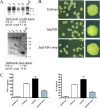
Complementation of the ΔrpfAB strain with combined expression of rpfA and rpfB restores the wt colony morphology and reduces the macrophage proinflammatory cytokine stimulation toward wt levels. (A) Southern blots demonstrating the presence of the rpfA and rpfB genes inserted at the attB site of the ΔrpfAB-complemented (compl) strain. Genomic DNAs prepared from putative ΔrpfAB-complemented strains (clones 1 to 4) or from Erdman wt were digested with EcoRI (top) or SacI (bottom) and probed with a 32P-labeled probe consisting of rpfA coding sequences (top) or rpfB coding sequences (bottom). The expected band sizes for the Erdman wt and for the ΔrpfAB-complemented strain are indicated below each blot. The positions of size markers (kb) are shown to the right of each blot. (B) Colonies of the ΔrpfAB-complemented (comp) strain (bottom) are rougher/more wrinkled than those of the ΔrpfAB parental strain (middle) and more closely resemble the rough colonies of the Erdman wt strain (top). Shown are plates with numerous colonies (similar in number) on the left and a single large colony from a sparsely inoculated plate containing only a few colonies in total on the right. (C) Production of TNF-α and IL-6 by macrophages infected with the Erdman wt, ΔrpfAB, and ΔrpfAB-complemented strains. C57BL/6 bone marrow-derived macrophages were left uninfected (vertically hatched bars) or were infected with the Erdman wt (white bars), ΔrpfAB (black bars), or ΔrpfAB-complemented (gray bars) strain at an MOI of 5; the supernatants collected at 24 h were assayed for TNF-α (left) or IL-6 (right) by ELISA. The results of a representative experiment are shown. The results are shown as means plus standard errors of two individual samples per treatment group (with each sample assayed in duplicate).

Similar articles

Cited by

References

    1. Bardarov, S., S. Bardarov, Jr., M. S. Pavelka, Jr., V. Sambandamurthy, M. Larsen, J. Tufariello, J. Chan, G. Hatfull, and W. R. Jacobs, Jr. 2002. Specialized transduction: an efficient method for generating marked and unmarked targeted gene disruptions in Mycobacterium tuberculosis, M. bovis BCG and M. smegmatis. Microbiology 1483007-3017. - PubMed
    1. Berg, C. M., N. B. Vartak, G. Wang, X. Xu, L. Liu, D. J. MacNeil, K. M. Gewain, L. A. Wiater, and D. E. Berg. 1992. The m gamma delta-1 element, a small gamma delta (Tn1000) derivative useful for plasmid mutagenesis, allele replacement and DNA sequencing. Gene 1139-16. - PubMed
    1. Biketov, S., V. Potapov, E. Ganina, K. Downing, B. D. Kana, and A. Kaprelyants. 2007. The role of resuscitation promoting factors in pathogenesis and reactivation of Mycobacterium tuberculosis during intra-peritoneal infection in mice. BMC Infect. Dis. 7146. - PMC - PubMed
    1. Chan, J., Y. Xing, R. S. Magliozzo, and B. R. Bloom. 1992. Killing of virulent Mycobacterium tuberculosis by reactive nitrogen intermediates produced by activated murine macrophages. J. Exp. Med. 1751111-1122. - PMC - PubMed
    1. Cohen-Gonsaud, M., P. Barthe, C. Bagneris, B. Henderson, J. Ward, C. Roumestand, and N. H. Keep. 2005. The structure of a resuscitation-promoting factor domain from Mycobacterium tuberculosis shows homology to lysozymes. Nat. Struct. Mol. Biol. 12270-273. - PubMed

Publication types

MeSH terms