

Evolution of metal(loid) binding sites in transcriptional regulators
- PMID: 18591244
- PMCID: PMC2533085
- DOI: 10.1074/jbc.M803209200
Evolution of metal(loid) binding sites in transcriptional regulators
Abstract
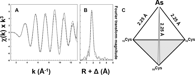
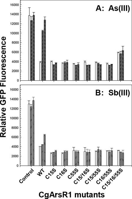
Expression of the genes for resistance to heavy metals and metalloids is transcriptionally regulated by the toxic ions themselves. Members of the ArsR/SmtB family of small metalloregulatory proteins respond to transition metals, heavy metals, and metalloids, including As(III), Sb(III), Cd(II), Pb(II), Zn(II), Co(II), and Ni(II). These homodimeric repressors bind to DNA in the absence of inducing metal(loid) ion and dissociate from the DNA when inducer is bound. The regulatory sites are often three- or four-coordinate metal binding sites composed of cysteine thiolates. Surprisingly, in two different As(III)-responsive regulators, the metalloid binding sites were in different locations in the repressor, and the Cd(II) binding sites were in two different locations in two Cd(II)-responsive regulators. We hypothesize that ArsR/SmtB repressors have a common backbone structure, that of a winged helix DNA-binding protein, but have considerable plasticity in the location of inducer binding sites. Here we show that an As(III)-responsive member of the family, CgArsR1 from Corynebacterium glutamicum, binds As(III) to a cysteine triad composed of Cys(15), Cys(16), and Cys(55). This binding site is clearly unrelated to the binding sites of other characterized ArsR/SmtB family members. This is consistent with our hypothesis that metal(loid) binding sites in DNA binding proteins evolve convergently in response to persistent environmental pressures.
Figures


Similar articles
-
Structures of two ArsR As(III)-responsive transcriptional repressors: Implications for the mechanism of derepression.J Struct Biol. 2019 Aug 1;207(2):209-217. doi: 10.1016/j.jsb.2019.05.009. Epub 2019 May 25. J Struct Biol. 2019. PMID: 31136796 Free PMC article.
-
The SmtB/ArsR family of metalloregulatory transcriptional repressors: Structural insights into prokaryotic metal resistance.FEMS Microbiol Rev. 2003 Jun;27(2-3):131-43. doi: 10.1016/S0168-6445(03)00054-8. FEMS Microbiol Rev. 2003. PMID: 12829264 Review.
-
Elucidation of primary (alpha(3)N) and vestigial (alpha(5)) heavy metal-binding sites in Staphylococcus aureus pI258 CadC: evolutionary implications for metal ion selectivity of ArsR/SmtB metal sensor proteins.J Mol Biol. 2002 Jun 7;319(3):685-701. doi: 10.1016/S0022-2836(02)00299-1. J Mol Biol. 2002. PMID: 12054863
-
A zinc(II)/lead(II)/cadmium(II)-inducible operon from the Cyanobacterium anabaena is regulated by AztR, an alpha3N ArsR/SmtB metalloregulator.Biochemistry. 2005 Jun 21;44(24):8673-83. doi: 10.1021/bi050450+. Biochemistry. 2005. PMID: 15952774
-
Structural determinants of metal selectivity in prokaryotic metal-responsive transcriptional regulators.Biometals. 2005 Aug;18(4):413-28. doi: 10.1007/s10534-005-3716-8. Biometals. 2005. PMID: 16158234 Review.
Cited by
-
Structures of two ArsR As(III)-responsive transcriptional repressors: Implications for the mechanism of derepression.J Struct Biol. 2019 Aug 1;207(2):209-217. doi: 10.1016/j.jsb.2019.05.009. Epub 2019 May 25. J Struct Biol. 2019. PMID: 31136796 Free PMC article.
-
Arsenate reductase, mycothiol, and mycoredoxin concert thiol/disulfide exchange.J Biol Chem. 2009 May 29;284(22):15107-16. doi: 10.1074/jbc.M900877200. Epub 2009 Mar 13. J Biol Chem. 2009. PMID: 19286650 Free PMC article.
-
Arsenic binding and transfer by the ArsD As(III) metallochaperone.Biochemistry. 2010 May 4;49(17):3658-66. doi: 10.1021/bi100026a. Biochemistry. 2010. PMID: 20361763 Free PMC article.
-
Purification, crystallization and preliminary X-ray diffraction studies of the arsenic repressor ArsR from Corynebacterium glutamicum.Acta Crystallogr Sect F Struct Biol Cryst Commun. 2011 Dec 1;67(Pt 12):1616-8. doi: 10.1107/S1744309111038966. Epub 2011 Nov 26. Acta Crystallogr Sect F Struct Biol Cryst Commun. 2011. PMID: 22139180 Free PMC article.
-
Arsenic binding to proteins.Chem Rev. 2013 Oct 9;113(10):7769-92. doi: 10.1021/cr300015c. Epub 2013 Jun 28. Chem Rev. 2013. PMID: 23808632 Free PMC article. Review. No abstract available.
References
Publication types
MeSH terms
Substances
Grants and funding
LinkOut - more resources
Full Text Sources
Research Materials
