. 2008 Sep;28(17):5494-506.
doi: 10.1128/MCB.00265-08. Epub 2008 Jun 30.

Inhibition of apoptosome formation by suppression of Hsp90beta phosphorylation in tyrosine kinase-induced leukemias

Affiliations

Inhibition of apoptosome formation by suppression of Hsp90beta phosphorylation in tyrosine kinase-induced leukemias

Manabu Kurokawa et al. Mol Cell Biol. 2008 Sep.

Abstract

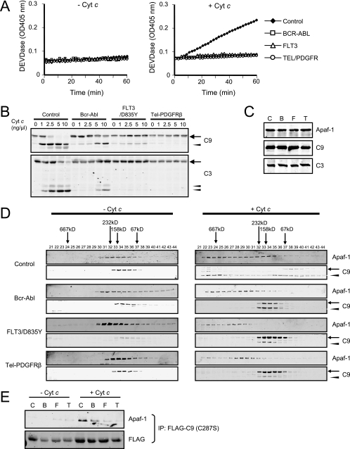
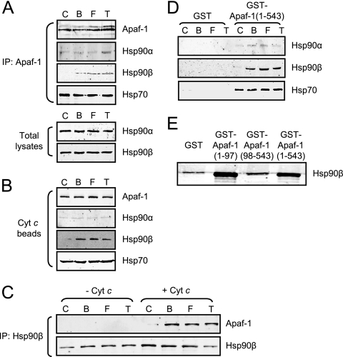
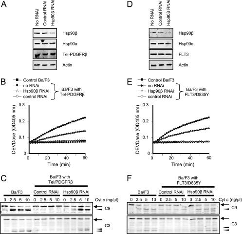
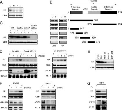
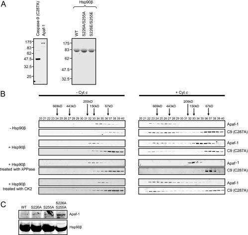
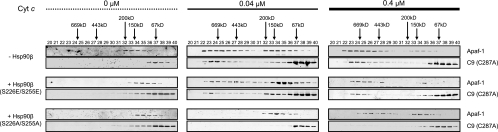
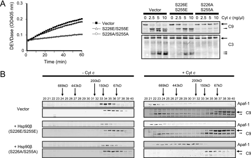
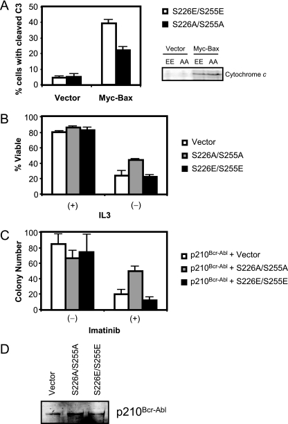
Constitutively active tyrosine kinases promote leukemogenesis by increasing cell proliferation and inhibiting apoptosis. However, mechanisms underlying apoptotic inhibition have not been fully elucidated. In many settings, apoptosis occurs by mitochondrial cytochrome c release, which nucleates the Apaf-1/caspase-9 apoptosome. Here we report that the leukemogenic kinases, Bcr-Abl, FLT3/D835Y, and Tel-PDGFRbeta, all can inhibit apoptosome function. In cells expressing these kinases, the previously reported apoptosome inhibitor, Hsp90beta, bound strongly to Apaf-1, preventing cytochrome c-induced Apaf-1 oligomerization and caspase-9 recruitment. Hsp90beta interacted weakly with the apoptosome in untransformed cells. While Hsp90beta was phosphorylated at Ser 226/Ser 255 in untransformed cells, phosphorylation was absent in leukemic cells. Expression of mutant Hsp90beta (S226A/S255A), which mimics the hypophosphorylated form in leukemic cells, conferred resistance to cytochrome c-induced apoptosome activation in normal cells, reflecting enhanced binding of nonphosphorylatable Hsp90beta to Apaf-1. In Bcr-Abl-positive mouse bone marrow cells, nonphosphorylatable Hsp90beta expression conferred imatinib (Gleevec) resistance. These data provide an explanation for apoptosome inhibition by activated leukemogenic tyrosine kinases and suggest that alterations in Hsp90beta-apoptosome interactions may contribute to chemoresistance in leukemias.


Figures

FIG. 1.
FIG. 1.
Leukemogenic tyrosine kinases inhibit recruitment of caspase-9 to Apaf-1. (A) Cell lysates were prepared from control Ba/F3 cells or Ba/F3 cells expressing Bcr-Abl, FLT3/D835Y, or Tel-PDGFRβ and incubated with 0 or 2.5 ng/μl cytochrome c (Cyt c) and 1 mM dATP. Caspase-3 activity was assayed by measuring cleavage of DEVD-pNA. (B) Lysates were incubated with 1 mM dATP and various concentrations of cytochrome c, and immunoblotting was performed for caspase-9 (C9) and caspase-3 (C3). Procaspases and cleaved caspases are indicated by arrows and arrowheads, respectively. (C) Total cell lysates from control Ba/F3 cells (C) or Ba/F3 cells expressing Bcr-Abl (B), FLT3/D835Y (F), or Tel-PDGFRβ (T) were immunoblotted with anti-Apaf-1, anti-caspase-9 (C9), and anti-caspase-3 (C3) antibodies. (D) Cell lysates were separated on a Superdex 200 column before and after incubation with 2.5 ng/μl cytochrome c and 1 mM dATP for 30 min. Immunoblotting was performed for Apaf-1 and caspase-9. Procaspase-9 and cleaved caspase-9 are indicated by arrows and arrowheads, respectively. (E) Ba/F3 cells were transfected with FLAG-tagged caspase-9 (C287S), and lysates were prepared. Immunoprecipitation (IP) was performed with or without addition of cytochrome c (2.5 ng/μl) and dATP (1 mM). Pellets were analyzed by immunoblotting with anti-Apaf-1 and anti-FLAG antibodies.
FIG. 2.
FIG. 2.
Apaf-1 binds to Hsp90β in cells expressing leukemogenic tyrosine kinases. (A) Control Ba/F3 cells (C) and those expressing Bcr-Abl (B), FLT3/D835Y (F), and Tel-PDGFRβ (T) were subjected to immunoprecipitation (IP) with anti-Apaf-1 antibody. The IP pellets were analyzed for Hsp90α, Hsp90β, and Hsp70 and Apaf-1 (top). Total Ba/F3 cell lysates were immunoblotted with anti-Hsp90α and -β antibodies (bottom). (B) Lysates were incubated with cytochrome c (Cyt c) beads, and pellets were subjected to immunoblotting with anti-Apaf-1, anti-Hsp90α and -β, and anti-Hsp70 antibodies. (C) IP with anti-Hsp90β antibody was carried out for the Ba/F3 cell lysates before and after cytochrome c (2.5 ng/μl) and dATP (1 mM) addition. The pellets were analyzed by immunoblotting with anti-Apaf-1 antibody. (D) Cell lysates were incubated with GST-Apaf-1 (1-543) or GST alone. Protein complexes were retrieved by using glutathione beads, and immunoblotting was performed for Hsp90α and -β or Hsp70. (E) Ba/F3 lysates expressing Tel-PDGFRβ were incubated with GST, GST-Apaf-1 (1-97), GST-Apaf-1 (98-543), or GST-Apaf-1 (1-543). Protein complexes were retrieved by using glutathione beads, and immunoblotting was performed for Hsp90β.
FIG. 3.
FIG. 3.
Hsp90β knockdown partially restores sensitivity to cytochrome c in Ba/F3 cells expressing Tel-PDGFRβ (A to C) or FLT3/D835Y (D to F). (A and D) Ba/F3 cells expressing Tel-PDGFRβ (A) or FLT3/D835Y (D) were treated with Hsp90β-specific siRNA or control siRNA. Total cell lysates were immunoblotted for Hsp90β, Hsp90α, Tel-PDGFRβ, FLT3, and actin. (B and E) Caspase activity was assayed by measuring cleavage of DEVD-pNA following incubation of the cell lysates with 5 ng/μl cytochrome c and 1 mM dATP. (C and F) Immunoblotting was performed for caspase-9 and caspase-3 upon addition of various amounts of exogenous cytochrome c (Cyt c) to the lysates. Procaspase-9/procaspase-3 and cleaved caspase-9/caspase-3 are indicated by arrows and arrowheads, respectively. RNAi, RNA interference.
FIG. 4.
FIG. 4.
Phosphorylation of Hsp90β at Ser 226/255 is suppressed in cells expressing the tyrosine kinases. (A) Recombinant His-tagged Hsp90β protein on nickel beads was incubated with the Ba/F3 cell lysates in the presence of [γ-32P]ATP; control Ba/F3 cells (C); and those expressing Bcr-Abl (B), FLT3/D835Y (F), and Tel-PDGFRβ (T). (B) Various deletion mutants of Hsp90β were made as GST fusion proteins and incubated with control Ba/F3 lysates (C) or lysates expressing Bcr-Abl (B) in the presence of [γ-32P]ATP. (C) Two point mutations (S226A and S255A) were introduced into GST-Hsp90β (178-300). The GST fusion proteins were incubated with control Ba/F3 lysates in the presence of [γ-32P]ATP. WT, wild type. (D) Ba/F3 cells expressing either wild-type Bcr-Abl or Bcr-Abl carrying the T315I mutation were treated with the Abl kinase inhibitor imatinib (1 μM) for the time indicated (left panel). Ba/F3 cells expressing FLT3/D835Y were treated with the FLT3 kinase inhibitor PKC412 (20 nM) over time (right panel). After each treatment, cell lysates were prepared and incubated with GST-Hsp90β (178-300) in the presence of [γ-32P]ATP. (E) GST-Hsp90β (178-300) was incubated with lysates from U-937, TF-1, MV4-11, Ku812, and SUP-B15 cells in the presence of [γ-32P]ATP. (F) Ku812 and MV4-11 cells were treated with imatinib (1 μM) and PKC412 (20 nM), respectively, for the indicated time. The cell lysates were incubated with GST-Hsp90β (178-300) in the presence of [γ-32P]ATP. (G) THP-1 cells were treated with or without recombinant human FLT3 ligand (FL; 50 ng/ml) for 2 h. The cell lysates were incubated with GST-Hsp90β (178-300) in the presence of [γ-32P]ATP. 32P incorporation and Coomassie blue staining (CBB) are shown. FLT3 was immunoprecipitated from whole-cell lysates with anti-FLT3 antibody. Western blotting was performed for Bcr-Abl, FLT3, and phosphotyrosine.
FIG. 5.
FIG. 5.
The phosphorylation of Hsp90β controls its inhibitory effect on Apaf-1 oligomerization. (A) Recombinant human caspase-9 (C287A) and human Apaf-1 proteins were produced and purified from BL21(DE3) and Sf9 cells, respectively, as described in Materials and Methods (left). Likewise, recombinant human Hsp90β proteins (wild type [WT] and mutants) were generated and purified from Sf9 cells (right). Shown is a sodium dodecyl sulfate-polyacrylamide gel stained with Coomassie blue; 5 μg of caspase-9 (C287A), 3 μg of Apaf-1, and 20 μg of Hsp90β were loaded per lane. Molecular masses (kDa) are indicated on the left side. (B) Purified recombinant Apaf-1 (0.4 μM) was mixed with catalytically inactive caspase-9 (C287A) (0.8 μM). After incubation with or without 1 mM dATP and 0.4 μM cytochrome c, the samples were loaded onto a Superdex 200 column, and each column fraction was analyzed for Apaf-1 and caspase-9 (C287A) by immunoblotting (top panel). The same experiment was performed in the presence of recombinant Hsp90β (1 μM) that was untreated (second panel) or pretreated with lambda phosphatase (λPPase; third panel) or CK2 (bottom panel). (C) His-Hsp90β wild type (WT), Hsp90β (S226A), Hsp90β (S255A), or Hsp90β (S226/255A) was incubated with control Ba/F3 cell lysates in the presence of 5 ng/μl cytochrome c. His-tagged proteins were retrieved on nickel beads, and the resultant pellets were analyzed for the presence of Apaf-1.
FIG. 6.
FIG. 6.
Hsp90β (S226A/S255A) inhibits Apaf-1 oligomerization and caspase-9 recruitment. Recombinant Apaf-1 (0.4 μM) was mixed with catalytically inactive caspase-9 [C9 (C287A)] (0.8 μM) and recombinant Hsp90β (1 μM; S226E/S255E and S226A/S255A) and then incubated with dATP (1 mM) and 0.1 μM or 0.4 μM of cytochrome c. After incubation, each sample was loaded onto a Superdex 200 column, and each column fraction was analyzed for Apaf-1 and C9 (C287A) by immunoblotting.
FIG. 7.
FIG. 7.
Hsp90β (S226A/S255A) causes post-cytochrome c protection in normal Ba/F3 cell lysates. (A) Control Ba/F3 cells were infected with a retroviral vector encoding Hsp90β (S226E/S255E) or Hsp90β (S226A/S255A) or empty vector. GFP-positive cells were sorted by FACS. Cell lysates were prepared and incubated with 5 ng/μl cytochrome c and 1 mM dATP. Caspase-3 activity was assayed by measuring the cleavage of DEVD-pNA over time (left). Likewise, the lysates were incubated with 1 mM dATP and various concentrations of cytochrome c (Cyt c), and immunoblotting was performed for caspase-9 (C9) and caspase-3 (C3) (right). Procaspase-9/procaspase-3 and cleaved caspase-9/caspase-3 are indicated by arrows and arrowheads, respectively. (B) Ba/F3 cell lysates expressing the empty vector, Hsp90β (S226E/S255E), or Hsp90β (S226A/S255A) were incubated with or without cytochrome c (5 ng/μl) and dATP (1 mM) and loaded onto a Superdex 200 column. Each column fraction was analyzed for Apaf-1 and caspase-9 by immunoblotting.
FIG. 8.
FIG. 8.
Expression of Hsp90β (S226A/S255A) renders cells resistant to proapoptotic stimuli. (A) Ba/F3 cells stably expressing Hsp90β (S226E/S255E) or Hsp90β (S226A/S255A) were transfected with myc-Bax. Five hours after transfection, the cells were fixed and stained with cleaved caspase-3 (C3) antibody and an Alexa 647-conjugated secondary antibody. The population of the cleaved caspase-3-positive cells was analyzed by FACS. (B) Ba/F3 cells were transfected with 2 μg of FLAG-tagged Hsp90β (S226E/S255E) or Hsp90β (S226A/S255A). Twenty-four hours after transfection, cells were transferred to IL-3-free medium (or IL-3-containing medium as a control) and further cultured for 20 h. The percentage of viable cells was analyzed by PI exclusion with FACS. (C) Mouse bone marrow KLS cells were coinfected with retroviral vectors encoding p210Bcr-Abl and Hsp90β (S226E/S255E) or Hsp90β (S226A/S255A). Coinfected cells were further selected by FACS and plated in methylcellulose medium. The colony numbers were counted 7 days after plating. Averages with standard errors of the means are shown. (D) Ba/F3 cells stably expressing p210Bcr-Abl were infected with a retroviral vector encoding Hsp90β (S226E/S255E) or Hsp90β (S226A/S255A) or empty vector. GFP-positive cells were selected by FACS and further cultured for 2 weeks. Cells were harvested and analyzed for expression of p210Bcr-Abl.

Similar articles

Cited by

References

    1. Abu-Duhier, F. M., A. C. Goodeve, G. A. Wilson, R. S. Care, I. R. Peake, and J. T. Reilly. 2001. Identification of novel FLT-3 Asp835 mutations in adult acute myeloid leukaemia. Br. J. Haematol. 113983-988. - PubMed
    1. Amarante-Mendes, G. P., A. J. McGahon, W. K. Nishioka, D. E. Afar, O. N. Witte, and D. R. Green. 1998. Bcl-2-independent Bcr-Abl-mediated resistance to apoptosis: protection is correlated with up regulation of Bcl-xL. Oncogene 161383-1390. - PubMed
    1. An, W. G., T. W. Schulte, and L. M. Neckers. 2000. The heat shock protein 90 antagonist geldanamycin alters chaperone association with p210bcr-abl and v-src proteins before their degradation by the proteasome. Cell Growth Differ. 11355-360. - PubMed
    1. Beere, H. M. 2005. Death versus survival: functional interaction between the apoptotic and stress-inducible heat shock protein pathways. J. Clin. Investig. 1152633-2639. - PMC - PubMed
    1. Bhatia, R., M. Holtz, N. Niu, R. Gray, D. S. Snyder, C. L. Sawyers, D. A. Arber, M. L. Slovak, and S. J. Forman. 2003. Persistence of malignant hematopoietic progenitors in chronic myelogenous leukemia patients in complete cytogenetic remission following imatinib mesylate treatment. Blood 1014701-4707. - PubMed

Publication types

MeSH terms