. 2008 Sep;28(17):5420-31.
doi: 10.1128/MCB.00717-08. Epub 2008 Jun 30.

GATA4 is a direct transcriptional activator of cyclin D2 and Cdk4 and is required for cardiomyocyte proliferation in anterior heart field-derived myocardium

Affiliations

GATA4 is a direct transcriptional activator of cyclin D2 and Cdk4 and is required for cardiomyocyte proliferation in anterior heart field-derived myocardium

Anabel Rojas et al. Mol Cell Biol. 2008 Sep.

Abstract

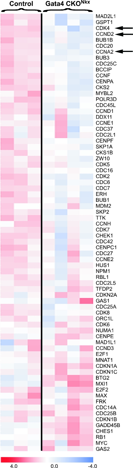
The anterior heart field (AHF) comprises a population of mesodermal progenitor cells that are added to the nascent linear heart to give rise to the majority of the right ventricle, interventricular septum, and outflow tract in mammals and birds. The zinc finger transcription factor GATA4 functions as an integral member of the cardiac transcription factor network in the derivatives of the AHF. In addition to its role in cardiac differentiation, GATA4 is also required for cardiomyocyte replication, although the transcriptional targets of GATA4 required for proliferation have not been previously identified. In the present study, we disrupted Gata4 function exclusively in the AHF and its derivatives. Gata4 AHF knockout mice die by embryonic day 13.5 and exhibit hypoplasia of the right ventricular myocardium and interventricular septum and display profound ventricular septal defects. Loss of Gata4 function in the AHF results in decreased myocyte proliferation in the right ventricle, and we identified numerous cell cycle genes that are dependent on Gata4 by microarray analysis. We show that GATA4 is required for cyclin D2, cyclin A2, and Cdk4 expression in the right ventricle and that the Cyclin D2 and Cdk4 promoters are bound and activated by GATA4 via multiple consensus GATA binding sites in each gene's proximal promoter. These findings establish Cyclin D2 and Cdk4 as direct transcriptional targets of GATA4 and support a model in which GATA4 controls cardiomyocyte proliferation by coordinately regulating numerous cell cycle genes.


Figures

FIG. 1.
FIG. 1.
Inactivation of Gata4 in the AHF results in lethality due to right ventricular hypoplasia and VSDs. (A and B) Whole-mount in situ hybridization showing expression of Gata4 mRNA in control (A) and Gata4 AHF knockout (B) hearts at E10.5. The excision of the Gata4 floxed allele by Mef2c-AHF-Cre results in loss of Gata4 mRNA in the right ventricle (RV) and outflow tract (OFT) in Gata4 AHF knockout embryos. LV, left ventricle. (C and D) Gata4 AHF knockout embryos (D) display obvious vascular hemorrhage (arrowheads) compared to littermate controls (C) at E13.5. (E to H) Hematoxylin- and eosin-stained transverse sections of littermate control (E) and Gata4 AHF knockout (F) embryos show that the formation of the ventricular septum (arrowheads) is aberrant at E13.5 in Gata4 AHF knockout embryos compared to controls. LA, left atrium; RA, right atrium. (G and H) The compact wall myocardium of the right ventricle (asterisks) is thinner at E13.5 in Gata4 AHF knockout embryos (H) than in littermate control embryos (G). Bars, 100 μm. Genotypes for control (Gata4flox/flox) and Gata4 AHF knockout (CreTg/0; Gata4flox/flox) embryos are indicated. n was 4 for each genotype.
FIG. 2.
FIG. 2.
Gata4 AHF knockout embryos have profound myocardial proliferation defects. (A to H) Immunohistochemical analyses of proliferation markers on transverse sections show that Gata4 AHF knockout embryos (B, D, F, and H) have reduced proliferation compared to control embryos (A, C, E, and G) at E10.5. (A and B) Gata4 AHF knockout embryos display decreased staining of the nuclear antigen Ki67 (brown) in the right ventricular myocardium and interventricular septum compared to control embryos (asterisks). (C and D) Closer view of the right ventricle (RV) shows that Ki67 staining in Gata4 AHF knockout hearts is reduced in the myocardium (myo) but not in other regions where Gata4 was not inactivated, such as the epicardium (epi). (E and F) BrdU incorporation is diminished in the myocardium of the right ventricle of Gata4 AHF knockout embryos compared to control embryos (asterisks). (G and H) Expression of the mitotic marker phospho-histone H3 (pHH3) is reduced in the right ventricle in Gata4 AHF knockout embryos compared to control littermates (arrowheads). No differences in the staining of any of these proliferation markers between Gata4 AHF knockout and control embryos was observed in the left ventricle (LV). (I and J) TUNEL staining on transverse sections of embryonic hearts shows no difference in apoptosis between Gata4 AHF knockout and control embryos at E10.5. Genotypes for control (Gata4flox/flox) and Gata4 AHF knockout (Gata4flox/flox; Mef2c-AHF-CreTg/0) embryos are indicated. (K and L) Quantification of BrdU (K)- and pHH3 (L)-labeled cells shows a significant decrease in proliferation in the right ventricle of conditional knockout (CKO) embryos compared to littermate controls. The total number of DAPI-labeled cells and the number of BrdU- or pHH3-labeled cells were determined by counting cells in a series of sections from three CKO and three control hearts. Data are presented as the mean percentages of cells labeled with BrdU or pHH3 plus standard errors of the means from three hearts of each genotype. P values were calculated using a two-tailed, unpaired t test.
FIG. 3.
FIG. 3.
GATA4 regulates multiple cell cycle control genes. Affymetrix gene expression data were analyzed by gene set enrichment (72). Several sets of genes with known roles in cell cycle regulation showed statistically significant, concordant differences between control (Gata4flox/+) and Gata4 CKONkx (Gata4flox/flox; Nkx2-5Cre/+) hearts. The heat map of genes comprising the cell cycle gene set with the most significant statistical score (Brentani cell cycle gene set) is shown (10). Color indicates degree of upregulation (red) or downregulation (blue) relative to the mean expression across all samples (see the color scale at the bottom). Numerous cell cycle control genes were significantly downregulated in Gata4 CKONkx hearts compared to controls, including Cyclin D2 (CCND2), Cyclin A2 (CCNA2), and Cdk4, which are denoted by arrows.
FIG. 4.
FIG. 4.
Gata4 inactivation leads to decreased expression of cell cycle proteins. Immunohistochemical staining of transverse sections with anti-cyclin D2 (A and B), anti-Cdk4 (C and D), and anti-cyclin A2 (E and F) antibodies shows that the expression of all three cell cycle proteins is dramatically reduced in the right ventricular myocardium in Gata4 AHF knockout embryos (B, D, and F) compared to that in littermate control embryos (A, C, and E) at E10.5 (asterisks). In panels A to D, staining for cyclin D2 and Cdk4 is red and nuclei have been counterstained with DAPI (blue). In panels E and F, cyclin A2 is stained in brown. No differences in cyclin D2, Cdk4, or cyclin A2 protein expression between knockout and control embryos were observed in regions outside the Mef2c-AHF-Cre domain, such as the left ventricle (LV). Genotypes for control (Gata4flox/flox) and Gata4 AHF knockout (CreTg/0;Gata4flox/flox) embryos are indicated. RV, right ventricle.
FIG. 5.
FIG. 5.
GATA4 binds directly to the Cyclin D2 and Cdk4 promoters in vivo and in vitro. (A and B) Schematic representations of the mouse Cyclin D2 and Cdk4 promoters. The Cyclin D2 construct encompasses nucleotides −671 to +260 relative to the transcriptional start site (bent arrow). The Cdk4 construct encompasses nucleotides −771 to +56 relative to the transcriptional start site (bent arrow). Boxes denote consensus GATA binding sites in the Cyclin D2 (Gata I [GI], Gata II, and Gata III) and Cdk4 (Gata I/II and Gata III) promoters. Arrowheads indicate the locations of primers used to amplify regions of the Cyclin D2 and Cdk4 promoters, containing consensus GATA sites, in ChIP assays. (C and D) Recombinant GATA4 proteins were transcribed and translated in vitro and used in EMSA with radiolabeled double-stranded oligonucleotides encompassing the CyclinD2 Gata I (C, lanes 1 to 4), Gata II (C, lanes 5 to 8), and Gata III (C, lanes 9 to 12) sites and the Cdk4 Gata I/II (D, lanes 1 to 4) and Gata III (D, lanes 5 to 8) sites. Lanes 1, 5, and 9 (C) and lanes 1 and 5 (D) contain reticulocyte lysate without recombinant GATA4 (−). GATA4 efficiently bound to all GATA sites in the Cyclin D2 and Cdk4 promoters in vitro. mI, mutant version of the Gata I site. (E) GATA4 binds to the endogenous Cyclin D2 and Cdk4 promoters in vivo. Differentiated P19CL6 cardiomyocytes were subjected to ChIP to detect endogenous GATA4 bound to the Cyclin D2 and Cdk4 promoters using anti-GATA4 antibody. Following ChIP, the Cyclin D2 promoter was detected using primers P1 and P2 (lanes 1 to 3) and the Cdk4 promoter was detected using primers P1 and P2 (lanes 4 to 6) and primers P3 and P4 (lanes 7 to 9). In addition, primers were used to detect the second exon of Cyclin D2 as a nonspecific control (lanes 10 to 12). PCR products were analyzed by agarose gel electrophoresis. Lanes 3, 6, 9, and 12 contain PCR products obtained following ChIP using anti-GATA4 antibody (α-G4). Lanes 1, 4, 7, and 10 contain PCR products obtained following ChIP using a nonspecific anti-IgG (α-IgG). Lanes 2, 5, 8, and 11 contain PCR products from input DNA (Inp) amplified prior to immunoprecipitation. ChIP products were detected only from promoter regions in samples where anti-GATA4 antibody was used. Sizes in bp are shown at the left.
FIG. 6.
FIG. 6.
The GATA sites in the Cyclin D2 and Cdk4 promoters are required for activation. (A and B) The Cyclin D2 (A) and Cdk4 (B) promoters were significantly activated by endogenous GATA factors in differentiated P19CL6 cardiomyocytes (lane 2) compared to the activity of the parent reporter construct, pAUG-β-gal (lane 1), and mutation of the GATA sites in each promoter significantly attenuated activity (lane 3). RLU, relative light units. (C) The Cyclin D2 promoter was significantly activated in C3H10T1/2 fibroblasts (lane 2) compared to the activity of the parent reporter (lane 1), and mutation of the GATA sites significantly attenuated promoter activation (lane 3). (D) GATA4-EnR inhibits activation of the Cyclin D2 promoter in C3H10T1/2 cells. Cotransfection of GATA4-EnR expression plasmid resulted in potent repression of the Cyclin D2 reporter construct (compare lanes 3 and 4). (E) Cotransfection of a GATA4-VP16 expression plasmid with the Cyclin D2-lacZ reporter plasmid resulted in potent transactivation of the Cyclin D2 promoter in C3H10T1/2 cells (lane 6). Mutation of the GATA sites (mGATA) in the Cyclin D2 promoter disrupted transactivation by GATA4-VP16 (lane 4). In all cases, the total amount of transfected plasmid DNA was held constant by addition of the appropriate amount of the parent expression plasmid. Error bars represent the standard errors of the means for at least three independent triplicate sets of transfections and analyses for each panel. P values were calculated by two-tailed, unpaired t test.

References

    1. Abu-Issa, R., and M. L. Kirby. 2007. Heart field: from mesoderm to heart tube. Annu. Rev. Cell Dev. Biol. 2345-68. - PubMed
    1. Adams, P. D. 2001. Regulation of the retinoblastoma tumor suppressor protein by cyclin/cdks. Biochim. Biophys. Acta 1471M123-M133. - PubMed
    1. Adams, P. D., and W. G. Kaelin, Jr. 1995. Transcriptional control by E2F. Semin. Cancer Biol. 699-108. - PubMed
    1. Ai, D., X. Fu, J. Wang, M. F. Lu, L. Chen, A. Baldini, W. H. Klein, and J. F. Martin. 2007. Canonical Wnt signaling functions in second heart field to promote right ventricular growth. Proc. Natl. Acad. Sci. USA 1049319-9324. - PMC - PubMed
    1. Arceci, R. J., A. A. King, M. C. Simon, S. H. Orkin, and D. B. Wilson. 1993. Mouse GATA-4: a retinoic acid-inducible GATA-binding transcription factor expressed in endodermally derived tissues and heart. Mol. Cell. Biol. 132235-2246. - PMC - PubMed

Publication types

MeSH terms