. 2008 Sep;28(17):5337-47.
doi: 10.1128/MCB.00563-08. Epub 2008 Jun 30.

The pentatricopeptide repeat protein PPR5 stabilizes a specific tRNA precursor in maize chloroplasts

Affiliations

The pentatricopeptide repeat protein PPR5 stabilizes a specific tRNA precursor in maize chloroplasts

Susanne Beick et al. Mol Cell Biol. 2008 Sep.

Abstract

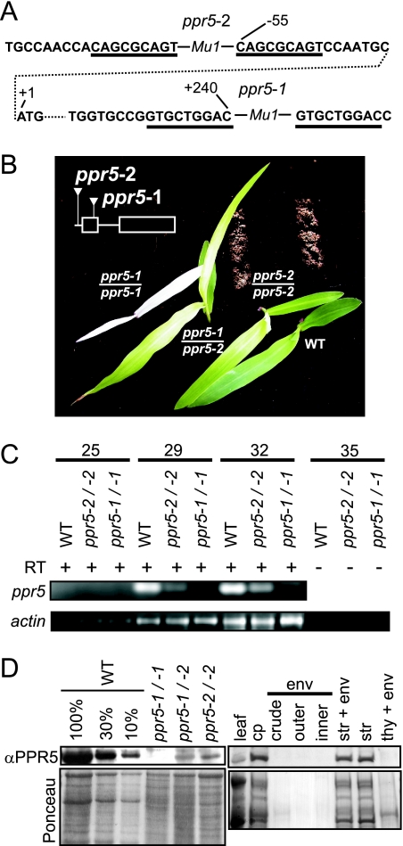
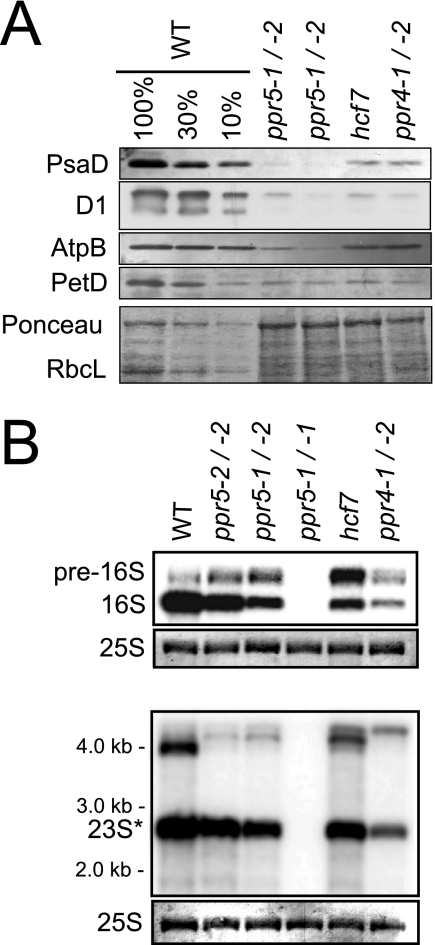
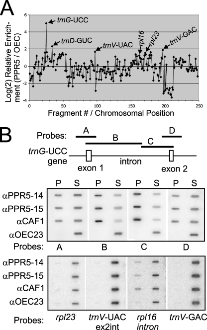
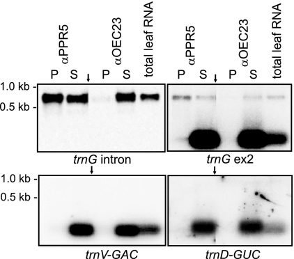
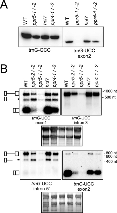
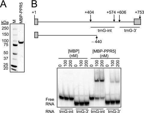
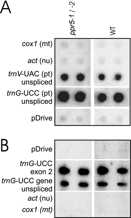
Genes for pentatricopeptide repeat (PPR) proteins are found in all eukaryotic genomes analyzed but are particularly abundant in land plants. The majority of analyzed PPR proteins play a role in the processing or translation of organellar RNAs. Few PPR proteins have been studied in detail, and the functional repertoire and mechanisms of action of proteins in the PPR family are poorly understood. Here we analyzed a maize ortholog of the embryo-essential Arabidopsis thaliana gene AtPPR5. A genome-wide analysis of chloroplast RNAs that coimmunoprecipitate with Zea mays PPR5 (ZmPPR5) demonstrated that ZmPPR5 is bound in vivo to the unspliced precursor of trnG-UCC. Null and hypomorphic Zmppr5 insertion mutants are embryo viable but are deficient for chloroplast ribosomes and die as seedlings. These mutants show a dramatic decrease in both spliced and unspliced trnG-UCC RNAs, while the transcription of trnG-UCC is unaffected. These results, together with biochemical data documenting the sequence-specific binding of recombinant PPR5 to the trnG-UCC group II intron, suggest that PPR5 stabilizes the trnG-UCC precursor by directly binding and protecting an endonuclease-sensitive site. These findings add to the evidence that chloroplast-localized PPR proteins that are embryo essential in Arabidopsis typically function in the biogenesis of the plastid translation machinery.
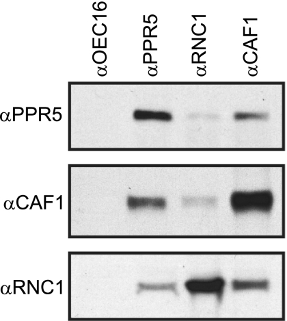

Figures

FIG. 1.
FIG. 1.
Maize ppr5 mutants. (A) Positions of the Mu transposon insertions in ppr5-1 and ppr5-2 mutants. The DNA sequence flanking the Mu insertions is shown. Residue numbers with respect to the start codon in the genomic sequence are indicated. The start codon is shown in the second row, connected to upstream and downstream noncontiguous sequences by a dotted line. The type of Mu element at each site (Mu1) was deduced from polymorphisms in the terminal inverted repeat sequences. The direct nonamer repeats that were duplicated during the insertion process are underlined. (B) Phenotypes of ppr5 mutant seedlings grown on soil for 14 days. The positions of the Mu insertions with respect to the ppr5 protein coding sequence are diagrammed in the insert. (C) ppr5 mRNA is decreased in ppr5 mutants. RT-PCR was used to assay ppr5 mRNA levels using primers located in exon 1 and exon 2 that flank the Mu1 insertion in ppr5-1 mutants. The number of PCR cycles is indicated at the top. The bottom panel shows the RT-PCR amplification of actin mRNA in the same RNA samples. The 35-cycle PCRs on the same samples without the inclusion of a RT step demonstrate that the signals do not arise from contaminating genomic DNA. Furthermore, no intron-containing amplification products were detected, demonstrating that the bands observed arose from RNA templates. (D) PPR5 localizes to chloroplast stroma. For the left panel, total leaf proteins from wild-type and ppr5 mutants were separated by SDS-polyacrylamide gel electrophoresis, blotted, and probed with the PPR5 antibody. A portion of the same blot stained with Ponceau S is shown below. Dilutions of the wild-type sample were included to aid in quantification. For the right panel, an immunoblot of the subcellular fractions described and verified previously (1) was probed with the PPR5 antibody. The chloroplast and subchloroplast samples are derived from the same quantity of chloroplasts (cp). Replicate blots were probed previously to detect markers for the stroma (str; Cp60), the thylakoid membrane (thy; PsbA), and the inner envelope membrane (env; IM35) (1). WT, wild type.
FIG. 2.
FIG. 2.
Loss of plastid rRNAs and plastid-encoded proteins in ppr5 mutants. (A) Immunoblot analysis of photosynthetic enzyme accumulation in ppr5 mutants. Total leaf proteins (5 μg or the indicated dilution of the wild-type sample) were analyzed by probing immunoblots with antisera to representative subunits of photosystem I (PsaD), photosystem II (D1), ATP synthase (AtpB), and the cytochrome b6f complex (PetD). The same filter was stained with Ponceau S to visualize total proteins (bottom panel). RbcL, the large subunit of Rubisco, is marked. The hcf7 and ppr4 mutants were shown previously to have a global loss of plastid-encoded proteins due to a plastid ribosome deficiency (3, 42). (B) RNA gel blot hybridizations showing plastid rRNA defects in ppr5 mutants. Total leaf RNA (0.5 μg) was analyzed by hybridization to probes for the plastid 16S (top panel) or 23S (bottom panel) rRNA. Excerpts of the same filters stained with methylene blue show the cytosolic 25S rRNA as a loading control. Previously described mutants with lesions in 16S rRNA maturation (hcf7 and ppr4-1/ppr4-2) are shown for comparison (3, 42). The maize chloroplast 23S rRNA exists as two cleavage products in vivo, one of which overlaps the probe used here (23S*). WT, wild type.
FIG. 3.
FIG. 3.
Identification of RNAs associated with PPR5 in chloroplast stroma. (A) Summary of RIP-chip data. The log2-transformed enrichment ratios (F635:F532) were normalized between two assays involving PPR5 antisera from different immunized rabbits and two control experiments with antibodies to OEC23 and OEC16. The median normalized values for replicate spots from the control assays were subtracted from those from the PPR5 assays and plotted according to fragment number, which reflects their chromosomal position. Fragments for which at least three replicate spots per array did not meet our background cutoff in the supernatant (F532) channel were not used in this plot and appear as gaps in the curve. (B) Validation of RIP-chip data by slot blot hybridization of RNAs that coimmunoprecipitate with PPR5. A schematic map of the trnG-UCC gene and the probes used for slot blot hybridization (A through D) are shown at the top. One-sixth of the RNA recovered from each immunoprecipitation pellet (P) and 1/12 of the RNA recovered from each supernatant (S) were applied to replicate slot blots and hybridized with probes A through D. Antibodies from two immunized rabbits were used in replicate assays (αPPR5-14 and αPPR5-15). An immunoprecipitation with antibody to OEC23 was used as a negative control. An immunoprecipitation with antibody against the splicing factor CAF1, which includes unspliced trnG-UCC among its ligands (34), served as a positive control. The same blots were reprobed after the decay of the previous signal to detect four RNAs for which the RIP-chip data suggested possible minor enrichment (bottom panel).
FIG. 4.
FIG. 4.
Coimmunoprecipitation of unspliced trnG-UCC precursors but not spliced trnG-UCC with PPR5. RNA purified from a PPR5 immunoprecipitation supernatant (S) and pellet (P) was analyzed on duplicate RNA gel blots by hybridization to the indicated exon-specific and intron-specific probes from the trnG-UCC locus (top panels). An immunoprecipitation with antibody to OEC23 served as a negative control. The probes were removed, and the blots were reprobed for the presence of trnD-GUC and trnV-GAC (bottom panels). Arrows point to where irrelevant lanes have been removed from images.
FIG. 5.
FIG. 5.
RNA gel blot hybridizations of trnG-UCC transcripts in ppr5 mutants. (A) Total leaf RNA (4 μg/lane) from seedlings with the indicated genotypes was fractionated on a 20% polyacrylamide gel and transferred to a nylon membrane. The membrane was cut into two strips, which were hybridized with the indicated oligonucleotide probes. (B) Total leaf RNA (4 μg/lane) was fractionated on agarose gels, transferred to nylon membranes, and hybridized to the probes indicated. The filter shown in the upper panels was first hybridized to a trnG-UCC exon 1 probe and, after removal of this probe, reprobed with a probe from the 3′ region of the trnG-UCC intron. The filter shown in the lower panels was first hybridized to a trnG-UCC exon 2 probe and, after removal of this probe, reprobed with a probe from the 5′ region of the trnG-UCC intron. Shown below are the same blots stained with methylene blue to illustrate equal sample loading. The diagrams on the left depict the three transcripts detected (top, unspliced tRNA; middle, truncated precursor tRNA; bottom, spliced tRNA). WT, wild type; nt, nucleotides.
FIG. 6.
FIG. 6.
RNA binding activity of recombinant PPR5. (A) Purified MBP-PPR5 used in RNA binding assays. The protein recovered after amylose-affinity and gel filtration chromatography was resolved by SDS-polyacrylamide gel electrophoresis and stained with Coomassie brilliant blue. M, molecular mass marker. (B) Gel mobility shift assays demonstrating that MBP-PPR5 binds with high affinity to the central region of the trnG-UCC intron. The trnG-UCC locus is diagrammed at the top, with exons shown as rectangles, the intron as a line, and nucleotide positions with respect to the 5′ end (+1) indicated. The sequences represented in RNAs used for the gel mobility shift assays (trnG-int and trnG-3′) and the truncated RNA of ∼440 nucleotides that accumulates in a PPR5-dependent manner (see Fig. 5) are diagrammed below. MBP was used in parallel binding assays as a negative control.
FIG. 7.
FIG. 7.
Run-on transcription assay of trnG-UCC transcription in ppr5 mutants. (A) RNAs purified from run-on transcription assays involving plastids isolated from ppr5-1/ppr5-2 seedlings or from their normal siblings (wild-type) were hybridized to the indicated DNA fragments on nylon membranes. Both the trnV-UAC and trnG-UCC loci include a group II intron, and these probes encompass exon and intron sequences. DNA fragments from the mitochondrial (mt) cox1 gene, nuclear (nu) actin, and the pDrive vector into which these probes were cloned were included as controls for nonspecific hybridization. The signals were quantified and used to calculate the mean trnG-to-trnV ratio of two replicate experiments. This was similar for the wild-type (2.6) and ppr5 mutants (1.7). pt, plastid. (B) Run-on transcripts were hybridized to a single-stranded antisense oligonucleotide specific for trnG-UCC exon 2 and to a PCR product spanning the entire trnG-UCC gene. The enhanced signal observed with the oligonucleotide probe relative to the PCR product is typical for experiments of this nature (data not shown) and likely represents the inefficient denaturation of double-stranded DNAs on the filter. WT, wild type.
FIG. 8.
FIG. 8.
Sucrose gradient sedimentation analysis of PPR5-containing particles in stromal extract. Stromal extract was treated with RNase A or incubated under the same conditions in the absence of RNase and sedimented through sucrose gradients. An equal volume of each gradient fraction was analyzed on immunoblots probed with PPR5 antibody. Blots stained with Ponceau S are shown below. The last lane in each panel contains material that pelleted in the gradients. The position of ribosomes was determined by the pattern of Ponceau S-stained bands and by the appearance of rRNAs in these fractions (data not shown).
FIG. 9.
FIG. 9.
Coimmunoprecipitation of PPR5 with CAF1 and RNC1 from chloroplast extract. Chloroplast stroma was incubated with the affinity-purified antiserum indicated above each lane. Immunoprecipitates were analyzed on immunoblots to detect PPR5 (top panel), CAF1 (middle panel), or RNC1 (bottom panel). Antiserum to OEC16 was used as a negative control.

Similar articles

Cited by

References

    1. Asakura, Y., and A. Barkan. 2007. A CRM domain protein functions dually in group I and group II intron splicing in land plant chloroplasts. Plant Cell 193864-3875. - PMC - PubMed
    1. Barkan, A. 1998. Approaches to investigating nuclear genes that function in chloroplast biogenesis in land plants. Methods Enzymol. 29738-57.
    1. Barkan, A. 1993. Nuclear mutants of maize with defects in chloroplast polysome assembly have altered chloroplast RNA metabolism. Plant Cell 5389-402. - PMC - PubMed
    1. Barkan, A., and M. Goldschmidt-Clermont. 2000. Participation of nuclear genes in chloroplast gene expression. Biochimie 82559-572. - PubMed
    1. Barkan, A., L. Klipcan, O. Ostersetzer, T. Kawamura, Y. Asakura, and K. P. Watkins. 2007. The CRM domain: an RNA binding module derived from an ancient ribosome-associated protein. RNA 1355-64. - PMC - PubMed

Publication types

MeSH terms