Review
. 2008 Aug;29(5):513-34.
doi: 10.1210/er.2008-0003. Epub 2008 Jun 30.

Clinical, agricultural, and evolutionary biology of myostatin: a comparative review

Affiliations
Review

Clinical, agricultural, and evolutionary biology of myostatin: a comparative review

Buel D Rodgers et al. Endocr Rev. 2008 Aug.

Abstract

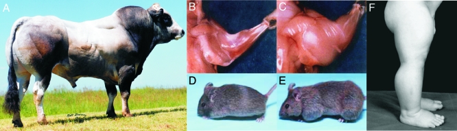
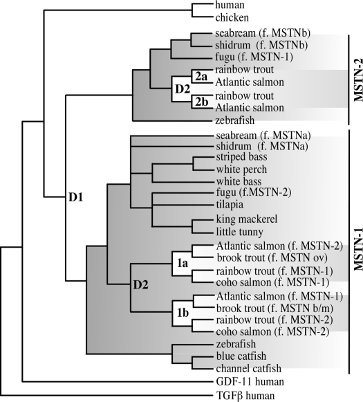
The discovery of myostatin and our introduction to the "Mighty Mouse" over a decade ago spurred both basic and applied research and impacted popular culture as well. The myostatin-null genotype produces "double muscling" in mice and livestock and was recently described in a child. The field's rapid growth is by no means surprising considering the potential benefits of enhancing muscle growth in clinical and agricultural settings. Indeed, several recent studies suggest that blocking myostatin's inhibitory effects could improve the clinical treatment of several muscle growth disorders, whereas comparative studies suggest that these actions are at least partly conserved. Thus, neutralizing myostatin's effects could also have agricultural significance. Extrapolating between studies that use different vertebrate models, particularly fish and mammals, is somewhat confusing because whole genome duplication events have resulted in the production and retention of up to four unique myostatin genes in some fish species. Such comparisons, however, suggest that myostatin's actions may not be limited to skeletal muscle per se, but may additionally influence other tissues including cardiac muscle, adipocytes, and the brain. Thus, therapeutic intervention in the clinic or on the farm must consider the potential of alternative side effects that could impact these or other tissues. In addition, the presence of multiple and actively diversifying myostatin genes in most fish species provides a unique opportunity to study adaptive molecular evolution. It may also provide insight into myostatin's nonmuscle actions as results from these and other comparative studies gain visibility in biomedical fields.


Figures

Figure 1
Figure 1
“Double muscling” and the myostatin-null phenotype. A, Muscle hypertrophy in Piedmontese cattle breeds is due to a missense mutation within the third exon of the bovine myostatin gene (4). [Picture reproduced with permission from the North American Piedmontese Cattle Association (NAPA, www.piedmontese.org).] B and C, Forearm musculature of wild-type (B) and myostatin “knock-out” (C) mice. [Reprinted by permission from Macmillan Publishers Ltd: Nature (Ref. 1), copyright 1997.] D and E, Wild-type (D) and follistatin transgenic (E) mice (31). [Copyright 2001 National Academy of Sciences, U.S.A.] F, Leg musculature of a 7-month-old infant boy with a null mutation within the splice donor site on exon 1 of the myostatin gene (26). [Copyright 2004 Massachusetts Medical Society. All rights reserved.]
Figure 2
Figure 2
Proteolytic processing of mature myostatin (MSTN) and conserved bioactivity. Left, Pro-myostatin is cleaved by a furin class protease at a conserved RXXR (R, arginine; X, any amino acid) epitope, and the resulting peptides dimerize via disulfide linkages at the indicated cysteines producing mature myostatin. The dominant-negative LAP sequesters myostatin dimers in a latent complex and can prohibit receptor activation. Right, Primary myosatellite cells from rainbow trout were incubated in 100-mm dishes with 50 ng/ml mouse myostatin (R&D Systems, Minneapolis, MN) or equimolar amounts of bovine serum albumin, and myostatin was added at 0 and 48 h. At each time point, cells were manually counted in each of six views. Mean values for each time point are shown (histogram), as are representative images.
Figure 3
Figure 3
Phylogenetic relationship of vertebrate myostatin (MSTN) homologs. The tree was constructed from previously published Maximum Likelihood and Bayesian Inference analyses (135,144,145). Clades for the two teleost fish paralogs, MSTN-1 and MSTN-2, are shaded. Within each clade are the additional salmonid paralogs (1a, MSTN-1a; etc.), which are indicated by reverse shading. Genome duplication events within bony fish and salmonid lineages are indicated as D1 and D2, respectively. The nomenclature for the fish homologs has been revised (146). Thus, the former (f.) names are indicated in parentheses (ov, ovarian; b/m, brain/muscle; GDF, growth/differentiating factor; TGF, transforming growth factor).
Figure 4
Figure 4
Comparative mapping of vertebrate myostatin genes and promoters. The genomic structure and organization of human (h), mouse (m), chicken (ckn) and rainbow trout (rt) myostatin (MSTN) genes are shown. Exons are boxed with open reading frames in white and untranslated regions in gray (if known). Exon sizes are indicated within the boxes, intron sizes in italics. Two in-frame stop codons within rtMSTN-2b are indicated by asterisks, and a 51-bp cassette missing from the second exon is indicated by dashed lines. The locations of putative cis elements within each gene’s promoter region (−2.4 and −1.5 kb for rtMSTN-2a and -2b, respectively; −2 kb for others) are indicated by the shapes in the key; only known myogenic elements are shown. Putative E-boxes are numbered and motifs determined to be functionally active in the human and mouse promoters are indicated by arrows.
Figure 5
Figure 5
Comparative mapping of myostatin coding domains. For each gene, sequential boxes represent coding domains from each of three exons. The first amino acid coded by each exon and the last of each protein are shown above. The sequences adjacent to each exonic boundary are shown inside the boxes. In all fish genes, the codon of the proline residue located at the first exonic boundary is partially coded by nucleotides in the first and second exons as shown. In-frame stop codons in the rainbow trout myostatin (MSTN)-2b paralog are indicated with asterisks. The location of each RXXR proteolytic processing domain is indicated in light gray, and the highly conserved domain that is eventually processed into the mature myostatin dimer is in dark gray.
Figure 6
Figure 6
Differential localization of myostatin and GDF-11 expression in mouse brains. Expression of both genes was determined by the Allen Brain Atlas project (202,203) using in situ hybridization. Brains from adult (56-d-old) male C56/B57 mice were hybridized with gene-specific probes as described (www.brain-map.org). Three-dimensional expression patterns were reconstructed using Brain Explorer 1.4 and sagittal section data files, both of which are available at the indicated website. A and B, Individual expression patterns of myostatin (MSTN) and GDF-11 expression, respectively. C, Overlay of A and B. D, Combined expression patterns on top of saggital nissl (left to right) with color-coded anatomical features (see website for key). E and F, Three-dimensional images of combined expression patterns with sagittal and horizontal nissls. Image and nissl orientation is indicated by the color-coded compass in upper right of panels D–F.
Figure 7
Figure 7
Expression levels of myostatin and GDF-11 in different brain regions. Expression of both genes was determined by the Allen Brain Atlas project (202,203) using in situ hybridization and gene-specific probes. Levels of expression were replotted from data available at www.brain-map.org and are scaled to normalized densities (number of expressing cells/anatomical space) and intensities (total level of expression/anatomical space) of 0 to 100. GDF, Growth/differentiation factor; RHP, retrohippocampal region; TH, thalamus; STRv, ventral striatum; STRd, dorsal striatum; STR, striatum; sAMY, striatum-like amydalar nuclei; PAL, pallidum; P, pons; OLF, olfactory bulb; MY, medulla; MB, midbrain; LSX, lateral septal complex; HY, hypothalamus; HPF, hippocampal formation; HIP, hippocampal region; CTX, cerebral cortex; CB, cerebellum.

References

    1. McPherron AC, Lawler AM, Lee SJ 1997 Regulation of skeletal muscle mass in mice by a new TGF-β superfamily member. Nature 387:83–90 - PubMed
    1. Grobet L, Martin LJ, Poncelet D, Pirottin D, Brouwers B, Riquet J, Schoeberlein A, Dunner S, Menissier F, Massabanda J, Fries R, Hanset R, Georges M 1997 A deletion in the bovine myostatin gene causes the double-muscled phenotype in cattle [see comments]. Nat Genet 17:71–74 - PubMed
    1. Kambadur R, Sharma M, Smith TP, Bass JJ 1997 Mutations in myostatin (GDF8) in double-muscled Belgian Blue and Piedmontese cattle. Genome Res 7:910–916 - PubMed
    1. McPherron AC, Lee SJ 1997 Double muscling in cattle due to mutations in the myostatin gene. Proc Natl Acad Sci USA 94:12457–12461 - PMC - PubMed
    1. Smith TP, Lopez-Corrales NL, Kappes SM, Sonstegard TS 1997 Myostatin maps to the interval containing the bovine mh locus. Mamm Genome 8:742–744 - PubMed

Publication types