. 2008 Jul 8;105(27):9355-60.
doi: 10.1073/pnas.0801294105. Epub 2008 Jun 30.

Syntrophin mutation associated with long QT syndrome through activation of the nNOS-SCN5A macromolecular complex

Affiliations

Syntrophin mutation associated with long QT syndrome through activation of the nNOS-SCN5A macromolecular complex

Kazuo Ueda et al. Proc Natl Acad Sci U S A. .

Abstract

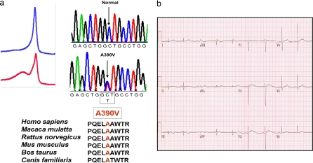
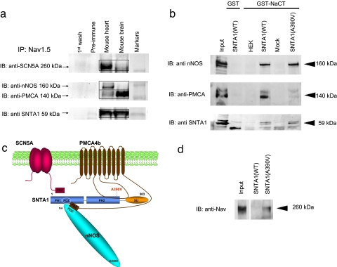
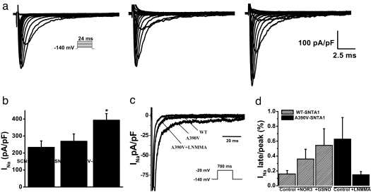
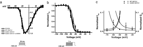
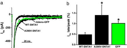
Mutations in 11 genes that encode ion channels or their associated proteins cause inherited long QT syndrome (LQTS) and account for approximately 75-80% of cases (LQT1-11). Direct sequencing of SNTA1, the gene encoding alpha1-syntrophin, was performed in a cohort of LQTS patients that were negative for mutations in the 11 known LQTS-susceptibility genes. A missense mutation (A390V-SNTA1) was found in a patient with recurrent syncope and markedly prolonged QT interval (QTc, 530 ms). SNTA1 links neuronal nitric oxide synthase (nNOS) to the nNOS inhibitor plasma membrane Ca-ATPase subtype 4b (PMCA4b); SNTA1 also is known to associate with the cardiac sodium channel SCN5A. By using a GST-fusion protein of the C terminus of SCN5A, we showed that WT-SNTA1 interacted with SCN5A, nNOS, and PMCA4b. In contrast, A390V-SNTA1 selectively disrupted association of PMCA4b with this complex and increased direct nitrosylation of SCN5A. A390V-SNTA1 expressed with SCN5A, nNOS, and PMCA4b in heterologous cells increased peak and late sodium current compared with WT-SNTA1, and the increase was partially inhibited by NOS blockers. Expression of A390V-SNTA1 in cardiac myocytes also increased late sodium current. We conclude that the A390V mutation disrupted binding with PMCA4b, released inhibition of nNOS, caused S-nitrosylation of SCN5A, and was associated with increased late sodium current, which is the characteristic biophysical dysfunction for sodium-channel-mediated LQTS (LQT3). These results establish an SNTA1-based nNOS complex attached to SCN5A as a key regulator of sodium current and suggest that SNTA1 be considered a rare LQTS-susceptibility gene.


Conflict of interest statement

The authors declare no conflict of interest.

Figures

Fig. 1.
Fig. 1.
An LQTS patient with the mutation A390V-SNTA1. (a) Sequence chromatogram of the proband and the amino acid conservation of A390 across species. (b) A 12-lead electrocardiogram from the LQTS index case with the A390V-SNTA1 missense mutation. The QTc exceeds 520 ms.
Fig. 2.
Fig. 2.
The nNOS complex is linked to SCN5A and disrupted by A390V-SNTA1. (a) SCN5A is associated with nNOS, PMCA, and SNTA1 in mouse heart homogenates. Homogenates were subjected to immunoprecipitation with anti-SCN5A antibody, and the precipitates were analyzed by immunoblotting. PMCA, nNOS, and SNTA1 were detected in the immunoprecipitates, whereas control preimmune serum did not immunoprecipitate the proteins. (b) The GST-labeled C terminus of SCN5A was incubated with lysates obtained from cells transiently expressing nNOS and PMCA4b and either WT-SNTA1 or A390V-SNTA1. Bound proteins were analyzed by SDS/PAGE and immunoblotted with anti-nNOS, anti-PMCA, and anti-SNTA antibodies. With WT-SNTA1, all three proteins were associated, but A390V-SNTA1 caused dissociation of PMCA4b. (c) Schematic representation of the sodium channel macromolecular complex with SCN5A, SNTA1, nNOS, and PMCA4b showing the location of A390V-SNTA1 in the PH2 domain. The PDZ domains of SCN5A, nNOS, SNTA1, and PMCA4b are indicated by boxes. The long intracellular linker between transmembrane domains 4–5 of PMCA4b interacts with the PH2 and SU domains of SNTA1. (d) A390V caused increased S-nitrosylation of SCN5A relative to WT by using the nitrosylation biotin switch assay. Cell lysates were prepared from cells stably expressing SCN5A and transiently expressing nNOS and PMCA4b and either WT-SNTA1 or A390V-SNTA1. Samples were biotinylated and subjected to SDS/PAGE and Western blot with the anti-SCN5A channel antibody.
Fig. 3.
Fig. 3.
Biophysical properties of SCN5A coexpressed with PMCA4b and nNOS without SNTA1 (labeled as SCN5A), with WT-SNTA1, or with A390V-SNTA1. (a) Whole-cell current traces from representative cells show increased peak INa associated with A390V-SNTA1. (Inset) INa was elicited by test depolarization to 24 ms from a holding potential of −140 mV. (b) Summary data of peak INa densities from each group (n = 9–16). (c) Representative traces showing increased late INa due to A390V-SNTA1 compared with WT-SNTA1 and A390V-SNTA1 plus l-NMMA. (Inset) Currents were elicited by a test depolarization pulse from −140 mV to −20 mV for 700 ms. (d) Summary data for late INa normalized to peak INa after leak subtraction. Late INa was measured as the mean between 600 and 700 ms after the initiation of the depolarization (n = 4–18). Symbols represent means, and bars represent the SEM. *, P < 0.05 versus WT-SNTA1.
Fig. 4.
Fig. 4.
Voltage-dependence relationships of steady-state activation, inactivation, and decay. INa was measured from cells transiently expressing SCN5A, PMCA4b, and nNOS without SNTA1 (squares), with WT-SNTA1 (circles), A390V-SNTA1 (triangles), and A390V-SNTA1 plus the nNOS blocker l-NMMA (inverted triangles). (a) A390V-SNTA1 did not affect steady-state activation. (b) A390V-SNTA1 shifted steady-state inactivation by +6 mV. (c) The peak-current activation data from a are replotted as a conductance curve with inactivation relationships from b to show that A390V-SNTA1 increases the overlap of these relationships. Lines represent fits to Boltzmann equations with parameters of the fit and n numbers in Table 1.
Fig. 5.
Fig. 5.
A390V-SNTA1 increased late INa in native cardiac cells compared with that of WT-SNTA1. (a) Whole-cell INa traces were elicited by step depolarization of 775 ms in duration to −20 mV from a holding potential of −140 mV and normalized to cell capacitance. Adenoviral recombinants of WT-SNTA1, A390V-SNTA1-Ires-GFP, and GFP alone were used to transduce neonatal cardiomyocytes for 48 h. (b) Summary data for the percentage of late INa at 775 ms normalized to peak current.

References

    1. Lehnart SE, et al. Inherited arrhythmias: A National Heart, Lung, and Blood Institute and Office of Rare Diseases workshop consensus report about the diagnosis, phenotyping, molecular mechanisms, and therapeutic approaches for primary cardiomyopathies of gene mutations affecting ion channel function. Circulation. 2007;116:2325–2345. - PubMed
    1. Abriel H, Kass RS. Regulation of the voltage-gated cardiac sodium channel Nav1.5 by interacting proteins. Trends Cardiovasc Med. 2005;15:35–40. - PubMed
    1. Vatta M, et al. Mutant caveolin-3 induces persistent late sodium current and is associated with long-QT syndrome. Circulation. 2006;114:2104–2112. - PubMed
    1. Medeiros-Domingo A, et al. SCN4B-encoded sodium channel β4 subunit in congenital long-QT syndrome. Circulation. 2007;116:134–142. - PMC - PubMed
    1. Chen L, et al. Mutation of an A-kinase-anchoring protein causes long-QT syndrome. Proc Natl Acad Sci USA. 2007;104:20990–20995. - PMC - PubMed

Publication types

MeSH terms