. 2008 Jul 8;105(27):9379-84.
doi: 10.1073/pnas.0712241105. Epub 2008 Jun 30.

Cytoplasmic lipid droplets are translocated into the lumen of the Chlamydia trachomatis parasitophorous vacuole

Affiliations

Cytoplasmic lipid droplets are translocated into the lumen of the Chlamydia trachomatis parasitophorous vacuole

Jordan L Cocchiaro et al. Proc Natl Acad Sci U S A. .

Abstract

The acquisition of host-derived lipids is essential for the pathogenesis of the obligate intracellular bacteria Chlamydia trachomatis. Current models of chlamydial lipid acquisition center on the fusion of Golgi-derived exocytic vesicles and endosomal multivesicular bodies with the bacteria-containing parasitophorous vacuole ("inclusion"). In this study, we describe a mechanism of lipid acquisition and organelle subversion by C. trachomatis. We show by live cell fluorescence microscopy and electron microscopy that lipid droplets (LDs), neutral lipid storage organelles, are translocated from the host cytoplasm into the inclusion lumen. LDs dock at the surface of the inclusion, penetrate the inclusion membrane and intimately associate with reticulate Bodies, the replicative form of Chlamydia. The inclusion membrane protein IncA, but not other inclusion membrane proteins, cofractionated with LDs and accumulated in the inclusion lumen. Therefore, we postulate that the translocation of LDs may occur at IncA-enriched subdomains of the inclusion membrane. Finally, the chlamydial protein Lda3 may participate in the cooption of these organelles by linking cytoplasmic LDs to inclusion membranes and promoting the removal of the LD protective coat protein, adipocyte differentiation related protein (ADRP). The wholesale transport of LDs into the lumen of a parasitophorous vacuole represents a unique mechanism of organelle sequestration and subversion by a bacterial pathogen.


Conflict of interest statement

The authors declare no conflict of interest.

Figures

Fig. 1.
Fig. 1.
Neutral lipid-rich reticular structures and Lipid Droplets (LDs) accumulate at the periphery of the C. trachomatis inclusion. (A) The neutral lipid dye BODIPY 493/503 labels C. trachomatis inclusions. HeLa cells were infected with C. trachomatis serovars L2, C and the mouse pneumonitis strain, MoPn, for 24–32 h and stained with the neutral lipid dye BODIPY 493/503. Note extensive BODIPY-positive reticular structures enveloping inclusions (arrowheads) and scattered bright lipid droplets (arrows). (B–D) LDs associate with the inclusion periphery. The formation of LDs was enhanced by addition of 100 μM oleic acid (B) or overexpression of EGFP-ADRP (C) and the degree of LD association with inclusions was assessed at various stages in the infectious cycle (D) (see Fig. S1 for details). Inclusion membranes were detected with anti-IncG antibodies. (B–C) Shown are fixed average projections of confocal stacks. Note the accumulation of distinct mature LDs at the periphery of the inclusions. (D) Data represent the mean ± SD from three independent experiments. N, nuclei
Fig. 2.
Fig. 2.
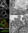
Intact LDs are present in the chlamydial inclusion lumen. (A) Neutral lipid-rich droplets are found within inclusions. Nonlipid loaded HeLa cells were infected with L2 for 20 h and the interaction between inclusions and neutral lipid-rich droplets assessed as in Fig. 1B. Note the presence of distinct droplets (arrowheads) within IncG-positive membranes in xy (Top) and zy (Lower) laser scanning confocal sections. (B and C) Ultrastructural analysis of inclusions reveals intact LDs in the inclusion lumen. HeLa cells were infected with L2 for 18 h, fixed in the presence of malachite green to preserve lipid structures, and processed for electron microscopy. LDs, internal membrane structures, and LD-like structures (black arrows) accumulated inside the inclusion. Note membrane blebs associated with intrainclusion LDs (arrowheads) and contacts between LDs, RBs, and the inclusion membrane (C). N, HeLa nuclei; M, mitochondria.
Fig. 3.
Fig. 3.
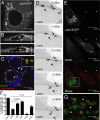
The inclusion membrane protein IncA copurifies with LDs and accumulates in the inclusion lumen. (A) Translocation of LDs across the inclusion membrane. Representative electron micrographs of L2 inclusions show LDs at various stages of crossing the inclusion membrane. Note membranes and blebs (arrowheads) associated with translocating LDs. CYT, cytoplasm. (B) IncA cofractionates with LDs. HeLa cells were infected with L2 for 40 h and treated with 100 μM OA 12–14 h before purification of LDs by density gradient ultracentrifugation. The fractionation of a host LD protein (Nsdhl), chlamydial outer membrane protein (Omp2), host proteins associated with the inclusion membrane (Rab11 and 14–3-3β), and Inc proteins (CT223, CT229, IncA, IncG and Cap1) were assessed by immunoblots. Note cofractionation of IncA with purified LDs and lack of other Inc proteins. (C and D) IncA-positive structures accumulate in the inclusion lumen. HeLa cells were infected with L2 for 18 and 24 h and immunostained with polyclonal anti-IncG or anti-IncA antibodies. Note the accumulation of IncA-positive material in the inclusion lumens (C). The frequency of IncA and IncG-positive intrainclusion vesicles (D) was determined by LSCM as in C, except that an anti-IncA mAb was used. Inclusions (150–300 per time point per experiment) were binned in categories according to the number of vesicles per inclusion. Data represent the mean ± SD of three independent experiments. The number of inclusions with intralumenal IncA-positive vesicles was significantly higher than IncG-positive (P < 0.001). (E) Partial colocalization of IncA-positive membranes with intrainclusion LDs. HeLa cells were infected with L2 and processed as in Fig. 2A with anti-IncA antibodies and BODIPY.
Fig. 4.
Fig. 4.
Exogenously expressed Lda3 binds to LDs and inclusion membranes and induces loss of ADRP. (A–C) Lda3-EGFP localizes to inclusion membranes and LDs. HeLa cells expressing Lda3-EGFP were infected with L2 for 20 h (A and B) and imaged by LSCM. Lda3-EGFP localized prominently to LDs at the periphery of the inclusion and inclusion membranes (A). Intrainclusion Lda3- positive material (arrows) was apparent in xz confocal sections (B). These intrainclusion structures are likely LDs as Lda3-DsRed positive structures in OA-treated cells also stain with BODIPY. (Inset) Magnification of Lda3-positive LDs (C). (D) Live cell analysis of LD translocation into the inclusion lumen. HeLa cells expressing Lda3-EGFP were infected with L2 for 30 h and imaged for 30 min. Representative frames show Lda3-EGFP tagged LDs (arrows) docked at the inclusion lumen in the process of translocation (see Movie S2). (E–G) Lda3-EGFP expressing cells display reduced levels of ADRP. HeLa cells were transiently transfected with Lda3-EGFP, treated with 100 μM OA for 12 h, and fixed. ADRP on LDs was detected by indirect immunofluorescence. Prominent localization of ADRP to LDs was only observed in untransfected cells (*). (E). The loss of ADRP from LDs was most pronounced in Lda3 compared with Lda1, Lda2LD, and a catalytically inactive ATGL (ATGL*). Overexpression of wild-type ATGL led to a loss ADRP. Data represent the mean ± SD of triplicates (F). Close up of Lda3-EGFP (green) positive LDs revealed a displacement of endogenous ADRP (red) to distinct puncta on the surface of LDs (arrowheads) (G). IM, inclusion membrane.
Fig. 5.
Fig. 5.
A model for LD interaction with the Chlamydia inclusion. (I) LDs are engaged by secreted Lda3 at the surface of the inclusion. (II) Lda3-tagged LDs are captured at the inclusion membrane by an unidentified inclusion membrane protein(s) (IncX). (III) The inclusion membrane invaginates to deliver the LD to the inclusion lumen. (IV) RBs intimately bind to the intrainclusion LD and associated inclusion membranes. Lda3 may participate in initiating LD lipolysis by promoting the removal of ADRP.

References

    1. Schachter J. In: Chlamydia: Intracellular Biology, Pathogenesis, and Immunity. Stephens RS, editor. Washington, D.C.: ASM; 1999. pp. 139–169.
    1. Belland R, Ojcius DM, Byrne GI. Chlamydia. Nat Rev Microbiol. 2004;2:530–531. - PubMed
    1. Fields KA, Hackstadt T. The chlamydial inclusion: Escape from the endocytic pathway. Annu Rev Cell Dev Biol. 2002;18:221–245. - PubMed
    1. Wylie JL, Hatch GM, McClarty G. Host cell phospholipids are trafficked to and then modified by Chlamydia trachomatis. J Bacteriol. 1997;179:7233–7242. - PMC - PubMed
    1. Hackstadt T, Scidmore MA, Rockey DD. Lipid metabolism in Chlamydia trachomatis-infected cells: Directed trafficking of Golgi-derived sphingolipids to the chlamydial inclusion. Proc Natl Acad Sci USA. 1995;92:4877–4881. - PMC - PubMed

Publication types

Substances