. 2008 Jul 8;105(27):9349-54.
doi: 10.1073/pnas.0711382105. Epub 2008 Jun 30.

A sonic hedgehog signaling domain in the arterial adventitia supports resident Sca1+ smooth muscle progenitor cells

Affiliations

A sonic hedgehog signaling domain in the arterial adventitia supports resident Sca1+ smooth muscle progenitor cells

Jenna N Passman et al. Proc Natl Acad Sci U S A. .

Abstract

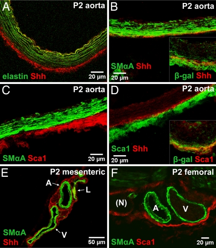
We characterize a sonic hedgehog (Shh) signaling domain restricted to the adventitial layer of artery wall that supports resident Sca1-positive vascular progenitor cells (AdvSca1). Using patched-1 (Ptc1(lacZ)) and patched-2 (Ptc2(lacZ)) reporter mice, adventitial Shh signaling activity was first detected at embryonic day (E) 15.5, reached the highest levels between postnatal day 1 (P1) and P10, was diminished in adult vessels, and colocalized with a circumferential ring of Shh protein deposited between the media and adventitia. In Shh(-/-) mice, AdvSca1 cells normally found at the aortic root were either absent or greatly diminished in number. Using a Wnt1-cre lineage marker that identifies cells of neural crest origin, we found that neither the adventitia nor AdvSca1 cells were labeled in arteries composed of neural crest-derived smooth muscle cells (SMCs). Although AdvSca1 cells do not express SMC marker proteins in vivo, they do express transcription factors thought to be required for SMC differentiation, including serum response factor (SRF) and myocardin family members, and readily differentiate to SMC-like cells in vitro. However, AdvSca1 cells also express potent repressors of SRF-dependent transcription, including Klf4, Msx1, and FoxO4, which may be critical for maintenance of the SMC progenitor phenotype of AdvSca1 cells in vivo. We conclude that a restricted domain of Shh signaling is localized to the arterial adventitia and may play important roles in maintenance of resident vascular SMC progenitor cells in the artery wall.


Conflict of interest statement

The authors declare no conflict of interest.

Figures

Fig. 1.
Fig. 1.
Hh signaling in artery wall. (A, B, and D–F) Tissues were isolated from PtclacZ mice at P2 and stained for β-gal activity. All major arteries examined, including coronary arteries, aorta, pulmonary trunk (A), intercostal (D), mesenteric (E), and femoral arteries (F) were positive for Ptc1lacZ or Ptc2lacZ activity as shown. (B) Adult hearts displayed low levels of β-gal activity in coronary arteries, aorta, and pulmonary artery. (C) A cross-section of the aorta at P2 reveals Ptc1lacZ-positive cells are restricted to the adventitia. Ica, intercostal artery; A, artery; V, vein; N, nerve. (Magnification: A and D–F, ×7; B, ×3; C, × 40.)
Fig. 2.
Fig. 2.
Distribution of Shh and Sca1+ cells in the adventitia. (A) Shh protein is found outside the aortic external elastic lamina (marked by green autofluorescence) at P2. (B) Shh protein (red) is found between the media (SMαActin, green) and adventitia of descending aorta. (B Inset) Shh (red) is colocalized with β-gal in aortic sections from Ptc1lacZ mice. (C) Sca1+ cells (red) reside outside the media (SMαActin, green) in P2 aorta. (D) Sca1+ cells (green) are found in close proximity to Shh (red) and are embedded within a β-gal-positive domain (Inset) in aortic adventitia from Ptc1lacZ mice. (E) Mesenteric vessels stained for SMαActin (green) and Shh (red). (F) Femoral arteries stained for SMαActin (green) and Sca1 (red). Images are stacked Z-plane sections from confocal microscopy. Adventitial localization of Sca1+ cells correlates with the distribution of Shh protein and active Shh signaling in vivo.
Fig. 3.
Fig. 3.
Analysis of AdvSca1 cells. (A) RT-PCR analysis of AdvSca1 cells from adult arteries. (B) Freshly isolated AdvSca1 cells (red) contain SRF (green) colocalized with DAPI-stained nuclei (blue) (arrows). (C) Freshly isolated AdvSca1 cells (red) contain Klf4 (green) colocalized with DAPI-stained nuclei (blue) (arrows). (D) RT-PCR analysis of AdvSca1 cells cultured for 0, 12, or 28 days. M is total RNA from aortic media used as a positive control for SMC markers. This analysis was performed three times with similar results. (E) AdvSca1 cells cultured in serum-containing medium for 9 days. Many cells down-regulate ScaI (red) and up-regulate SMαActin (green). (Magnifications: B and C, ×40; E, ×10.)
Fig. 4.
Fig. 4.
AdvSca1 cells in developing aorta. (A–F) Tissues were obtained at the time points indicated. Cross-sections through the aortic root were stained for Sca1 (red), SMαActin (green), and DAPI (blue). AdvSca1 cells first appear between E15.5 and E18.5 in the adventitial space between aorta and pulmonary trunk (white arrows in C) and persist in this location into adulthood. (G) Cross-section from Wnt1-cre/R26R (5 weeks old) aortic root stained for β-gal activity and nuclear fast red. SMCs (blue) but not adventitial cells originate from neural crest. Dashed box is shown at higher magnification in H. (H) Cross-section through aortic root shows no overlap between β-gal-positive and AdvSca1+ cells. Ao, aorta; PT, pulmonary trunk. (Magnification scale bar in E applies to A–F; G, ×10.)
Fig. 5.
Fig. 5.
AdvSca1 cells in Shh−/− arteries. (A–D) Aortic tissues from WT (A and C) and Shh−/− (B and D) embryos at E18.5. Cross-sections through aortic root (A and B) and descending aorta (C and D) were immunostained for Sca1 (red), SMαActin (green), and DAPI (blue). (E) Solid lines in indicate relative positions of sections in A–D. (F) Dotted line boxes in A–D correspond to aortic segments shown here. AdvSca1 cells are absent in aortic root, greatly reduced in ascending and transverse aorta (termed ascending), and diminished in descending aorta of Shh−/− embryos.

References

    1. Heistad D, Marcus M, Larsen G, Armstrong M. Role of vasa vasorum in nourishment of the aortic wall. Am J Physiol. 1981;240:H781–H787. - PubMed
    1. Sartore S, et al. Contribution of adventitial fibroblasts to neointima formation and vascular remodeling: From innocent bystander to active participant. Circ Res. 2001;89:1111–1121. - PubMed
    1. Moulton K, et al. Inhibition of plaque neovascularization reduces macrophage accumulation and progression of advanced atherosclerosis. Proc Natl Acad Sci USA. 2003;100:4726–4741. - PMC - PubMed
    1. Haurani M, Pagano P. Adventitial fibroblast reactive oxygen species as autacrine and paracrine mediators of remodeling: Bellwether for vascular disease? Cardiovasc Res. 2007;75:679–689. - PubMed
    1. Ryan S, Koteliansky V, Gotwals P, Lindner V. Transforming growth factor-β-dependent events in vascular remodeling following arterial injury. J Vasc Res. 2003;40:37–46. - PubMed

Publication types

MeSH terms