. 2008 Oct 16;27(47):6120-30.
doi: 10.1038/onc.2008.207. Epub 2008 Jun 30.

HER2 regulates the mammary stem/progenitor cell population driving tumorigenesis and invasion

Affiliations

HER2 regulates the mammary stem/progenitor cell population driving tumorigenesis and invasion

H Korkaya et al. Oncogene. .

Abstract

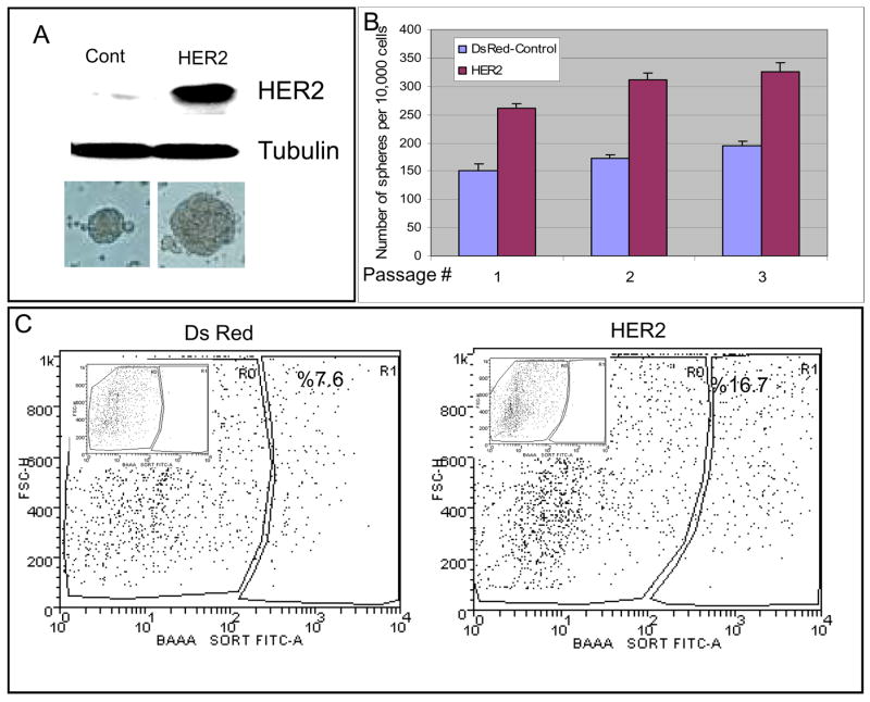
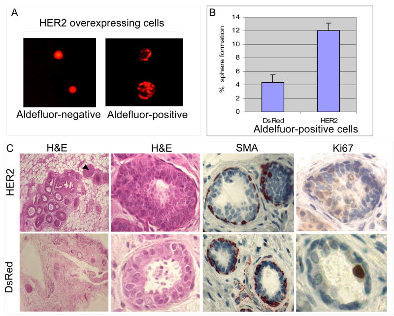
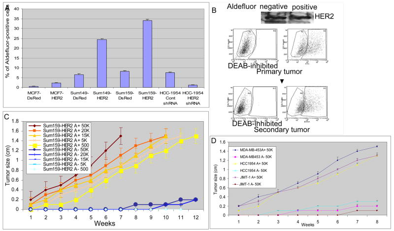
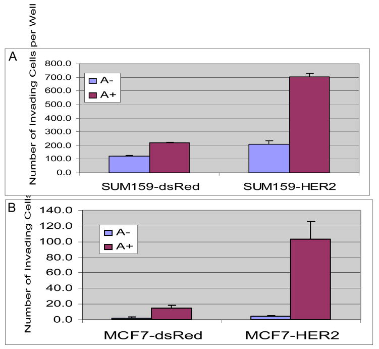
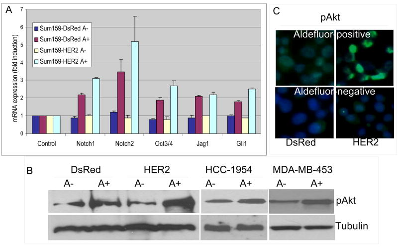
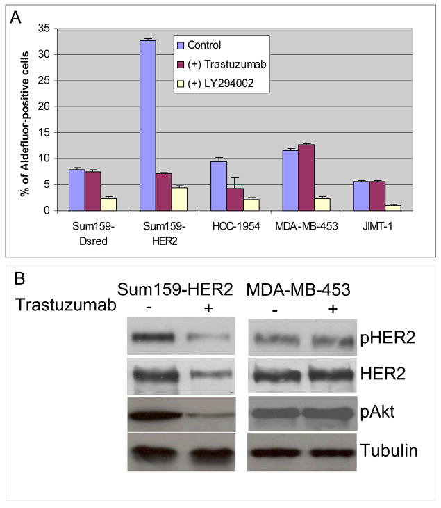
The cancer stem cell hypothesis proposes that cancers arise in stem/progenitor cells through disregulation of self-renewal pathways generating tumors, which are driven by a component of 'tumor-initiating cells' retaining stem cell properties. The HER2 gene is amplified in 20-30% of human breast cancers and has been implicated in mammary tumorigenesis as well as in mediating aggressive tumor growth and metastasis. We demonstrate that HER2 overexpression drives mammary carcinogenesis, tumor growth and invasion through its effects on normal and malignant mammary stem cells. HER2 overexpression in normal mammary epithelial cells (NMEC) increases the proportion of stem/progenitor cells as demonstrated by in vitro mammosphere assays and the expression of stem cell marker aldehyde dehydrogenase (ALDH) as well as by generation of hyperplastic lesions in humanized fat pads of NOD (nucleotide-binding oligomerization domain)/SCID (severe combined immunodeficient) mice. Overexpression of HER2 in a series of breast carcinoma cell lines increases the ALDH-expressing 'cancer stem cell' population which displays increased expression of stem cell regulatory genes, increased invasion in vitro and increased tumorigenesis in NOD/SCID mice. The effects of HER2 overexpression on breast cancer stem cells are blocked by trastuzumab in sensitive, but not resistant, cell lines, an effect mediated by the PI3-kinase/Akt pathway. These studies provide support for the cancer stem cell hypothesis by suggesting that the effects of HER2 amplification on carcinogenesis, tumorigenesis and invasion may be due to its effects on normal and malignant mammary stem/progenitor cells. Furthermore, the clinical efficacy of trastuzumab may relate to its ability to target the cancer stem cell population in HER2-amplified tumors.


Figures

Figure 1
Figure 1. HER2 expression increases mammosphere formation and the Aldelfuor-positive cell population
A. HER2 expression in normal mammary epithelial cells infected with HER2 or DsRed lentivirus. Increased size of mammospheres in HER2 expressing cells as compared to DsRed control. B. Serial passage of HER2 and DsRed control mammospheres. HER2 expression increases mammosphere forming capacity upon serial passage. P values for each passage are 5.6×10−37, 8.05×10−43, 3.4×10⁻³² respectively. C. HER2 or DsRed cells were assessed for ALDH activity utilizing the Aldefluor assay. HER2 expressing cells demonstrate greater than a two-fold increase in the Aldefluor-positive cell population compared to DsRed control cells. Insert show DEAB inhibited flow results for compensation. Data represented here are the mean of 5 independent experiments performed in triplicates.
Figure 2
Figure 2. HER2 expression increases the frequency of mammosphere formation and of ductal structures in NOD-SCID mice
A. When plated at clonal density, only the Aldefluor-positive cells are capable of mammosphere formation. B. A three-fold increase in mammosphere initiating single cells in the HER2 Aldefluor-positive cell population compared to the DsRed Aldefluor-positive population. C. Serial dilutions of HER2 or DsRed expressing NMECs were injected into the mammary fat pads of humanized NOD/SCID mice. HER2 overexpressing cells generated an increased number of mammary outgrowths which displayed hyperplasia (arrows) characterized by an 8 fold increase in Ki67 staining. Myoepithelial cells were demonstrated by smooth muscle actin (SMA) staining.
Figure 3
Figure 3. Effects of HER2 overexpression on Aldefluor-positivity and tumorigenicity of human mammary carcinoma cell lines
A. Overexpression of HER2 increased the Aldefluor-positive populations three- to five-fold compared to control DsRed cells in indicated breast cancer cell lines. Knockdown of HER2 in HCC1954 cells using HER2 siRNA decreased the Aldefluor-positive cell population. B. Aldefluor-positive but not Aldefluor-negative cells display properties of tumor stem cells despite equivalent HER2 expression in both populations as shown by Western blotting. Tumors generated from the Aldefluor-positive population are composed of both Aldefluor-positive and -negative populations recapitulating the initial tumor phenotype. C. Growth curves of serial dilutions of Aldefluor-positive and -negative SUM159-HER2 cells. Aldefluor-positive cells form tumors at all cell concentrations whereas the Aldefluor-negative cells failed to do so. D. In HER2 amplified cell lines MDA-MB453, HCC1954 and JIMT-1, only Aldefluor-positive cells were tumorigenic.
Figure 4
Figure 4. Effects of HER2 overexpression on invasive properties of Aldelfuor-positive and -negative populations of breast cancer cell lines
Aldefluor-positive populations from Sum159-DsRed, SUM159-HER2 (A), MCF7-DsRed or MCF7-HER2 cells (B) showed significantly higher invasiveness compared to Aldefluor-negative populations. Furthermore, HER2 overexpression resulted in a 5 to 6-fold increase in the invasive potential of Aldefluor-positive cells.
Figure 5
Figure 5. Expression of stem cell related genes and Akt phosphorylation
A. Aldefluor-positive cells expressed higher levels of all indicated stem cell related genes than Aldefluor-negative cells. The expression of these genes was further elevated by HER2 overexpression. B. Aldefluor-positive populations of indicated breast cancer cell lines showed higher Akt-phosphorylation when compared to Aldefluor-negative populations. C. Akt-phosphorylation as assessed by immunoflourescence in Aldefluor-positive and –negative SUM159-DsRed and SUM159-HER2 cells. Increased phospho-Akt is detected in Aldefluor-positive and HER2 overexpressing cells.
Figure 6
Figure 6. Effects of trastuzumab and LY294002 on the Aldefluor-positive population in breast cancer cell lines
A. Trastuzumab reduced the proportion of Aldefluor-positive cells by as much as 80% in SUM159-HER2 and HCC1954 cells. In contrast, trastuzumab had no effect on the Aldefluor-positive population in MDA-MB-453 and JIMT-1 trastuzumab resistant cell lines. In contrast, LY294002 reduced the proportion of Aldefluor-positive cells in all cell lines. (B) Trastuzumab reduced phospho-HER2 and phospho-AKT expression in Sum159-HER2 cells but has no effect on expression of these proteins in trastuzumab resistant MDA-MB-453 cells.

References

    1. Al-Hajj M, Wicha MS, Benito-Hernandez A, Morrison SJ, Clarke MF. Prospective identification of tumorigenic breast cancer cells. Proc Natl Acad Sci U S A. 2003;100:3983–8. - PMC - PubMed
    1. Allred DC, O’Connell P, Fuqua SA, Osborne CK. Immunohistochemical studies of early breast cancer evolution. Breast Cancer Res Treat. 1994;32:13–8. - PubMed
    1. Berns K, Horlings HM, Hennessy BT, Madiredjo M, Hijmans EM, Beelen K, et al. A Functional Genetic Approach Identifies the PI3K Pathway as a Major Determinant of Trastuzumab Resistance in Breast Cancer. Cancer Cell. 2007;12:395–402. - PubMed
    1. Beuzeboc P, Scholl S, Garau XS, Vincent-Salomon A, Cremoux PD, Couturier J, et al. [Herceptin, a monoclonal humanized antibody anti-HER2: a major therapeutic progress in breast cancers overexpressing this oncogene?] Bull Cancer. 1999;86:544–9. - PubMed
    1. Burstein HJ, Kuter I, Campos SM, Gelman RS, Tribou L, Parker LM, et al. Clinical activity of trastuzumab and vinorelbine in women with HER2-overexpressing metastatic breast cancer. J Clin Oncol. 2001;19:2722–30. - PubMed

Publication types

MeSH terms