

Loss of CHFR in human mammary epithelial cells causes genomic instability by disrupting the mitotic spindle assembly checkpoint
- PMID: 18592005
- PMCID: PMC2435002
- DOI: 10.1593/neo.08176
Loss of CHFR in human mammary epithelial cells causes genomic instability by disrupting the mitotic spindle assembly checkpoint
Abstract
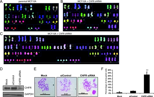
CHFR is an E3 ubiquitin ligase and an early mitotic checkpoint protein implicated in many cancers and in the maintenance of genomic stability. To analyze the role of CHFR in genomic stability, by siRNA, we decreased its expression in genomically stable MCF10A cells. Lowered CHFR expression quickly led to increased aneuploidy due to many mitotic defects. First, we confirmed that CHFR interacts with the mitotic kinase Aurora A to regulate its expression. Furthermore, we found that decreased CHFR led to disorganized multipolar mitotic spindles. This was supported by the finding that CHFR interacts with alpha-tubulin and can regulate its ubiquitination in response to nocodazole and the amount of acetylated alpha-tubulin, a component of the mitotic spindle. Finally, we found a novel CHFR interacting protein, the spindle checkpoint protein MAD2. Decreased CHFR expression resulted in the mislocalization of both MAD2 and BUBR1 during mitosis and impaired MAD2/CDC20 complex formation. Further evidence of a compromised spindle checkpoint was the presence of misaligned metaphase chromosomes, lagging anaphase chromosomes, and defective cytokinesis in CHFR knockdown cells. Importantly, our results suggest a novel role for CHFR regulating chromosome segregation where decreased expression, as seen in cancer cells, contributes to genomic instability by impairing the spindle assembly checkpoint.
Figures


Similar articles
-
CHFR binds to and regulates MAD2 in the spindle checkpoint through its cysteine-rich domain.Biochem Biophys Res Commun. 2011 Jun 10;409(3):389-93. doi: 10.1016/j.bbrc.2011.04.143. Epub 2011 May 7. Biochem Biophys Res Commun. 2011. PMID: 21575600 Free PMC article.
-
Enhanced genomic instabilities caused by deregulated microtubule dynamics and chromosome segregation: a perspective from genetic studies in mice.Carcinogenesis. 2009 Sep;30(9):1469-74. doi: 10.1093/carcin/bgp081. Epub 2009 Apr 16. Carcinogenesis. 2009. PMID: 19372138 Free PMC article. Review.
-
Bub1 and aurora B cooperate to maintain BubR1-mediated inhibition of APC/CCdc20.J Cell Sci. 2005 Aug 15;118(Pt 16):3639-52. doi: 10.1242/jcs.02487. Epub 2005 Jul 26. J Cell Sci. 2005. PMID: 16046481
-
The HECT E3 ligase Smurf2 is required for Mad2-dependent spindle assembly checkpoint.J Cell Biol. 2008 Oct 20;183(2):267-77. doi: 10.1083/jcb.200801049. Epub 2008 Oct 13. J Cell Biol. 2008. PMID: 18852296 Free PMC article.
-
Regulation of APC-Cdc20 by the spindle checkpoint.Curr Opin Cell Biol. 2002 Dec;14(6):706-14. doi: 10.1016/s0955-0674(02)00382-4. Curr Opin Cell Biol. 2002. PMID: 12473343 Review.
Cited by
-
The War on Cancer rages on.Neoplasia. 2009 Dec;11(12):1252-63. doi: 10.1593/neo.91866. Neoplasia. 2009. PMID: 20019833 Free PMC article.
-
CHFR: a key checkpoint component implicated in a wide range of cancers.Cell Mol Life Sci. 2012 May;69(10):1669-87. doi: 10.1007/s00018-011-0892-2. Epub 2011 Dec 13. Cell Mol Life Sci. 2012. PMID: 22159584 Free PMC article. Review.
-
Primary cilia and aberrant cell signaling in epithelial ovarian cancer.Cilia. 2012 Aug 10;1(1):15. doi: 10.1186/2046-2530-1-15. Cilia. 2012. PMID: 23351307 Free PMC article.
-
Deficiencies in Chfr and Mlh1 synergistically enhance tumor susceptibility in mice.J Clin Invest. 2009 Sep;119(9):2714-24. doi: 10.1172/JCI37405. Epub 2009 Aug 17. J Clin Invest. 2009. PMID: 19690386 Free PMC article.
-
CYLD negatively regulates cell-cycle progression by inactivating HDAC6 and increasing the levels of acetylated tubulin.EMBO J. 2010 Jan 6;29(1):131-44. doi: 10.1038/emboj.2009.317. Epub 2009 Nov 5. EMBO J. 2010. PMID: 19893491 Free PMC article.
References
-
- Chaturvedi P, Sudakin V, Bobiak ML, Fisher PW, Mattern MR, Jablonski SA, Hurle MR, Zhu Y, Yen TJ, Zhou BB. Chfr regulates a mitotic stress pathway through its RING-finger domain with ubiquitin ligase activity. Cancer Res. 2002;2:1797–1801. - PubMed
-
- Ogi K, Toyota M, Mita H, Satoh A, Kashima L, Sasaki Y, Suzuki H, Akino K, Nishikawa N, Noguchi M, et al. Small interfering RNA-induced CHFR silencing sensitizes oral squamous cell cancer cells to microtubule inhibitors. Cancer Biol Ther. 2005;4:73–780. - PubMed
-
- Privette LM, Gonzalez ME, Ding L, Kleer CG, Petty EM. Altered expression of the early mitotic checkpoint protein, CHFR, in breast cancers: implications for tumor suppression. Cancer Res. 2007;67:6064–6074. - PubMed
-
- Sakai M, Hibi K, Kanazumi N, Nomoto S, Inoue S, Takeda S, Nakao A. Aberrant methylation of the CHFR gene in advanced hepatocellular carcinoma. Hepatogastroenterology. 2005;52:1854–1857. - PubMed
-
- Satoh A, Toyota M, Itoh F, Sasaki Y, Suzuki H, Ogi K, Kikuchi T, Mita H, Yamashita T, Kojima T, et al. Epigenetic inactivation of CHFR and sensitivity to microtubule inhibitors in gastric cancer. Cancer Res. 2003;63:8606–8613. - PubMed
Publication types
MeSH terms
Substances
Grants and funding
LinkOut - more resources
Full Text Sources