

Endonasal ethmoidectomy and bifrontal craniotomy with craniofacial approach for resection of frontoethmoidal osteoma causing tension pneumocephalus
- PMID: 18592021
- PMCID: PMC2435469
- DOI: 10.1055/s-2007-993046
Endonasal ethmoidectomy and bifrontal craniotomy with craniofacial approach for resection of frontoethmoidal osteoma causing tension pneumocephalus
Abstract
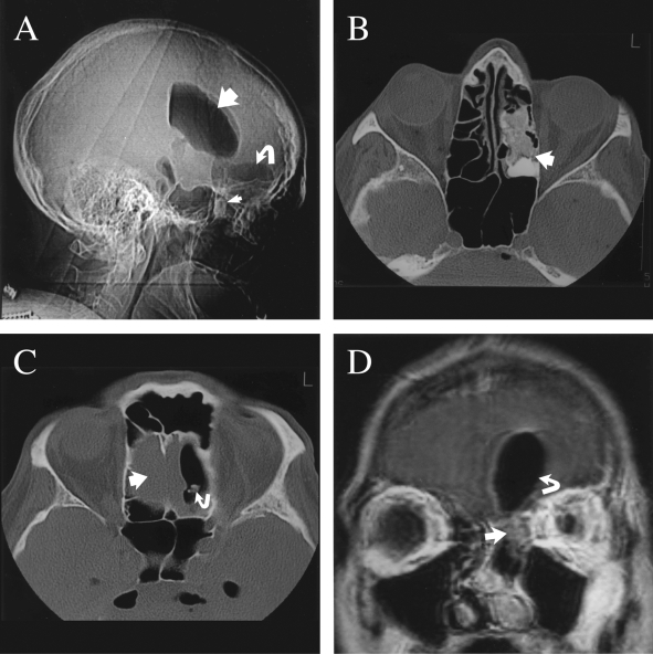
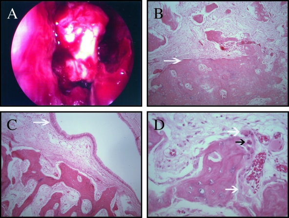
Tension pneumocephalus is an unusual, potentially life-threatening complication of frontal fossa tumors. We present an uncommon case of a frontoethmoidal osteoma causing a tension pneumocephalus and neurological deterioration prompting a combined endonasal ethmoidectomy and bifrontal craniotomy with craniofacial approach for resection. A 68-year-old man presented with a 1-week history of worsening headache, slowness of speech, and increasing confusion. Standard computed tomography scan revealed a marked tension pneumocephalus with ventricular air and 1-cm midline shift to the right. Further studies showed a calcified left ethmoid mass and a left anterior cranial-base defect. A team composed of neurosurgery and otolaryngology performed a combined endonasal ethmoidectomy and bifrontal craniotomy with craniofacial approach to resect a large frontoethmoid bony tumor. No abscess or mucocele was identified. The skull base defect was repaired with the aid of a transnasal endoscopy, a titanium mesh, and a pedunculated pericranial flap. Postoperatively, the pneumocephalus and the patient's symptoms completely resolved. Pathology was consistent with a benign osteoma. This is an uncommon case of a frontoethmoidal osteoma associated with tension pneumocephalus. Recognition of this entity and timely diagnosis and treatment, consisting of an endonasal ethmoidectomy and a bifrontal craniotomy with craniofacial approach, may prevent potential life-threatening complications.
Keywords: Bifrontal craniotomy; craniofacial; endonasal ethmoidectomy; frontoethmoidal osteoma; tension pneumocephalus.
Figures
Similar articles
-
Frontoethmoidal Osteoma with Secondary Intradural Mucocele Extension Causing Frontal Lobe Syndrome and Pneumocephalus: Case Report and Review of Literature.World Neurosurg. 2018 Jul;115:301-308. doi: 10.1016/j.wneu.2018.04.071. Epub 2018 Apr 19. World Neurosurg. 2018. PMID: 29679781 Review.
-
Management of frontoethmoidal osteoma causing pneumocephalus and cerebrospinal fluid leakage with minimally invasive techniques: illustrative cases.J Neurosurg Case Lessons. 2024 Feb 5;7(6):CASE23699. doi: 10.3171/CASE23699. Print 2024 Feb 5. J Neurosurg Case Lessons. 2024. PMID: 38315987 Free PMC article.
-
A rare association of tension pneumocephalus and a large frontoethmoidal osteoma: imaging features and surgical treatment.J Craniofac Surg. 2011 Jan;22(1):212-3. doi: 10.1097/SCS.0b013e3181f76031. J Craniofac Surg. 2011. PMID: 21233751
-
Tension Pneumocephalus: A Potentially Fatal Complication of Expanded Endoscopic Endonasal Approach.Indian J Otolaryngol Head Neck Surg. 2023 Sep;75(3):2523-2528. doi: 10.1007/s12070-023-03802-5. Epub 2023 Apr 27. Indian J Otolaryngol Head Neck Surg. 2023. PMID: 37636702 Free PMC article.
-
Tension pneumocephalus after skull base surgery. A case report and review of literature.J Clin Neurosci. 2020 May;75:218-220. doi: 10.1016/j.jocn.2020.03.041. Epub 2020 Apr 2. J Clin Neurosci. 2020. PMID: 32249175 Review.
Cited by
-
Giant osteomas of the ethmoid and frontal sinuses: Clinical characteristics and review of the literature.Oncol Lett. 2013 May;5(5):1724-1730. doi: 10.3892/ol.2013.1239. Epub 2013 Mar 8. Oncol Lett. 2013. PMID: 23759920 Free PMC article.
-
A rare case of childhood hemiparesis.Childs Nerv Syst. 2011 May;27(5):841-3. doi: 10.1007/s00381-010-1356-y. Epub 2011 Jan 5. Childs Nerv Syst. 2011. PMID: 21207042 No abstract available.
References
-
- Carmody T E. Osteoma of the nasal accessory sinuses. Ann Otol Rhinol Laryngol. 1935;44:626–643.
-
- Childrey J H. Osteoma of the sinuses, the frontal and the sphenoid bone: report of fifteen cases. Arch Otolaryngol. 1939;30:63–72.
-
- Bartlett J R. Intracranial neurological complications of frontal and ethmoidal osteomas. Br J Surg. 1971;58:607–613. - PubMed
-
- Boysen M. Osteomas of the paranasal sinuses. J Otolaryngol. 1978;7:366–370. - PubMed
-
- Summers L E, Mascott C R, Tompkins J R, Richardson D E. Frontal sinus osteoma associated with cerebral abscess formation: a case report. Surg Neurol. 2001;55:235–239. - PubMed
