

Adipose-derived stem cells: isolation, expansion and differentiation
- PMID: 18593609
- PMCID: PMC3668445
- DOI: 10.1016/j.ymeth.2008.03.006
Adipose-derived stem cells: isolation, expansion and differentiation
Abstract
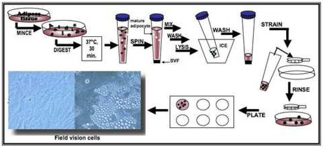
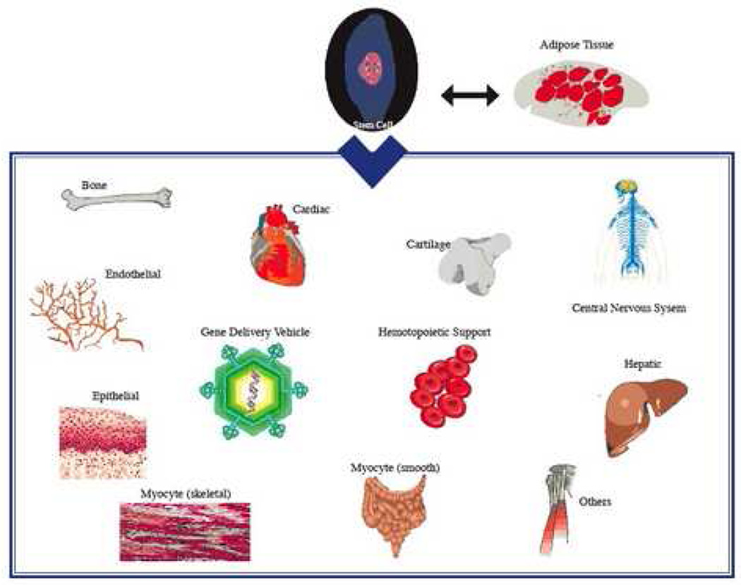
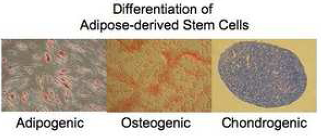
The emerging field of regenerative medicine will require a reliable source of stem cells in addition to biomaterial scaffolds and cytokine growth factors. Adipose tissue has proven to serve as an abundant, accessible and rich source of adult stem cells with multipotent properties suitable for tissue engineering and regenerative medical applications. There has been increased interest in adipose-derived stem cells (ASCs) for tissue engineering applications. Here, methods for the isolation, expansion and differentiation of ASCs are presented and described in detail. While this article has focused on the isolation of ASCs from human adipose tissue, the procedure can be applied to adipose tissues from other species with minimal modifications.
Figures
References
-
- Gimble JM. Expert Opin Biol Ther. 2003;3:705–713. - PubMed
-
- Wei G, Schubiger G, Harder F, Muller AM. Stem Cells. 2000;18:409–414. - PubMed
-
- Woodbury D, Reynolds K, Black IB. J. Neurosci. Res. 2002;96:908–917. - PubMed
-
- Beltrami AP, Urbanek K, Kajstura J, Yan SM, Finato N, Bussani R, Nadal Ginard B, Silvestri F, Leri A, Beltrami CA. New Engl. Jour. of Med. 2001;344:1750–1757. - PubMed
-
- Gussoni E, Soneoka Y, Strickland CD, Buzney EA, Khan MK, Flint AF, Kunkel LM, Mulligan RC. Nature. 1999;401:390–394. - PubMed
Publication types
MeSH terms
Grants and funding
LinkOut - more resources
Full Text Sources
Other Literature Sources
