

JC virus infects the enteric glia of patients with chronic idiopathic intestinal pseudo-obstruction
- PMID: 18593810
- PMCID: PMC2865195
- DOI: 10.1136/gut.2008.152512
JC virus infects the enteric glia of patients with chronic idiopathic intestinal pseudo-obstruction
Abstract
Background and aims: Chronic idiopathic intestinal pseudo-obstruction (CIIP) is characterised by severe impairment of intestinal propulsive motility that mimics bowel obstruction. JC virus (JCV) is a polyomavirus that can infect brain glial cells causing a fatal disease, but may also be found throughout the normal gastrointestinal tract. The hypothesis that JCV infects the myenteric plexuses of patients with CIIP was tested.
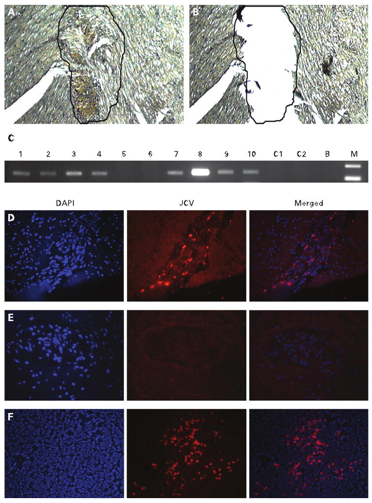
Methods: 10 patients with CIIP and 61 normal specimens (30 ascending colon and 31 ileum) from patients with uncomplicated colon cancer were studied. DNA was extracted from the myenteric plexuses, and JCV T antigen (TAg) DNA and the viral regulatory region were detected by PCR and sequencing. Immunohistochemistry was performed to detect JCV viral protein expression, neuronal and glial markers. Fluorescence in situ hybridisation was performed for cellular localisation of the JCV infection.
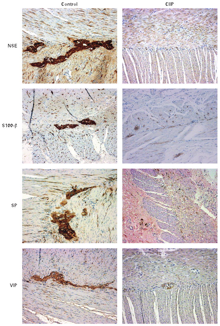
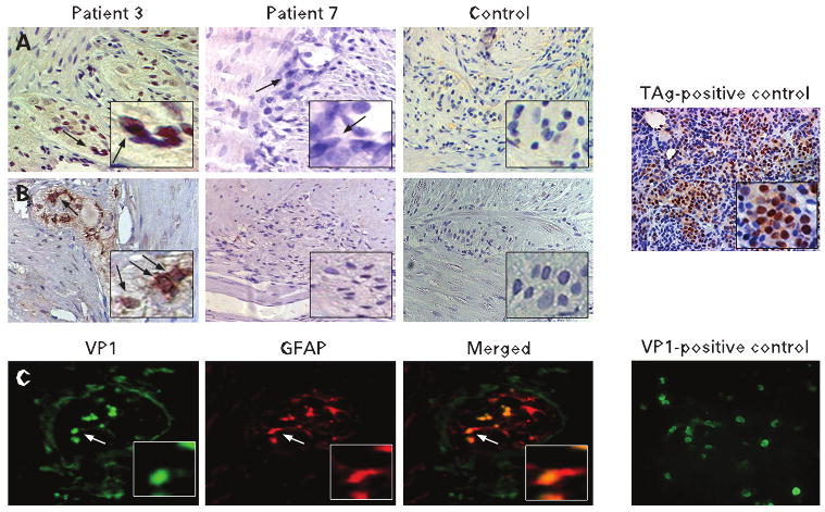
Results: Clinical studies demonstrated neurogenic impairment, and pathological analyses showed neuropathy in each patient with CIIP. JCV TAg DNA was found in the myenteric plexuses of 8/10 (80%) of the patients with CIIP and 3/31 (9.7%) of the control patients (p<0.001). All samples were JCV Mad-1 strains. Seven of the 10 CIIP specimens expressed both JCV TAg and the JCV viral protein VP1, while none of the controls expressed either. JCV infection co-localised with glial fibrillary acidic protein expression, a marker of enteric glial cells.
Conclusion: JCV infection occurs in the myenteric plexuses of patients with CIIP. The JCV localisation in enteroglial cells suggests a possible pathological role for this virus in enteric neuropathy.
Conflict of interest statement
Figures
Comment in
-
Chronic intestinal pseudo-obstruction: how important is JC virus infection?Gut. 2009 Jan;58(1):2-3. doi: 10.1136/gut.2008.160101. Gut. 2009. PMID: 19091825 Review. No abstract available.
Similar articles
-
Could JC virus be linked to chronic idiopathic intestinal pseudo-obstruction?Clin J Gastroenterol. 2020 Jun;13(3):377-381. doi: 10.1007/s12328-019-01069-4. Epub 2019 Nov 14. Clin J Gastroenterol. 2020. PMID: 31728918
-
Detection of JC virus DNA sequences and expression of viral T antigen and agnoprotein in esophageal carcinoma.Cancer. 2005 Feb 1;103(3):516-27. doi: 10.1002/cncr.20806. Cancer. 2005. PMID: 15630684
-
DNA viruses in the pathogenesis of sporadic chronic idiopathic intestinal pseudo-obstruction.Gut. 1997 Jul;41(1):100-6. doi: 10.1136/gut.41.1.100. Gut. 1997. PMID: 9274480 Free PMC article.
-
Chronic intestinal pseudo-obstruction: how important is JC virus infection?Gut. 2009 Jan;58(1):2-3. doi: 10.1136/gut.2008.160101. Gut. 2009. PMID: 19091825 Review. No abstract available.
-
[Chronic idiopathic intestinal pseudo-obstruction: visceral myopathy. Report of 4 cases].Acta Gastroenterol Latinoam. 1993;23(4):239-43. Acta Gastroenterol Latinoam. 1993. PMID: 8203187 Review. Spanish.
Cited by
-
The gut connectome: making sense of what you eat.J Clin Invest. 2015 Mar 2;125(3):888-90. doi: 10.1172/JCI81121. Epub 2015 Mar 2. J Clin Invest. 2015. PMID: 25729849 Free PMC article.
-
The management of adult patients with severe chronic small intestinal dysmotility.Gut. 2020 Dec;69(12):2074-2092. doi: 10.1136/gutjnl-2020-321631. Epub 2020 Aug 21. Gut. 2020. PMID: 32826308 Free PMC article.
-
Enteric glia in homeostasis and disease: From fundamental biology to human pathology.iScience. 2021 Jul 15;24(8):102863. doi: 10.1016/j.isci.2021.102863. eCollection 2021 Aug 20. iScience. 2021. PMID: 34401661 Free PMC article. Review.
-
Could Chronic Idiopatic Intestinal Pseudo-Obstruction Be Related to Viral Infections?J Clin Med. 2021 Jan 13;10(2):268. doi: 10.3390/jcm10020268. J Clin Med. 2021. PMID: 33450988 Free PMC article. Review.
-
Cytokine-induced alterations of gastrointestinal motility in gastrointestinal disorders.World J Gastrointest Pathophysiol. 2011 Oct 15;2(5):72-81. doi: 10.4291/wjgp.v2.i5.72. World J Gastrointest Pathophysiol. 2011. PMID: 22013552 Free PMC article.
References
-
- Coulie B, Camilleri M. Intestinal pseudo-obstruction. Annu Rev Med. 1999;50:37–55. - PubMed
-
- De Giorgio R, Guerrini S, Barbara G, et al. Inflammatory neuropathies of the enteric nervous system. Gastroenterology. 2004;126:1872–83. - PubMed
-
- Di Lorenzo C. Pseudo-obstruction: current approaches. Gastroenterology. 1999;116:980–7. - PubMed
-
- De Giorgio R, Guerrini S, Barbara G, et al. New insights into human enteric neuropathies. Neurogastroenterol Motil. 2004;16(Suppl 1):143–7. - PubMed
Publication types
MeSH terms
Substances
Grants and funding
LinkOut - more resources
Full Text Sources
Other Literature Sources