. 2008 Jul 1;22(13):1804-15.
doi: 10.1101/gad.1645808.

Nuclear HuR accumulation through phosphorylation by Cdk1

Affiliations

Nuclear HuR accumulation through phosphorylation by Cdk1

Hyeon Ho Kim et al. Genes Dev. .

Abstract

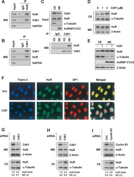
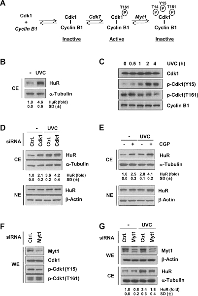
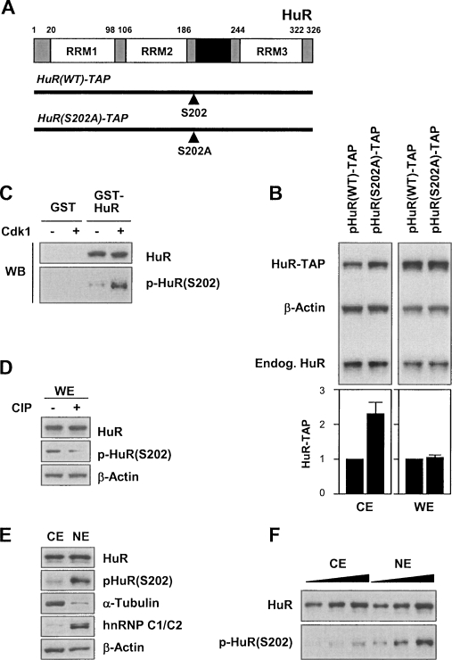
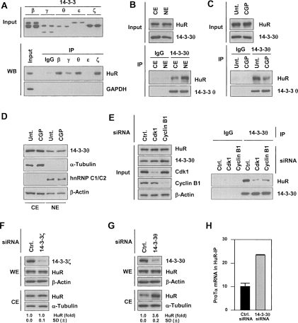
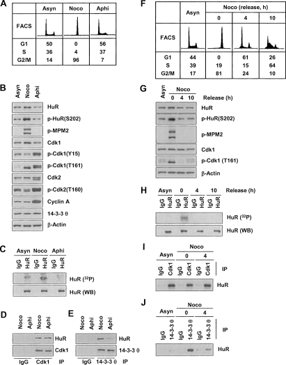
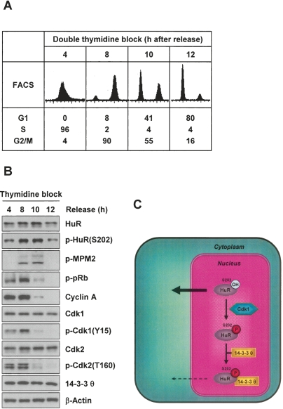
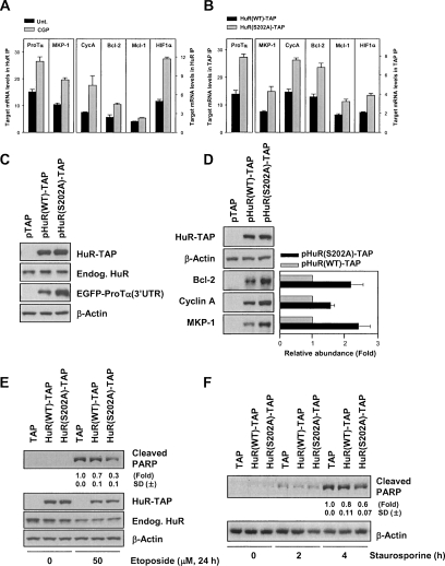
A predominantly nuclear RNA-binding protein, HuR translocates to the cytoplasm in response to stress and proliferative signals, where it stabilizes or modulates the translation of target mRNAs. Here, we present evidence that HuR phosphorylation at S202 by the G2-phase kinase Cdk1 influences its subcellular distribution. HuR was specifically phosphorylated in synchronous G2-phase cultures; its cytoplasmic levels increased by Cdk1-inhibitory interventions and declined in response to Cdk1-activating interventions. In keeping with the prominently cytoplasmic location of the nonphosphorylatable point mutant HuR(S202A), phospho-HuR(S202) was shown to be predominantly nuclear using a novel anti-phospho-HuR(S202) antibody. The enhanced cytoplasmic presence of unphosphorylated HuR was linked to its decreased association with 14-3-3 and to its heightened binding to target mRNAs. Our findings suggest that Cdk1 phosphorylates HuR during G2, thereby helping to retain it in the nucleus in association with 14-3-3 and hindering its post-transcriptional function and anti-apoptotic influence.


Figures

Figure 1.
Figure 1.
Inhibition or silencing of Cdk1, an HuR-interacting protein, elevates cytoplasmic HuR levels. (A) HeLa whole-cell lysates were used in immunoprecipitation (IP) assays with either control mouse IgG or anti-HuR antibodies, followed by Western blot (WB) detection of Cdk1, HuR, and the negative control GAPDH. (B) Assays were performed as in A except that control rabbit IgG or anti-Cdk1 antibodies were used for IP. (C) Cytosolic extracts (CE) and nuclear extracts (NE) were immunoprecipitated with either control rabbit IgG or anti-Cdk1 antibodies, followed by Western blot detection of HuR and Cdk1. The levels of Cdk1, HuR, α-Tubulin (cytosolic marker), and hnRNP C1/C2 (nuclear marker) were detected by Western blot analysis. (D) Following treatment for 2 h with the indicated CGP concentrations, HeLa cytoplasmic extracts (CE) were prepared and the levels of HuR and loading controls α-Tubulin and β-Actin were monitored by Western blot analysis. (E) After treatment with CGP (2 h, 10 μM), CE (10 μg) and NE (5 μg) were prepared and the levels of HuR, the cytoplasmic marker α-Tubulin, and the nuclear marker hnRNP C1/C2 were assessed by Western blot analysis. (F) Immunofluorescence microscopy to assess HuR distribution (green) in cells that were either left untreated (Unt.) or were treated with CGP (2 μM for 2 h). Nuclei were visualized using Topro-3 (blue) and the transcription factor SP1 (red). (Merged) Overlay of Topro-3, HuR, and SP1 signals. (GI) Cells were transfected with control siRNAs (Ctrl.) or with siRNAs targeting Cdk1, Cdk7 or cyclin B1; 48 h after transfection, the levels of HuR, Cdk1, Cdk7, Cyclin B1, and loading controls β-Actin and α-Tubulin in whole-cell extracts (WE, 5 μg) and CE were monitored by Western blot analysis. Shown are fold changes in HuR levels as measured by densitometry (±SD, standard deviation from at least three experiments).
Figure 2.
Figure 2.
UVC-mediated elevation of cytoplasmic HuR levels concomitant with Cdk1 inhibition. (A) Schematic of the modifications activating and inhibiting Cdk1. (B) HeLa cells were irradiated with UVC (25 J/m2) and collected 2 h later to assess the levels of cytosolic (CE) HuR by Western blot analysis. (C) At the times shown after UVC irradiation, the levels of total Cdk1, Cdk1 phosphorylated at Tyr 15 [p-Cdk1(Y15)] or Thr 161 [p-Cdk1(T161)], and Cyclin B1 were examined by Western blot analysis of WE. (D,E) After silencing Cdk1 for 48 h (D) or treating with CGP (2 μM) for 1 h (E), cells were either left without further treatment (−) or were irradiated with UVC (25 J/m2); 2 h later, HuR and α-Tubulin levels in CE were assessed by Western blot analysis. (F) Cells were transfected with Myt1 siRNA; 48 h after transfection, WE were prepared and the levels of Cdk1, p-Cdk1(Y15), and p-Cdk1(T161) were studied by Western blot analysis. (G) Following Myt1 silencing with a specific siRNA, cells were either left untreated (−) or were irradiated with UVC (25 J/m2) and collected 2 h later for the detection of HuR (and loading control α-Tubulin) in CE. HuR signals in A, C, D, and F were quantified by densitometry; ±SD from at least three experiments are indicated.
Figure 3.
Figure 3.
HuR phosphorylation at Ser-202 enhances its nuclear localization. (A) HuR schematic showing the position of S202, the residue phosphorylated by Cdk1, and the vectors that were constructed to express a chimeric wild-type HuR protein linked to TAP [HuR(WT)-TAP] and a chimeric protein with nonphosphorylatable S202 [HuR(S202A)-TAP]. (RRM) RNA-recognition motif; (HNS) HuR nucleocytoplasmic shuttling sequence. (B) HeLa cells were transfected with plasmids to express the chimeric proteins depicted in A and the levels of ectopic [HuR(WT)-TAP and HuR(S202A)-TAP] and endogenous HuR in CE and WE were monitored by Western blot analysis. Shown are fold changes in ectopic HuR-TAP levels from three independent experiments, as assessed by densitometry. (C) HuR phosphorylation was assessed after an in vitro kinase reaction using GST-HuR as substrate (Materials and Methods); the levels of total HuR-GST and phosphorylated HuR-GST were monitored by Western blotting using antibodies that recognized either total HuR or phospho-HuR [p-HuR(S202)], respectively. (D) WE were prepared from HeLa cells and treated with CIP for 1 h; the levels of HuR and p-HuR(S202) were detected by Western blotting using antibodies that recognized either total HuR or p-HuR(S202). (E) CE and NE were prepared and the levels of total HuR and p-HuR(S202), as well as those of cytoplasmic marker α-Tubulin and nuclear marker hnRNP C1/C2, were detected by Western blot analysis. (F) Increasing amounts of CE and NE lysates were tested in order to achieve comparable total HuR signals and then compare p-HuR(S202) signals.
Figure 4.
Figure 4.
Phosphorylated HuR binds 14–3–3θ and localizes in the nucleus. (A, top) Western blot analysis of the indicated 14–3–3 isoforms (paired samples). (Bottom) Various 14–3–3 isoforms were immunoprecipitated using specific antibodies, and the presence of HuR and negative control GAPDH in the IP materials was tested by Western blot analysis. (B) CE and NE were prepared from HeLa cells, used for IP with control rabbit IgG or anti-14–3–3θ antibodies, and HuR and 14–3–3θ in the IP samples was detected by Western blot analysis. (C) Following treatment with CGP (2 μM, 2 h), WE were prepared and assayed by IP using control rabbit IgG or anti-14–3–3θ antibodies; bound HuR and 14–3–3θ were assessed by Western blotting. (D) After CGP treatment (2 μM, 2 h), CE and NE were prepared and the levels of 14–3–3θ, the cytosolic marker α-Tubulin, the nuclear marker hnRNP C1/C2, and the loading control β-Actin were analyzed by Western blotting. (E) Forty-eight hours after transfection of cells with the siRNAs shown, WE were prepared and used for IP in the presence of rabbit IgG or anti-14–3–3θ antibodies. (Left) The levels of HuR, Cdk1, cyclin B1, and loading control β-Actin in the Input material were tested by Western blotting. (Right) The presence of HuR and 14–3–3θ in the IP samples was assessed by Western blotting. (F,G) Cells were transfected with either control (Ctrl.) siRNA or with an siRNA targeting 14–3–3ζ or 14–3–3θ. Forty-eight hours after transfection, CE and WE were prepared and the levels of HuR and 14–3–3ζ (F) and 14–3–3θ (G) were detected by Western blotting; β-Actin and α-Tubulin were used as loading controls; ±SD of HuR signals from at least three experiments are indicated. (H) In cells transfected with 14–3–3θ siRNA, ProTα mRNA levels in HuR IP were determined by RT–qPCR (Materials and Methods); graph depicts the means and SD from three independent experiments.
Figure 5.
Figure 5.
Specific HuR phosphorylation and binding to 14–3–3 during G2/M. (A) FACS analysis of HeLa cells that were either growing asynchronously (Asyn) or synchronized in G2/M using nocodazole (Noco, 100 ng/mL, 16 h) or in G1 and S using aphidicolin (Aphi, 2 μg/mL, 16 h). (B) WE lysates were prepared from the populations in A, and the levels of proteins and phosphoproteins (including G1 and S markers p-cdk1 [Y15], p-cdk2 [T160], and cyclin A; G2/M marker p-cdk1 [T161], and mitotic marker p-MPM2) were assessed by Western blot analysis. (C) In vivo HuR phosphorylation was studied by incubating the populations described in A for 2 h with 32Pi and assessing the incorporation of the radiolabel into HuR by IP using anti-HuR antibody (or IgG in control IP reactions). Total HuR in IP materials was detected by Western blot analysis (WB). Co-IP analysis was used to study the association of HuR with Cdk1 (D) and with 14–3–3θ (E). (F) FACS analysis of HeLa cells synchronized with nocodazole (100 ng/mL for 16 h), released by removing nocodazole, and examined 4 and 10 h later. (G) Using cells that were processed as explained in F, the levels of the proteins shown were studied by Western blot analysis of WE lysates. (H) In vivo HuR phosphorylation was studied by incubating the populations described in F for 2 h with 32Pi and assessing the incorporation of the radiolabel into HuR by IP using anti-HuR antibody (or IgG in control IP reactions). Total HuR in IP materials was detected by Western blotting (WB). WE lysates from the populations in F were used in co-IP assays to study the association of HuR with Cdk1 (I) and with 14–3–3θ (J).
Figure 6.
Figure 6.
Synchronous phosphorylation of HuR and model. (A) FACS analysis of HeLa cells at 4, 8, 10, and 12 h after release from double thymidine block (Supplemental Material). (B) WE lysates from cells that were treated as in A were collected for Western blot analysis of the proteins shown. (C) Schematic of the proposed influence of Cdk1 on HuR localization; see text for details.
Figure 7.
Figure 7.
S202 phosphorylation influences the levels of cytoplasmic HuR RNP complexes and the levels of proteins encoded by HuR target mRNAs. HeLa cells were treated with CGP (2 μM, 2 h) (A) or transfected with either pHuR-TAP or pHuR(S202A)-TAP and collected 48 h later (B); CE were prepared (Materials and Methods) and immunoprecipitated with mouse IgG or anti-HuR antibodies (A) or with IgG agarose beads (B). The levels of prothymosin α (ProTα), MKP-1, cyclin A (CycA), Mcl-1, Bcl-2, and HIF1α were determined by RT–qPCR; the data (means and SEM from three independent experiments) were normalized to the abundance of GAPDH mRNA (a housekeeping mRNA present at background levels) in each set of IP samples. (C) Cells were cotransfected with pTAP, pHuR(WT)-TAP, or pHuR(S202A)-TAP along with plasmid pEGFP-ProTα(3′UTR); 48 h after transfection, the levels of endogenous HuR, ectopic HuR-TAP, EGFP, and loading control β-Actin were detected in WE by Western blotting. (D) Cells were transfected as in C, WE were prepared, and the levels of MKP-1, Cyclin A, and Bcl-2 were detected by Western blotting, quantified by densitometry (graph), and normalized to the levels of loading control β-Actin. (E) After transfection with pTAP, pHuR(WT)-TAP, or pHuR(S202A)-TAP, cells were treated with etoposide for 24 h (50 μM) (left) or with staurosporine (1 μM) for the times shown (right) and WE were prepared for Western blot analysis and densitometric quantification of cleaved PARP. Data are representative of three independent experiments.

References

    1. Abdelmohsen K., Pullmann R., Lal A., Kim H.H., Galban S., Yang X., Blethrow J.D., Walker M., Shubert J., Gillespie D.A., et al. Phosphorylation of HuR by Chk2 regulates SIRT1 expression. Mol. Cell. 2007a;25:543–557. - PMC - PubMed
    1. Abdelmohsen K., Lal A., Kim H.H., Gorospe M. Posttranscriptional orchestration of an anti-apoptotic program by HuR. Cell Cycle. 2007b;6:1288–1292. - PubMed
    1. Anderson P., Kedersha N. Stressful initiations. J. Cell Sci. 2002;115:3227–3234. - PubMed
    1. Antic D., Lu N., Keene J.D. ELAV tumor antigen, Hel-N1, increases translation of neurofilament M mRNA and induces formation of neurites in human teratocarcinoma cells. Genes & Dev. 1999;13:449–461. - PMC - PubMed
    1. Atasoy U., Watson J., Patel D., Keene J.D. ELAV protein HuA (HuR) can redistribute between nucleus and cytoplasm and is upregulated during serum stimulation and T cell activation. J. Cell Sci. 1998;111:3145–3156. - PubMed

Publication types

LinkOut - more resources