. 2008 Jul 1;22(13):1828-37.
doi: 10.1101/gad.477908.

Pax3 regulation of FGF signaling affects the progression of embryonic progenitor cells into the myogenic program

Affiliations

Pax3 regulation of FGF signaling affects the progression of embryonic progenitor cells into the myogenic program

Mounia Lagha et al. Genes Dev. .

Abstract

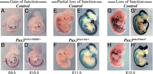
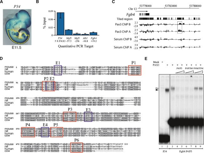
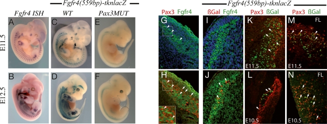
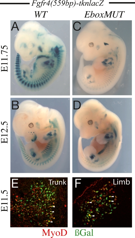
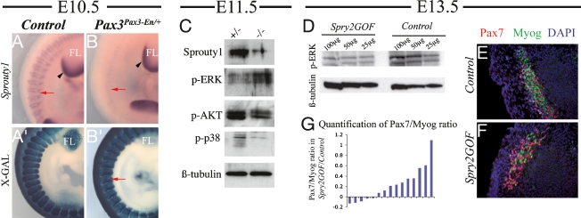
Pax3/7-dependent stem cells play an essential role in skeletal muscle development. We now show that Fgfr4 lies genetically downstream from Pax3 and is a direct target. In chromatin immunoprecipitation (ChIP)-on-chip experiments, Pax3 binds to a sequence 3' of the Fgfr4 gene that directs Pax3-dependent expression at sites of myogenesis in transgenic mouse embryos. The activity of this regulatory element is also partially dependent on E-boxes, targets of the myogenic regulatory factors, which are expressed as progenitor cells enter the myogenic program. Other FGF signaling components, notably Sprouty1, are also regulated by Pax3. In vivo manipulation of Sprouty expression reveals that FGF signaling affects the balance between Pax-positive progenitor cells and committed myoblasts. These results provide new insight into the Pax-initiated regulatory network that modulates stem cell maintenance versus tissue differentiation.


Figures

Figure 1.
Figure 1.
Fgfr4 lies genetically downstream from Pax3 during myogenesis. (A–H) Whole-mount in situ hybridization with a Fgfr4 probe, at the embryonic day (E) indicated. (A,C,E,G) Control embryos. (B,D) Pax3PAX3-FKHR-IRESnlacZ/+ (Pax3PAX3-FKHR/+) embryos expressing the gain-of-function allele of Pax3. (F) A Pax3Pax3-En-IRESnlacZ/+ (Pax3Pax3-En/+) embryo expressing a dominant-negative allele of Pax3 that in the heterozygote results in partially compromised Pax3 function. (H) Pax3nlacZ/nlacZ, a null mutant of Pax3. (E′–H′) X-Gal staining of control (E′,G′), Pax3Pax3-En-IRESnlacZ/+ (F′), and Pax3nlacZ/nlacZ (H′) embryos to show the presence of Pax3-positive cells at the stages indicated. Red arrows point to differences in Fgfr4 transcripts at sites of myogenesis in the somites. Black arrowheads indicate internal controls at sites where Fgfr4 expression is not affected and Pax3 is not expressed.
Figure 2.
Figure 2.
Identification of an in vivo Pax3-binding site in the Fgfr4 locus. (A) X-Gal staining at E11.5 of a P34 transgenic embryo in which nlacZ expression is regulated by five consensus Pax3-binding sites (Pax3)5-tk-nlacZ. The tissue used for ChIP is outlined. (B) Real-time quantitative PCR using primers (targets) for the P34 Pax3-binding sites, a functional Pax3 site at −57.5 kb from Myf5, a control Myf5 flanking sequence, a control Albumin sequence, and the Fgfr4 +19.2-kb sequence, containing sites P4/P5 identified by Chip-chip. Results are expressed as a percentage of input showing enrichment after Pax3 immunoprecipitation, with the serum control subtracted. (C) Results of the tiling arrays for Pax3 and for serum controls within the 3′ part of the Fgfr4 locus. Fgfr4 exons are indicated as black boxes. The region of strong Pax3 hybridization signal is outlined. (D) The nucleotide sequence of the Fgfr4 (+18,832 to +19,391 bp) distal element in mouse and comparison with a homologous region of the rat, human, and cow genomes, with conserved bases indicated on a gray background. Six putative Pax3 binding sites (P1–P6) are framed in red. E-box consensus sequences for myogenic regulatory factors are also indicated in blue (E1–E4). (E) Gel shift mobility assays for Pax3 binding, using a reticulocyte lysate without (lane 1) or with (lane 2-9) Pax3 protein. A labeled oligonucleotide (30 bp) containing a consensus Pax3 site of the P34 transgene (lanes 1,2) or an oligonucleotide (60 bp) from the Fgfr4 (559 bp) sequence containing sites P4 and P5 (lanes 3–9) shows Pax3 binding (3,8,9). Lanes 4–9 show competition experiments with 50-fold (lane 4) or 150-fold (lane 5) excess of wild-type sequence (P4/P5), with this sequence with P5 mutated (P4/P5M) (lanes 6,7) or with P4 and P5 mutated (P4M/P5M) (lanes 8,9).
Figure 3.
Figure 3.
The Fgfr4 distal element directs Pax3-dependent myogenic expression in vivo. (A,B) Whole-mount in situ hybridization for Fgfr4 transcripts on embryos at the stages indicated. (C–F) X-Gal staining of transient transgenic embryos in which a tk-nlacZ transgene is under the control of the wild-type (C,D) Fgfr4 distal element (559 bp) or this element with the six Pax3 sites, mutated (E,F). (G,H) Coimmunohistochemistry on DAPI-stained transverse sections in the myotome at interlimb level of E10.5 embryos, showing Fgfr4 (green) and DAPI staining (G) and Fgfr4 (green) and Pax3 (red) staining (H). Insert in H represents a higher magnification of the region outlined in G and H. Examples of colocalization where the plane of section includes a nucleus showing Pax3 staining are indicated by arrowheads. Arrows indicate dorsally located Pax3+ cells. (I,J) Coimmunohistochemistry on DAPI-stained sections (I) at trunk level of an E11.5 Fgfr4(559bp)-tk-nlacZ transgenic embryo using antibodies to Fgfr4 (green) (I,J) and to nuclear β-Gal (red) (J). Arrowheads as for H. (K,L) Coimmunohistochemistry on transverse sections at interlimb level on an E11.5 (K) or E10.5 (L) Fgfr4(559bp)-tk-nlacZ transgenic embryo showing expression of Pax3 (red) and β-Gal (green). (M,N) Sections of the forelimb (FL) at E11.5 (M) or E10.5 (N) similarly stained for Pax3 and β-Gal. Arrows point to nuclear coexpression.
Figure 4.
Figure 4.
Potential myogenic factor regulation of the Fgfr4 element. (A,B) Control embryos expressing the Fgfr4(559bp)-tk-nlacZ transgene at E11.75 (A) and E12.5 (B). (C,D) Embryos expressing this transgene with the four E-boxes mutated at E11.75 (C) and E12.5 (D). (E,F) Coimmunohistochemistry on DAPI-stained transverse sections of Fgfr4(559bp)-tk-nlacZ transgenic embryos at E11.5 in the interlimb region (Trunk) (E) and forelimb (Limb) (F) using antibodies to MyoD (red) and β-Gal (green). Arrowheads point to examples of colocalization.
Figure 5.
Figure 5.
Components of the FGF signaling pathway are regulated by Pax3; Sprouty modulates myogenesis in vivo. (A,B) Whole-mount in situ hybridization for Sprouty1 transcripts in control (A) andPax3Pax3-En/+ (B) embryos at E10.5. (A′,B′) X-Gal staining of Pax3nlacZ/+ (A′) and Pax3Pax3-En/+ (B′) embryos at E10.5. The red arrows indicate somites. Black arrowheads point to Pax3-independent Sprouty1 expression in the distal forelimb bud (FL). (C) Western blot of the same number of GFP-positive cells isolated by flow cytometry, from somites of Pax3GFP/+ (+/−) and Pax3GFP/nlacZ (−/−) embryos at E11.5, using the antibodies indicated; (p) phosphorylated. (D) Western blots on extracts (100 μg, 50 μg, and 25 μg of total protein) from limbs of control and Sprouty2 gain of function (Spry2 GOF) transgenic embryos at E13.5, using the antibodies indicated. (E,F) Coimmunohistochemistry on DAPI-stained transverse sections of equivalent muscles in the interlimb region of control (E) and Spry2 GOF (F) embryos at E13.5 using antibodies to Pax7 (red) and myogenin (MyoG) (green). (G) Quantitative analysis of the ratio of Pax7 to myogenin-positive cells on sections, counted using Metamorph software. Each bar represents the differential ratio [(Pax7/Myog)Spry2GOF − (Pax7/Myog)control] between Spry2GOF and control embryos quantified for equivalent sections from the same deep back muscles, with a minimum of 500 total cells counted per section. The ratio (Pax7/Myog) is significantly higher in Spry2GOF samples compared to the control (P < 0.05, with the two-sided Wilcoxon signed rank test).

Similar articles

Cited by

References

    1. Bajard L., Relaix F., Lagha M., Rocancourt D., Daubas P., Buckingham M.E. A novel genetic hierarchy functions during hypaxial myogenesis: Pax3 directly activates Myf5 in muscle progenitor cells in the limb. Genes & Dev. 2006;20:2450–2464. - PMC - PubMed
    1. Basson M.A., Echevarria D., Petersen Ahn C., Sudarov A., Joyner A.L., Mason I.J., Martinez S., Martin G.R. Specific regions within the embryonic midbrain and cerebellum require different levels of FGF signaling during development. Development. 2008;135:889–898. - PMC - PubMed
    1. Ben-Yair R., Kalcheim C. Lineage analysis of the avian dermomyotome sheet reveals the existence of single cells with both dermal and muscle progenitor fates. Development. 2005;132:689–701. - PubMed
    1. Bladt F., Riethmacher D., Isenmann S., Aguzzi A., Birchmeier C. Essential role for the c-met receptor in the migration of myogenic precursor cells into the limb bud. Nature. 1995;376:768–771. - PubMed
    1. Buckingham M. Myogenic progenitor cells and skeletal myogenesis in vertebrates. Curr. Opin. Genet. Dev. 2006;16:525–532. - PubMed

Publication types

MeSH terms

LinkOut - more resources