. 2008 Jul 1;68(13):5122-31.
doi: 10.1158/0008-5472.CAN-07-6123.

The p53 homologue DeltaNp63alpha interacts with the nuclear factor-kappaB pathway to modulate epithelial cell growth

Affiliations

The p53 homologue DeltaNp63alpha interacts with the nuclear factor-kappaB pathway to modulate epithelial cell growth

Kathryn E King et al. Cancer Res. .

Abstract

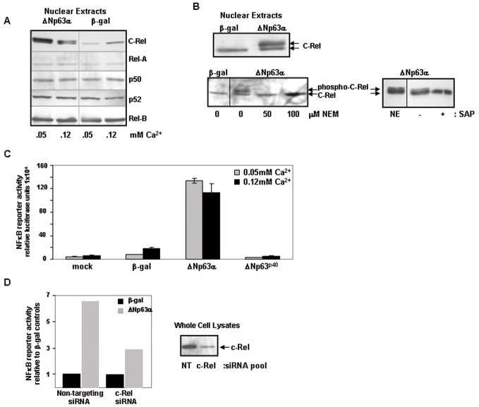
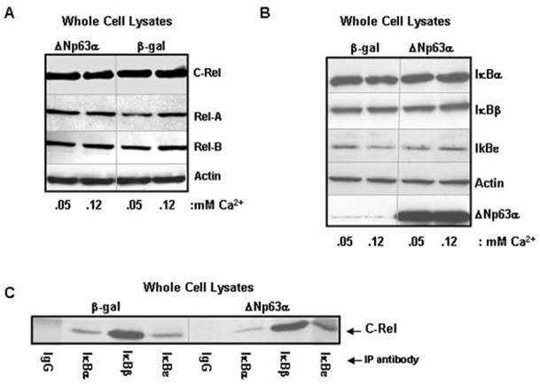
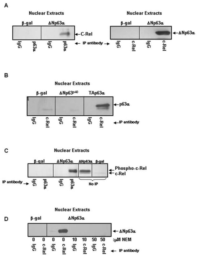
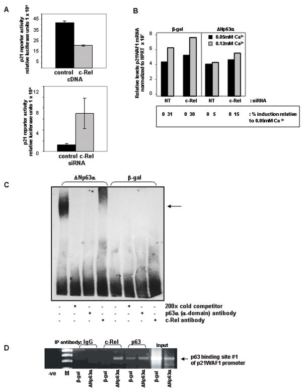
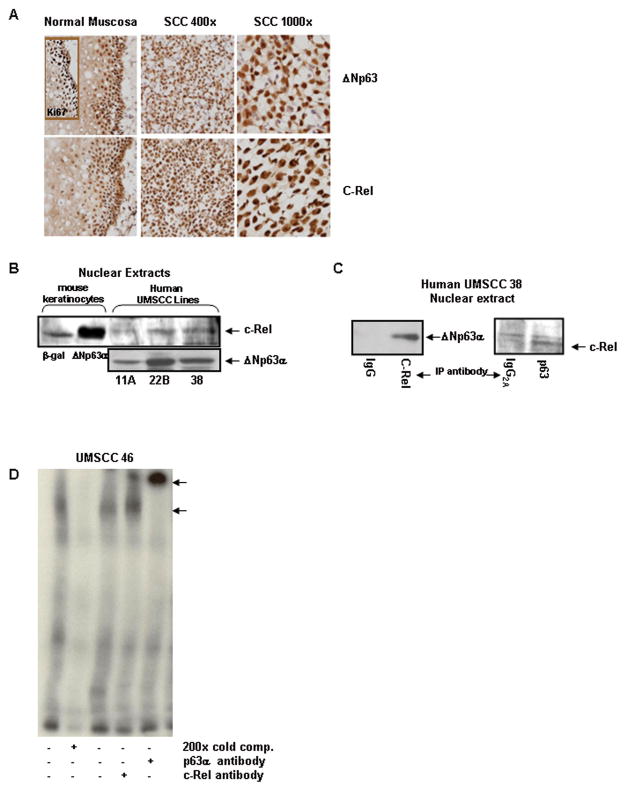
The p53 homologue DeltaNp63alpha is overexpressed and inhibits apoptosis in a subset of human squamous cell carcinomas (SCC). Here, we report that in normal keratinocytes overexpressing DeltaNp63alpha and in human squamous carcinoma cells, DeltaNp63alpha physically associates with phosphorylated, transcriptionally active nuclear c-Rel, a nuclear factor-kappaB family member, resulting in increased c-Rel nuclear accumulation. This accumulation and the associated enhanced proliferation driven by elevated DeltaNp63alpha are attenuated by c-Rel small interfering RNA or overexpression of mutant IkappaBalphaM, indicating that c-Rel-containing complex formation is critical to the ability of elevated DeltaNp63alpha to maintain proliferation in the presence of growth arresting signals. Consistent with a role in growth regulation, DeltaNp63alpha-c-Rel complexes bind a promoter motif and repress the cyclin-dependent kinase inhibitor p21WAF1 in both human squamous carcinoma cells and normal keratinocytes overexpressing DeltaNp63alpha. The relationship between DeltaNp63alpha and activated c-Rel is reflected in their strong nuclear staining in the proliferating compartment of primary head and neck SCC. This is the first report indicating that high levels of DeltaNp63alpha interact with activated c-Rel in keratinocytes and SCC, thereby promoting uncontrolled proliferation, a key alteration in the pathogenesis of cancers.


Figures

Figure 1
Figure 1. ΔNp63α overexpression results in increased nuclear levels of transcriptionally active phosphorylated c-Rel in murine keratinocytes
A) Elevated Δ Np63α expression specifically enhances nuclear levels of the NF-κ B subunit c-Rel. Western blots of nuclear extracts from primary murine keratinocytes harvested 21h post-adenoviral introduction of human ΔNp63α or β-gal. Cultures were maintained in medium containing 0.05mM Ca2+ throughout or exposed to 0.12 mM Ca2+ for the final 4h. C-Rel levels are increased in the nuclei of ΔNp63α-overexpressing keratinocytes. B) c-Rel is phosphorylated in response to elevated ΔNp63α. Western analysis of nuclear extracts from keratinocytes overexpressing ΔNp63α or β-gal cultured under control conditions (top) or in the presence of the thiol modifier N-ethylmaleimide (NEM) at concentrations noted for 21h following adenoviral introduction to block in vivo phosphorylation (bottom left). The upper species is eliminated in the presence of NEM. Phosphorylation was confirmed by western analysis of nuclear extracts derived from keratinocytes overexpressing ΔNp63α incubated in the presence or absence of Shrimp Alkaline Phosphatase (SAP) for 3h at 37°C (bottom right). A non-incubated control nuclear extract (NE) is included for visual reference. SAP treatment results in the loss of the upper phosphorylated species. C) ΔNp63α overexpression results in NF-κ B-mediated transactivation; α-domain of ΔNp63α required. NF-κB responsive reporter gene activity in keratinocytes overexpressing ΔNp63α, ΔNp63p40 (ΔNp63 lacking α-domain) or β-gal. Samples were harvested 24h post-transfection. Results depict mean +/− s.d. of triplicate samples of a representative experiment performed thrice. D) c-Rel is required for NF-κ B-mediated transactivation following ΔNp63α overexpression. Keratinocytes were co-transfected with NF-κB reporter construct and c-Rel-targeting siRNA or control siRNA, 24h prior to adenoviral infection with ΔNp63α or β-gal. Samples were harvested 24h post adenoviral introduction. Right side: Western blot reveals depletion of c-Rel in whole cell lysates at time of harvest (NT: non targeting siRNA). Left side: Fold increase in NF-κB reporter gene activity in ΔNp63α overexpressing cultures compared to β-gal control cultures in the presence or absence of c-Rel siRNA. Triplicate wells were averaged and are presented as fold increase relative to β-gal controls that are normalized to 1.0. Experiment was performed twice with consistent results; representative experiment shown.
Figure 2
Figure 2. Enhanced nuclear c-Rel is required for ΔNp63α-mediated loss of keratinocyte growth regulation, but not differentiation defects
A–C: Flow cytometry analysis of BrdU incorporation in keratinocytes overexpressing ΔNp63α or β-gal under proliferating (0.05mM Ca2+) or differentiating (0.12mM Ca2+) conditions. A) Blocking NF-κB nuclear translocation with the IκBαM super-repressor restores normal Ca2+-mediated growth regulation to ΔNp63α-overexpressing keratinocytes. Primary murine keratinocytes (1.5 days post plating) were co- infected with adenovirus encoding ΔNp63α or β-gal in combination with IκBαM super-repressor or empty vector control. 17h post-infection the medium was changed; cells were maintained for a further 24h in 0.05mM or 0.12mM Ca2+ and pulsed with BrdU (10μM) for the final 4h. Results depict mean +/− s.d. of triplicate samples from a representative experiment performed thrice. Right: Western blot of corresponding nuclear extracts confirms that co-infection with the IκBαM super-repressor effectively reduces levels of nuclear c-Rel in ΔNp63α overexpressing keratinocytes. B) c-Rel siRNA knockdown partially restores normal growth arrest in ΔNp63α-overexpressing keratinocytes. Keratinocyte cultures were transfected with c-Rel or non-targeting siRNA 12 hours prior to introduction of ΔNp63α or β-gal by adenovirus. Decreasing c-Rel levels by siRNA results in an overall reduction of proliferation in all cultures (right panel vs left panel of histogram), and partially restores Ca2+-mediated growth arrest to ΔNp63α-overexpressing keratinocytes (right side, right panel of histogram). Results depict mean +/− s.d. of triplicate samples from a representative experiment. Right: Western blot of whole cell lysates confirms that c-Rel expression is reduced in keratinocytes transfected with c-Rel siRNA. C) Rel-A does not contribute to the aberrant growth arrest response observed in ΔNp63α overexpressing keratinocytes. Keratinocytes were transfected with Rel-A targeted siRNA to deplete Rel-A levels, or with non-targeting siRNA as control. Depleting Rel-A by siRNA has no effect on keratinocyte proliferation under these conditions. Results depict mean +/− s.d. of triplicate samples from a representative experiment. Right: Western blot of whole cell lysates confirms that Rel-A siRNA effectively reduces Rel-A expression in these cultures. D) Blocking NF-κB nuclear translocation does not restore induction of markers of terminal differentiation in ΔNp63α-overexpressing keratinocytes. Western blot of whole cell lysates from keratinocytes overexpressing ΔNp63α or β-gal +/− IκBαM super-repressor and exposed to 0.12mM Ca2+ for 24h. Blocking NF-κB nuclear translocation does not restore the Ca2+-mediated induction of the early marker of keratinocyte differentiation, keratin 10, or the late marker, filaggrin.
Figure 3
Figure 3. Mechanism of nuclear c-Rel enhancement is not dependent on disruption of cytoplasmic IκB:c-Rel interactions
A) ΔNp63 overexpression does not alter total levels of cellular NF-κB. Western blots of whole cell lysates from primary mouse keratinocytes harvested 21h post-adenoviral introduction of human ΔNp63α or β-gal. Cultures were maintained in medium containing 0.05mM Ca2+ or exposed to 0.12 mM Ca2+ for the final 4h. B) Levels of IκB regulatory proteins are not decreased in ΔNp63α-overexpressing keratinocytes that accumulate nuclear c-Rel. Western blot of whole cell lysates of keratinocytes overexpressing β-gal or ΔNp63α. C) ΔNp63α overexpression does not inhibit normal cytoplasmic interactions between c-Rel and the IκB proteins. Co-immunoprecipitation analysis of whole cell lysates from keratinocytes overexpressing ΔNp63α or β-gal.
Figure 4
Figure 4. ΔNp63α and c-Rel physically interact in a phosphorylation, p63 α-domain dependent manner
A) ΔNp63α and c-Rel physically interact in the nuclei of ΔNp63α overexpressing keratinocytes. Co-immunoprecipitation analysis of nuclear extracts from keratinocytes overexpressing ΔNp63α or β-gal. Nuclear extracts were immunoprecipitated with antibody to p63 (left panel) or c-Rel (right panel) and probed for c-Rel or p63, as noted. B) The α-domain but not the ΔN-domain of p63 is required for the interaction between p63 and c-Rel. Nuclear extracts from keratinocytes overexpressing ΔNp63p40 (a truncated form of ΔNp63 lacking the α-COOH-terminus), TAp63α, or β-gal were immunoprecipitated with antibody to c-Rel and probed for p63. C) ΔNp63α associates with phosphorylated c-Rel. Co-immunoprecipitation analysis of nuclear extracts from keratinocytes overexpressing ΔNp63α or β-gal resolved alongside ΔNp63α or β-gal nuclear extracts (no-IP). Co-migration and western blot reveals that the upper, phosphorylated species of c-Rel interacts with ΔNp63α. D) Protein phosphorylation is required for association between ΔNp63α and c-Rel. Co-immunoprecipitation analysis of nuclear extracts from keratinocytes overexpressing ΔNp63α or β-gal. Culture with NEM for one day following adenoviral infection to block in vivo phosphorylation eliminates the interaction between ΔNp63α and c-Rel.
Figure 5
Figure 5. ΔNp63α and c-Rel both negatively regulate the cyclin dependent kinase inhibitor p21WAF1, and interact in vitro and in vivo at a p63 binding site on the p21WAF1 promoter
A) Modulating c-Rel levels alters p21WAF1 reporter gene activity. p21WAF1 reporter gene activity following co-transfection in combination with a human c-Rel cDNA construct, c-Rel targeted siRNA, or controls. Overexpression of c-Rel represses p21WAF1 reporter gene activity (Figure 5A, top), while reducing c-Rel expression levels with targeted siRNA enhances expression of a p21WAF1 luciferase reporter construct (bottom). B) Incomplete silencing of c-Rel by targeted siRNA allows slight restoration of induction of endogenous p21WAF1. Semi-quantitative RT-PCR analysis of p21WAF1. Ca2+-mediated induction of p21WAF1 is unaffected by siRNA knockdown of c-Rel in control keratinocytes overexpressing β-gal. Consistent with previous results, Ca2+-mediated p21WAF1 induction is blocked by the overexpression of ΔNp63α. siRNA knockdown of c-Rel results in a small but reproducible induction of p21WAF1 in ΔNp63α overexpressing keratinocytes in response to 0.12mM Ca2+ (15% in c-Rel targeted vs 5% induction in non-targeted siRNA controls), as determined by spot densitometry analysis using an Alpha Innotech imaging system. Experiment was repeated with consistent results. C) Δ Np63α and c-Rel physically associate on the p21WAF1 promoter in vitro. EMSA analysis of nuclear extracts from keratinocytes overexpressing ΔNp63α or β-gal. The p63 binding site #1 from the p21WAF1 promoter used in the reporter gene assays was biotin-labeled and used in the binding reactions. A protein:DNA complex seen only in the presence of overexpressed ΔNp63α is supershifted with a c-Rel antibody, and interrupted with a p63-specific antibody. The experiment was performed 4 times with consistent results; representative experiment is presented. D) ΔNp63α and c-Rel physically associate on the p21WAF1 promoter in vivo. ChIP analysis was performed on samples derived from keratinocytes overexpressing ΔNp63α or β-gal using the antibodies noted. PCR primers were designed to flank the p63 binding site #1 from the p21WAF1 promoter. Association of the C-Rel:ΔNp63α complex with p63 binding site #1 is observed. Input DNA: PCR products generated using DNA template from total genomic DNA. Lane labeled “-ve” indicates an absence of DNA in the PCR reaction. M: molecular weight marker. Results shown are representative of 2 independent experiments.
Figure 6
Figure 6. Endogenous ΔNp63α and c-Rel expression are correlated and expanded in primary human cancers, and associate in the nuclei of human squamous cell carcinoma cells
A) Nuclear expression patterns of ΔNp63α and c-Rel are expanded and associated in primary human squamous cell carcinomas. Immunostaining of normal mucosa and squamous cell carcinoma tissue sections with p63 and c-Rel. The proliferative compartment of normal mucosa is identified by Ki67 immunoreactivity. B) Endogenous Δ Np63α and c-Rel are present in nuclei of human HNSCC lines. Western blots of nuclear extracts prepared from SCC squamous cell carcinoma lines. Mouse keratinocytes overexpressing ΔNp63α or β-gal are included as controls. C) Endogenous nuclear ΔNp63α and c-Rel physically interact in squamous cell carcinoma cells. Co-immunoprecipitation analysis of UMSCC-38 nuclear extracts. Nuclear extracts were immunoprecipitated with antibody to c-Rel (left panel) or p63 (right panel) and probed for c-Rel or p63, as noted. D) Endogenous nuclear ΔNp63α and c-Rel derived from squamous carcinoma cell lines are associated on the p21WAF1 promoter in vitro. EMSA analyses of nuclear extracts derived from the HNSCC cell line UMSCC 46. A 32P labeled probe using the p63 binding site #1 from the p21WAF1 promoter was used in these reactions. A protein:DNA complex is seen and can be partially supershifted with a c-Rel antibody. Use of the smaller 32P tag in the HNSCC experiments allowed for a supershift band to be seen with the p63 antibody as well.

References

    1. King KE, Weinberg WC. p63: defining roles in morphogenesis, homeostasis, and neoplasia of the epidermis. Mol Carcinog. 2007;46:716–24. - PubMed
    1. Hibi K, Trink B, Patturajan M, et al. AIS is an oncogene amplified in squamous cell carcinoma. Proc Natl Acad Sci USA. 2000;97:5462–67. - PMC - PubMed
    1. Wrone DA, Yoo S, Chipps LK, et al. The expression of p63 in actinic keratoses, seborrheic keratosis, and cutaneous squamous cell carcinomas. Dermatol Surg. 2004;30:1299–302. - PubMed
    1. Lin Z, Liu M, Li Z, et al. DeltaNp63 protein expression in uterine cervical and endometrial cancers. J Cancer Res Clin Oncol. 2006;132:811–16. - PMC - PubMed
    1. Yang A, Kaghad M, Wang Y, et al. p63, a p53 homolog at 3q27–29, encodes multiple products with transactivating, death-inducing, and dominant-negative activities. Mol Cell. 1998;2:305–16. - PubMed

Publication types

MeSH terms