. 2008 Jul 1;14(13):4275-83.
doi: 10.1158/1078-0432.CCR-08-0168.

EML4-ALK fusion gene and efficacy of an ALK kinase inhibitor in lung cancer

Affiliations

EML4-ALK fusion gene and efficacy of an ALK kinase inhibitor in lung cancer

Jussi P Koivunen et al. Clin Cancer Res. .

Abstract

Purpose: The EML4-ALK fusion gene has been detected in approximately 7% of Japanese non-small cell lung cancers (NSCLC). We determined the frequency of EML4-ALK in Caucasian NSCLC and in NSCLC cell lines. We also determined whether TAE684, a specific ALK kinase inhibitor, would inhibit the growth of EML4-ALK-containing cell lines in vitro and in vivo.

Experimental design: We screened 305 primary NSCLC [both U.S. (n = 138) and Korean (n = 167) patients] and 83 NSCLC cell lines using reverse transcription-PCR and by exon array analyses. We evaluated the efficacy of TAE684 against NSCLC cell lines in vitro and in vivo.

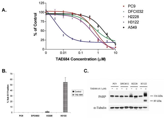
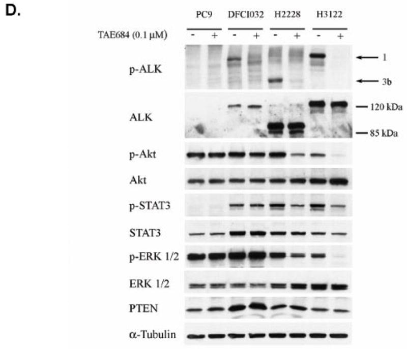
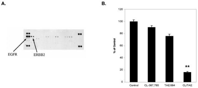
Results: We detected four different variants, including two novel variants, of EML4-ALK using reverse transcription-PCR in 8 of 305 tumors (3%) and 3 of 83 (3.6%) NSCLC cell lines. All EML4-ALK-containing tumors and cell lines were adenocarcinomas. EML4-ALK was detected more frequently in NSCLC patients who were never or light (<10 pack-years) cigarette smokers compared with current/former smokers (6% versus 1%; P = 0.049). TAE684 inhibited the growth of one of three (H3122) EML4-ALK-containing cell lines in vitro and in vivo, inhibited Akt phosphorylation, and caused apoptosis. In another EML4-ALK cell line, DFCI032, TAE684 was ineffective due to coactivation of epidermal growth factor receptor and ERBB2. The combination of TAE684 and CL-387,785 (epidermal growth factor receptor/ERBB2 kinase inhibitor) inhibited growth and Akt phosphorylation and led to apoptosis in the DFCI032 cell line.

Conclusions: EML4-ALK is found in the minority of NSCLC. ALK kinase inhibitors alone or in combination may nevertheless be clinically effective treatments for NSCLC patients whose tumors contain EML4-ALK.


Figures

Figure 1
Figure 1
EML4-ALK in NSCLC cell lines and tumors. A. Detection of ALK fusion genes in lung cancer cell lines using exon arrays. In the screen of 83 lung cancer cell lines (81/83 NSCLCs), exon arrays showed that H3122 and H2228 cell lines had significantly higher signal (log2 difference) for ALK probes #80–140 corresponding to exons 20–29 of ALK compared with other 81 cell lines. Probes were assigned into three categories based on their labeling intensity; non-responsive probes (purple), low-intensity probes (blue), high-intensity probes (black). Only high-intensity probes were used in breakpoint detection. B. RT-PCR detection of EML4-ALK fusion in NSCLC cell lines and tumors. Primer set 2 amplifies EML4-ALK fusion genes from H3122, H2228, and DFCI032 cell lines but not from A549 line. Primer sets 1 and 2 also detected EML4-ALK fusion from 8 primary NSCLCs. H3122; positive control, A549; negative control. C. Schematic representation of the four different EML4-ALK variants in NSCLC.
Figure 2
Figure 2
Detection of EML4-ALK using FISH. A. PC-9 (EGFR del E746_A750) cell line; signals for ALK (red dot) and EML4 (green dot) are seen separately. B. H2228 cell line, the fusion signal of EML4-ALK (arrow) is detected in a small extra-chromosomal fragment (yellow). C. DFCI032 cell line; the EML4-ALK fusion signal in yellow (arrow) is heterozygous. Similar findings were observed for H3122 (data not shown). D. Interphase FISH for EML4-ALK from the formalin fixed paraffin embedded tumor specimen obtained from the pleura of the patient whose pleural effusion was used to establish the DFCI032 cell line in C. The tumor is heterozygous for the EML4-ALK fusion signal (yellow dot; arrow).
Figure 3
Figure 3
Effect of TAE684 on growth and signaling in EML4-ALK containing NSCLC cell lines. A. NSCLC cells were treated with TAE-684 at the indicated concentrations, and viable cells were measured after 72 hours of treatment. The percentage of viable cells is shown relative to untreated controls. A549 (KRAS G12S); PC9 (EGFR delE746_A750); H2228 (EML4-ALK variant 3); H3122 (EML4-ALK variant 1); DFCI032 (EML4-ALK variant 1). B. FACS analysis of sub G1 fraction without treatment (left bar) and after treatment with 0.1μM TAE684 for 72h (right bar). Significant apoptosis following TAE-684 treatment is only observed in the H3122 cell line. C. Western analysis of PARP following treatment with 0.1μM TAE684 for 72h. The 89 kDa cleaved PARP products is observed only in the H3122 cell line consistent with the effects of TAE-684 on cell growth in A. D. Western analysis following TAE684 treatment in wild type and EML4-ALK positive NSCLC cell lines. Total and phosphorylated ALK are only detected in EML4-ALK positive cell lines (H3122, H2228, DFCI032) but not in wild type control (PC-9). In H3122 and DFCI032 cell lines, ALK positive band migrates at ~120 kDa corresponding to predicted molecular weight (117kDa) of the variant 1 (arrow 1) while in H2228, the band migrates at ~90kDa which also corresponds to the predicted molecular weight (90/91kDa) of the variant 3 (arrow 3). ALK phosphorylation is completely inhibited following 0.1μM TAE684 treatment (6 hours) in all the cell lines. Phosphorylation of Akt, STAT3, and ERK1/2 decrease in H3122 and H2228 cell lines with TAE684 but remain unchanged in DFCI032 and PC-9 lines. All the cell lines show presence of PTEN. α-tubulin is used as a loading control.
Figure 3
Figure 3
Effect of TAE684 on growth and signaling in EML4-ALK containing NSCLC cell lines. A. NSCLC cells were treated with TAE-684 at the indicated concentrations, and viable cells were measured after 72 hours of treatment. The percentage of viable cells is shown relative to untreated controls. A549 (KRAS G12S); PC9 (EGFR delE746_A750); H2228 (EML4-ALK variant 3); H3122 (EML4-ALK variant 1); DFCI032 (EML4-ALK variant 1). B. FACS analysis of sub G1 fraction without treatment (left bar) and after treatment with 0.1μM TAE684 for 72h (right bar). Significant apoptosis following TAE-684 treatment is only observed in the H3122 cell line. C. Western analysis of PARP following treatment with 0.1μM TAE684 for 72h. The 89 kDa cleaved PARP products is observed only in the H3122 cell line consistent with the effects of TAE-684 on cell growth in A. D. Western analysis following TAE684 treatment in wild type and EML4-ALK positive NSCLC cell lines. Total and phosphorylated ALK are only detected in EML4-ALK positive cell lines (H3122, H2228, DFCI032) but not in wild type control (PC-9). In H3122 and DFCI032 cell lines, ALK positive band migrates at ~120 kDa corresponding to predicted molecular weight (117kDa) of the variant 1 (arrow 1) while in H2228, the band migrates at ~90kDa which also corresponds to the predicted molecular weight (90/91kDa) of the variant 3 (arrow 3). ALK phosphorylation is completely inhibited following 0.1μM TAE684 treatment (6 hours) in all the cell lines. Phosphorylation of Akt, STAT3, and ERK1/2 decrease in H3122 and H2228 cell lines with TAE684 but remain unchanged in DFCI032 and PC-9 lines. All the cell lines show presence of PTEN. α-tubulin is used as a loading control.
Figure 4
Figure 4
TAE-684 effectively inhibits the growth of H3122 in vivo. Xenografts on H3122 in nu/nu mice were generated as described in Methods. Erlotinib and TAE-684 treatments were administered by oral gavage and tumors were measured three times weekly. The control and erlotinib treated mice reached a median tumor size of 2000 mm3 by 15 days of treatment and were sacrificed. In contrast the median tumor size of mice treated with TAE684 at either 10 mg/kg/day or 25 mg/kg/day did not reach 2000 mm3 even after 53 days of treatment.
Figure 5
Figure 5
Co-activation of EGFR and ERBB2 in DFCI032 cell line. A. A phospho-receptor tyrosine kinase (RTK) array reveals that the DFCI032 cells contain strong activation of both EGFR and ERBB2. Cells were grown in media and the cell lysates were hybridized to a phospho-RTK array. In the array, each RTK is spotted in duplicate. Hybridization signals at the corners serve as controls. B. The combination of TAE684 and CL-387,785 effectively inhibits growth of DFCI032 cells. DFCI032 cells were treated with either CL-387,785 (1 μM) alone, TAE684 (100 nM) alone or the two in combination for 72 hours. Growth was assayed by MTS (Methods). The combination of TAE684 and CL-387,785 led to significant inhibition of growth compared to untreated (p < 0.001; paired t-test) or treatment with either agent alone (p < 0.001; paired t-test for both comparisons, respectively). **; p < 0.001 C. The combination of CL-387,785 and TAE-684 leads to significant apoptosis. Cells were treated as in B. and apoptosis was estimated from sub-G1 fraction using FACS (Methods). The combination of CL-387,785 and TAE-684 led to significant increase in apoptosis compared with untreated (p < 0.05; paired t-test) or treatment with either agent alone (p < 0.05; paired t-test for both comparisons, respectively). *; p < 0.05. D. Combination of CL-387,785 and TAE-684 leads to inhibition of Akt and ERK 1/2 phosphorylation. Cells were treated as in B. for 6 hours or 48 hours. Cells were lysed and the indicated proteins were detected by immunoblotting. Only the combination of CL-387,785 and TAE-684 leads to significant downregulation of Akt and ERK 1/2 signaling and to apoptosis as measured by appearance of cleaved (89 kDa) PARP fragment.
Figure 5
Figure 5
Co-activation of EGFR and ERBB2 in DFCI032 cell line. A. A phospho-receptor tyrosine kinase (RTK) array reveals that the DFCI032 cells contain strong activation of both EGFR and ERBB2. Cells were grown in media and the cell lysates were hybridized to a phospho-RTK array. In the array, each RTK is spotted in duplicate. Hybridization signals at the corners serve as controls. B. The combination of TAE684 and CL-387,785 effectively inhibits growth of DFCI032 cells. DFCI032 cells were treated with either CL-387,785 (1 μM) alone, TAE684 (100 nM) alone or the two in combination for 72 hours. Growth was assayed by MTS (Methods). The combination of TAE684 and CL-387,785 led to significant inhibition of growth compared to untreated (p < 0.001; paired t-test) or treatment with either agent alone (p < 0.001; paired t-test for both comparisons, respectively). **; p < 0.001 C. The combination of CL-387,785 and TAE-684 leads to significant apoptosis. Cells were treated as in B. and apoptosis was estimated from sub-G1 fraction using FACS (Methods). The combination of CL-387,785 and TAE-684 led to significant increase in apoptosis compared with untreated (p < 0.05; paired t-test) or treatment with either agent alone (p < 0.05; paired t-test for both comparisons, respectively). *; p < 0.05. D. Combination of CL-387,785 and TAE-684 leads to inhibition of Akt and ERK 1/2 phosphorylation. Cells were treated as in B. for 6 hours or 48 hours. Cells were lysed and the indicated proteins were detected by immunoblotting. Only the combination of CL-387,785 and TAE-684 leads to significant downregulation of Akt and ERK 1/2 signaling and to apoptosis as measured by appearance of cleaved (89 kDa) PARP fragment.

References

    1. Morris SW, Kirstein MN, Valentine MB, et al. Fusion of a kinase gene, ALK, to a nucleolar protein gene, NPM, in non-Hodgkin’s lymphoma. Science. 1994;263:1281–4. - PubMed
    1. Amin HM, Lai R. Pathobiology of ALK+ anaplastic large-cell lymphoma. Blood. 2007;110:2259–67. - PMC - PubMed
    1. Soda M, Choi YL, Enomoto M, et al. Identification of the transforming EML4-ALK fusion gene in non-small-cell lung cancer. Nature. 2007;448:561–6. - PubMed
    1. Piva R, Chiarle R, Manazza AD, et al. Ablation of oncogenic ALK is a viable therapeutic approach for anaplastic large-cell lymphomas. Blood. 2006;107:689–97. - PMC - PubMed
    1. Li R, Morris SW. Development of anaplastic lymphoma kinase (ALK) small-molecule inhibitors for cancer therapy. Med Res Rev. 2007 - PubMed

Publication types