. 2008 Jul 24;454(7203):486-91.
doi: 10.1038/nature07101. Epub 2008 Jun 25.

Structure of a beta1-adrenergic G-protein-coupled receptor

Affiliations

Structure of a beta1-adrenergic G-protein-coupled receptor

Tony Warne et al. Nature. .

Abstract

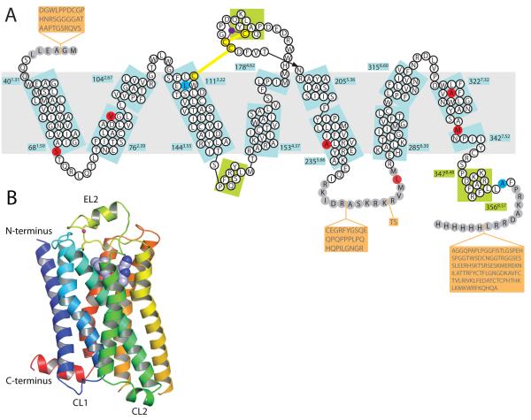
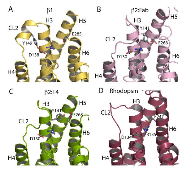
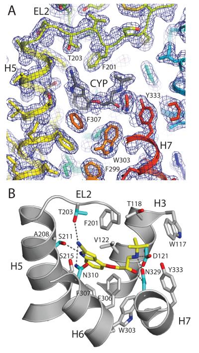
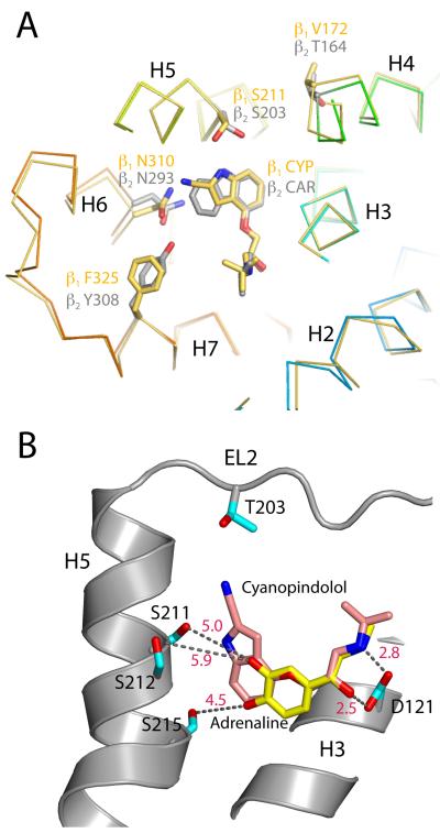
G-protein-coupled receptors have a major role in transmembrane signalling in most eukaryotes and many are important drug targets. Here we report the 2.7 A resolution crystal structure of a beta(1)-adrenergic receptor in complex with the high-affinity antagonist cyanopindolol. The modified turkey (Meleagris gallopavo) receptor was selected to be in its antagonist conformation and its thermostability improved by earlier limited mutagenesis. The ligand-binding pocket comprises 15 side chains from amino acid residues in 4 transmembrane alpha-helices and extracellular loop 2. This loop defines the entrance of the ligand-binding pocket and is stabilized by two disulphide bonds and a sodium ion. Binding of cyanopindolol to the beta(1)-adrenergic receptor and binding of carazolol to the beta(2)-adrenergic receptor involve similar interactions. A short well-defined helix in cytoplasmic loop 2, not observed in either rhodopsin or the beta(2)-adrenergic receptor, directly interacts by means of a tyrosine with the highly conserved DRY motif at the end of helix 3 that is essential for receptor activation.


Figures

Figure 1
Figure 1
Schematic representations of the turkey β1AR structure. (A) Diagram of the turkey β1AR sequence in relation to secondary structure elements. Amino sequence in white circles indicates regions that are well ordered, but sequences in grey circles were not resolved in the structure. Sequences on an orange background were deleted to make the β1AR construct for expression. Thermostabilising mutations are in red and two other mutations C116L (increases functional expression) and C358A (eliminates palmitoylation site) are in blue. The Na+ ion is in purple. Numbers refer to the first and last amino acid residues in each helix (blue boxes), with the Ballesteros-Weinstein numbering in superscript. Helices were defined using the Kabasch & Sander algorithm, with helix distortions being defined as residues that have main chain torsion angles that differ by more than 40° from standard α-helix values (−60°,−40°). (B) Ribbon representation of the β1AR structure in rainbow colouration (N-terminus blue, C-terminus red), with the Na+ ion in pink, the two disulphide bonds in yellow and cyanopindolol as a space-filling model. Extracellular loop 2 (EL2) and cytoplasmic loops 1 and 2 (CL1, CL2) are labelled.
Figure 2
Figure 2
Comparison of the CL2 loop regions in four GPCR structures. (A) β1AR, (B) β2AR:T4 lysozyme fusion, (C) β2:Fab complex and (D) rhodopsin. Residues DR from the highly conserved conserved D3.48R3.49Y3.50 motif are shown. Residue E6.30, which is half of the putative ionic lock, is also shown as E247 in rhodopsin, and E285 and E268 in β1AR and β2AR respectively: E2476.30 was thought to form a salt bridge with R1353.49 in rhodopsin, but the evidence is weak. Finally, Y149 in β1 forms a hydrogen bond with D1383.49 in β1AR.
Figure 3
Figure 3
Structure of the ligand binding pocket. (A) 2Fo-Fc map prior to inclusion of cyanopindolol (CYP) in the model showing the interaction of CYP with Thr203 and Phe201 in EL2. (B) Amino acid residues that interact with the ligand cyanopindolol (yellow) by polar interactions (aquamarine) or non-polar interactions (grey).
Figure 4
Figure 4
Comparisons between β receptor ligand binding pockets and the binding of different ligands. (A) Superposition of β1AR molecule B with β2AR (PDB code 2RH110) in the region surrounding the ligand binding site. Shown are side chains that have different rotamer conformations (N3106.55 and S2115.42) along with two residues that are conserved yet consistently different between β1 and β2 receptors (F325/Y3087.35 and V172/T1644.56). Cyanopindolol (CYP) is in the ligand binding pocket of the β1 receptor and carazolol (CAR) is in the β2 receptor. The biggest backbone deviation is seen at the V172/T1644.56 position. (B) Superposition of a model of the agonist, adrenaline (yellow), with the structure of the antagonist, cyanopindolol (pink), as it binds to β1AR, showing the distances (Å, red) to the nearest side chains known to interact with the hydoxyl groups on the catechol ring of the agonist. It is clear that a 2-3 Å tightening of the pocket around the ligand must occur on agonist binding.

Similar articles

Cited by

References

    1. Fredriksson R, Schioth HB. The repertoire of G-protein-coupled receptors in fully sequenced genomes. Mol. Pharmacol. 2005;67:1414–1425. - PubMed
    1. Hubbell WL, Altenbach C, Hubbell CM, Khorana HG. Rhodopsin structure, dynamics, and activation: A perspective from crystallography, site-directed spin labeling, sulfhydryl reactivity, and disulfide cross-linking. Adv. Protein Chem. 2003;63:243–290. - PubMed
    1. Altenbach C, et al. High resolution distance mapping in rhodopsin reveals the pattern of helix movement due to activation. Proc. Natl. Acad. Sci. USA. 2008 In Press. - PMC - PubMed
    1. Foord SM, et al. International Union of Pharmacology. XLVI. G protein-coupled receptor list. Pharmaco.l Rev. 2005;57:279–288. - PubMed
    1. Baldwin JM, Schertler GF, Unger VM. An alpha-carbon template for the transmembrane helices in the rhodopsin family of G-protein-coupled receptors. J. Mol. Biol. 1997;272:144–164. - PubMed

Publication types

MeSH terms

Associated data