. 2008:4:199.
doi: 10.1038/msb.2008.36. Epub 2008 Jul 1.

Gene network dynamics controlling keratinocyte migration

Affiliations

Gene network dynamics controlling keratinocyte migration

Hauke Busch et al. Mol Syst Biol. 2008.

Abstract

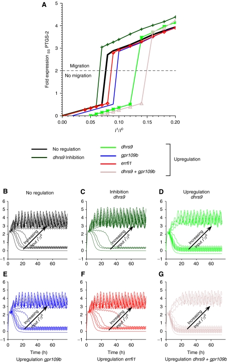
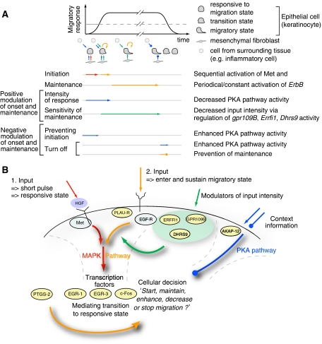
Translation of large-scale data into a coherent model that allows one to simulate, predict and control cellular behavior is far from being resolved. Assuming that long-term cellular behavior is reflected in the gene expression kinetics, we infer a dynamic gene regulatory network from time-series measurements of DNA microarray data of hepatocyte growth factor-induced migration of primary human keratinocytes. Transferring the obtained interactions to the level of signaling pathways, we predict in silico and verify in vitro the necessary and sufficient time-ordered events that control migration. We show that pulse-like activation of the proto-oncogene receptor Met triggers a responsive state, whereas time sequential activation of EGF-R is required to initiate and maintain migration. Context information for enhancing, delaying or stopping migration is provided by the activity of the protein kinase A signaling pathway. Our study reveals the complex orchestration of multiple pathways controlling cell migration.


Figures

Figure 1
Figure 1
Identification of genes mediating HGF-induced cellular transition to migration. (A) Workflow of data analysis, inverse modeling and experimental validation. (B) Histogram of rank scores for all 20 188 measured genes after HGF stimulation (cf. Materials and methods). The ranks numbered 1–20 correspond to the 20 highest ranked genes (cf. gene list in (C)). (C) Top: mean and peak fold expression of the 20 top-ranked genes upon HGF stimulation. Bottom: expression of the same genes upon FGF-7 stimulation. Data points were normalized to the maximal mean and peak fold expression of the respective experiments (cf. Materials and methods).
Figure 2
Figure 2
An HGF pulse is able to stimulate a sustained migratory response. (A) Hypothetical input functions to the gene regulatory network. Blue: pulse; black: constant; green: oscillatory; yellow: nonlinear increase; red: exponential decay. (B) A short HGF input pulse is able to induce a sustained migratory response. HGF-induced migration distance of HaCaT cells for untreated (control), prolonged incubation (30 h), prolonged incubation (30 h) with actinomycin D inhibition (5 μg/ml) and short time (1.5 h) incubation with 10 ng/ml recombinant human HGF is shown (from left to right). For short time treatment, cultures were washed extensively after stimulation and media were replaced. Error bars denote ±s.d. of the mean migration distance from three independent experiments. Note that migratory responses for a constant and pulsed stimulus are almost indistinguishable.
Figure 3
Figure 3
Gene regulatory network topology of genes mediating HGF-induced migration predicts multiple points of interference. (A) Spline-interpolated gene fold expression time series of all genes considered for the in silico analysis of the nine-node network. Error bars denote the standard deviation of the probe sets' fold expression values for each gene. The minimum error is ±0.08-fold expression, calculated from the mean fold expression of all genes. (B) Interaction weights Wij for the CTRNNs obtained by inverse modeling for networks of size 3, 5, 7 and 9 genes (from top to bottom). (C) Offsets θ, delays Δτ, time constant τ and input amplitudes I for the nine-gene network. The interaction weights (B) and the parameter values (C) are color-coded. Note that parameter values are mostly robust with increasing network size. (D) Maximal interaction strengths Wijmax (equation (6)) for the nine-gene network shown in (E). (E) Network diagram for a nine-gene network with interaction weights taken from (B). Note that only the strongest interactions are drawn for better illustration. (F) Histogram of the linear Pearson correlation coefficients of parameter estimates from a nine-gene network using the original (pink), time-randomized (green) and additionally normalized experimental data (blue), having the correlation mean and s.d. values of 0.97/0.01, 0.25/0.24 and 0.13/0.20, respectively (cf. Materials and methods section). (G) Modulation of HGF-induced migration by PKA or EGF-R activity, simultaneous to HGF pulse. To induce a migratory response, HaCaT cells were incubated (30 h) with or without 10 ng/ml recombinant human HGF. PKA activity was modulated by the addition of 1 μM H-89 (Calbiochem) or 200 μM 8-(4-chlorophenylthio)adenosine 3′,5′-cyclic monophosphate sodium salt (Sigma). Inhibition of PKA by the addition of 10 μM myristoylated PKI (14–22) amide, cell-permeable PKA inhibitor (Biomol), gave rise to similar results (data not shown). EGF-R activity was blocked by the addition of 1 μM GW2974 (Sigma). Error bars denote ±s.d. of the mean migration distance from three independent experiments.
Figure 4
Figure 4
Sustained migration depends on a second input mediated by the EGF receptor. System response to varied input strengths and shapes is shown. (A) System response to increasing HGF input strength. Input functions are depicted in the small insets. The learned maximal input amplitudes are scaled with the factors {0.0, 0.7, 0.8, 0.9, 1.0, 1.5, 2.0, 3.0} from dark green to pink. (B) System response to a combined initial HGF input and subsequent second periodic input. Simulations were performed with a nine-gene network. The ratio of maximal input amplitudes between the first and the second input increases from 0.0 (black) via 0.04, 0.06, 0.07, 0.08 and 0.1 to 0.12 (purple). Results in (A, B) are shown for egr3 (left), ptgs2 (middle) and akap12 (right). (C) Blocking the second, periodic input at (I) T=7.5 h, (II) T=22 h and (III) T=42.5 h. The red arrows denote the time point of blocking. A sample input function normalized to one is shown in the small insets. (D) Relative migration upon EGF-R blocking or PTGS-2 inhibition 22 h after HGF stimulation. HaCaT cells were stimulated for 1.5 h with 10 ng/ml recombinant human HGF. Cells were washed extensively and further incubated (30 h). Functional perturbation of PTGS-2 (or EGF-R) activity was introduced by the addition of 50 μM meloxicam (4-hydroxy-2-methyl-N-(5-methyl-2-thiazolyl)-2H-1,2-benzothiazine -2-carbox-amide-1,1-dioxide), 5 μM GW 2974 (Sigma) or 150 nM tyrphostin AG1473 (Biomol) 20.5 h after HGF pulse stimulation to culture media. Migratory response was determined in the time window of 22–30 h beginning with the time point of perturbation. Pretreatment of cells with recombinant EGF for 2 h (1 ng/ml) before HGF stimulation is not able to replace EGF-R activity directly blocked after HGF stimulation. Error bars denote ±s.d. of the mean migration distance from three independent experiments.
Figure 5
Figure 5
A group of modulator genes regulate the minimum threshold intensity of the second input for sustained migration. (A) Steady-state behavior of ptgs2 as a function of the increasing input amplitude of the second pulsed input. ptgs2 as the highest ranked gene has been shown to be important over the whole period of cell migration. Hence its expression level can be considered as a marker for migration. (BG) Simulation of ptgs2 expression under increasing second input amplitude with regulated modulator genes. Input amplitudes of the second input I1 are measured in relation to the initial input strength of the learned amplitude I0 of the first input. Ratios are increased from 0 to 0.2 in steps of 0.02. Each line corresponds to the simulated expression level of ptgs2 for a given ratio I1/I0.
Figure 6
Figure 6
Enhanced PKA pathway activity is able to prevent the onset of migration or stop a developed migratory response at any time. (A, B) Effect of akap12 on simulated gene expression for the nine-gene network. The insets depict the normalized input into the network. (A) A short upshot of akap12 expression at T=5/20/40 h (I/II/III) switches off gene expression and thus cell migration despite continued input signaling. (B) Inhibition of akap12 rescues the system from switching off (I), and (II, III) slightly upregulates ptgs2 (top green line) and downregulates egr3, egr1 and fos expression (bottom, pink, blue and green lines). The strength of downregulation correlates with the transient length for ptgs2 (depicted by gray area) to reach the new steady state. (C) Activation of the PKA pathway at different time points with respect to HGF pulse stimulation (10 ng/ml for 1.5 h) by supplementing culture media with 200 μM 8-(4-chlorophenylthio)adenosine 3′,5′-cyclic monophosphate (Sigma) strongly decreases HGF-induced HaCaT migration. Inhibition of PKA activity by the addition of 0.5 μM H-89 at the time of HGF stimulation slightly increases migration. Inhibition of PKA-activity by H-89 (0.5 μM) directly after HGF pulse stimulation (1.5 h) does not alter development of a migratory response. Inhibition of PKA activity after 22 h, when cells already developed a full migratory response, delays further migration. Error bars denote ±s.d. of the mean migration distance from three independent experiments.
Figure 7
Figure 7
Time ordered sequential events control cellular decision of migration. Schematic representation of events regulating the initiation, maintenance, modulation and stopping of cell migration. (A) The migratory response depends on a certain threshold of gene activity, as indicated by the dashed line, and proceeds as long as no context information is provided via a stop signal or the sustained activation is depleted. (B) Summary scheme of the model for migration. A first pulse-like HGF stimulus is required to transform the cell into a sensitive state for migration. To initiate and sustain migration, a second input preferably through the EGF-R signaling pathway is required. This second input can be modulated by a number of genes. Context information is integrated through the PKA pathway, which can stop migration at any point in time.

Comment in

Similar articles

Cited by

References

    1. Aldridge BB, Haller G, Sorger PK, Lauffenburger DA (2006) Direct lyapunov exponent analysis enables parametric study of transient signalling governing cell behaviour. IEE Proc Syst Biol 153: 425–432 - PubMed
    1. Amit I, Citri A, Shay T, Lu Y, Katz M, Zhang F, Tarcic G, Siwak D, Lahad J, Jacob-Hirsch J, Amariglio N, Vaisman N, Segal E, Rechavi G, Alon U, Mills GB, Domany E, Yarden Y (2007) A module of negative feedback regulators defines growth factor signaling. Nat Genet 39: 503–512 - PubMed
    1. Asthagiri AR, Lauffenburger DA (2000) Bioengineering models of cell signaling. Annu Rev Biomed Eng 2: 31–53 - PubMed
    1. Bansal M, Belcastro V, Ambesi-Impiombato A, di Bernardo D (2007) How to infer gene networks from expression profiles. Mol Syst Biol 3: 78. - PMC - PubMed
    1. Banu N, Buda A, Chell S, Elder D, Moorghen M, Paraskeva C, Qualtrough D, Pignatelli M (2007) Inhibition of cox-2 with ns-398 decreases colon cancer cell motility through blocking epidermal growth factor receptor transactivation: possibilities for combination therapy. Cell Prolif 40: 768–779 - PMC - PubMed

Publication types

MeSH terms