

Heterogeneity and clinical significance of ETV1 translocations in human prostate cancer
- PMID: 18594527
- PMCID: PMC2480965
- DOI: 10.1038/sj.bjc.6604472
Heterogeneity and clinical significance of ETV1 translocations in human prostate cancer
Abstract
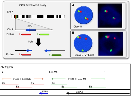
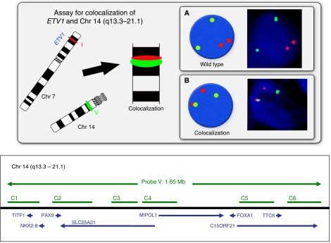
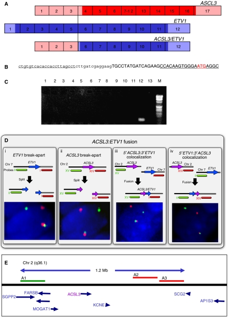
A fluorescence in situ hybridisation (FISH) assay has been used to screen for ETV1 gene rearrangements in a cohort of 429 prostate cancers from patients who had been diagnosed by trans-urethral resection of the prostate. The presence of ETV1 gene alterations (found in 23 cases, 5.4%) was correlated with higher Gleason Score (P=0.001), PSA level at diagnosis (P=<0.0001) and clinical stage (P=0.017) but was not linked to poorer survival. We found that the six previously characterised translocation partners of ETV1 only accounted for 34% of ETV1 re-arrangements (eight out of 23) in this series, with fusion to the androgen-repressed gene C15orf21 representing the commonest event (four out of 23). In 5'-RACE experiments on RNA extracted from formalin-fixed tissue we identified the androgen-upregulated gene ACSL3 as a new 5'-translocation partner of ETV1. These studies report a novel fusion partner for ETV1 and highlight the considerable heterogeneity of ETV1 gene rearrangements in human prostate cancer.
Figures
Similar articles
-
Gene fusions between TMPRSS2 and ETS family genes in prostate cancer: frequency and transcript variant analysis by RT-PCR and FISH on paraffin-embedded tissues.Mod Pathol. 2007 Sep;20(9):921-8. doi: 10.1038/modpathol.3800903. Epub 2007 Jul 13. Mod Pathol. 2007. PMID: 17632455
-
Novel 5' fusion partners of ETV1 and ETV4 in prostate cancer.Neoplasia. 2013 Jul;15(7):720-6. doi: 10.1593/neo.13232. Neoplasia. 2013. PMID: 23814484 Free PMC article.
-
Distinct classes of chromosomal rearrangements create oncogenic ETS gene fusions in prostate cancer.Nature. 2007 Aug 2;448(7153):595-9. doi: 10.1038/nature06024. Nature. 2007. PMID: 17671502
-
ETV1, 4 and 5: an oncogenic subfamily of ETS transcription factors.Biochim Biophys Acta. 2012 Aug;1826(1):1-12. doi: 10.1016/j.bbcan.2012.02.002. Epub 2012 Mar 8. Biochim Biophys Acta. 2012. PMID: 22425584 Free PMC article. Review.
-
ETS factors in prostate cancer.Cancer Lett. 2022 Apr 1;530:181-189. doi: 10.1016/j.canlet.2022.01.009. Epub 2022 Jan 14. Cancer Lett. 2022. PMID: 35033589 Free PMC article. Review.
Cited by
-
Promoter capture Hi-C-based identification of recurrent noncoding mutations in colorectal cancer.Nat Genet. 2018 Oct;50(10):1375-1380. doi: 10.1038/s41588-018-0211-z. Epub 2018 Sep 17. Nat Genet. 2018. PMID: 30224643 Free PMC article.
-
Expression of Long-chain Fatty Acyl-CoA Synthetase 4 in Breast and Prostate Cancers Is Associated with Sex Steroid Hormone Receptor Negativity.Transl Oncol. 2010 Apr;3(2):91-8. doi: 10.1593/tlo.09202. Transl Oncol. 2010. PMID: 20360933 Free PMC article.
-
Integrative Molecular Analyses of the MD Anderson Prostate Cancer Patient-derived Xenograft (MDA PCa PDX) Series.Clin Cancer Res. 2024 May 15;30(10):2272-2285. doi: 10.1158/1078-0432.CCR-23-2438. Clin Cancer Res. 2024. PMID: 38488813 Free PMC article.
-
Targeting Oct1 genomic function inhibits androgen receptor signaling and castration-resistant prostate cancer growth.Oncogene. 2016 Dec 8;35(49):6350-6358. doi: 10.1038/onc.2016.171. Epub 2016 Jun 6. Oncogene. 2016. PMID: 27270436
-
Hsp-27 expression at diagnosis predicts poor clinical outcome in prostate cancer independent of ETS-gene rearrangement.Br J Cancer. 2009 Oct 6;101(7):1137-44. doi: 10.1038/sj.bjc.6605227. Epub 2009 Aug 25. Br J Cancer. 2009. PMID: 19707199 Free PMC article.
References
-
- Albertsen PC, Hanley JA, Barrows GH, Penson DF, Kowalczyk PD, Sanders MM, Fine J (2005) Prostate cancer and the Will Rogers phenomenon. J Natl Cancer Inst 97: 1248–1253 - PubMed
-
- Attard G, Clark J, Ambroisine L, Fisher G, Kovacs G, Flohr P, Berney D, Foster C, Fletcher A, Gerald WL, Moller H, Reuter V, de Bono J, Scardino P, Cooper CS, on behalf of the Transatlantic Prostate Group (2008) Duplication of the fusion of TMPRSS2 to ERG sequences identifies fatal human prostate cancer. Oncogene 27: 253–263 - PMC - PubMed
-
- Clark J, Attard G, Jhavar S, Flohr P, Reid A, De-Bono J, Eeles R, Scardino P, Cuzick J, Fisher G, Parker MD, Foster CS, Berney D, Kovacs G, Cooper CS (2008) Complex patterns of ETS gene alteration arise during cancer development in the human prostate. Oncogene 27: 1993–2003 - PubMed
-
- Clark J, Merson S, Jhavar S, Flohr P, Edwards S, Foster CS, Eeles R, Martin FL, Phillips DH, Crundwell M, Christmas T, Thompson A, Fisher C, Kovacs G, Cooper CS (2006) Diversity of TMPRSS2-ERG fusion transcripts in the human prostate. Oncogene 26: 2667–2673 - PubMed
Publication types
MeSH terms
Substances
Grants and funding
LinkOut - more resources
Full Text Sources
Other Literature Sources
Medical
Research Materials
Miscellaneous
