. 2008 Jul 8;99(1):118-25.
doi: 10.1038/sj.bjc.6604465.

Attenuated p53 activation in tumour-associated stromal cells accompanies decreased sensitivity to etoposide and vincristine

Affiliations

Attenuated p53 activation in tumour-associated stromal cells accompanies decreased sensitivity to etoposide and vincristine

A C Dudley et al. Br J Cancer. .

Abstract

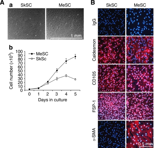
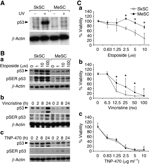
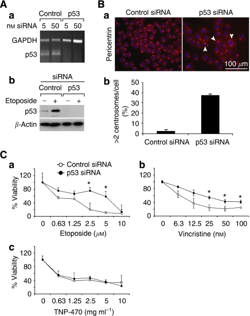
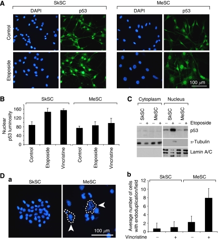
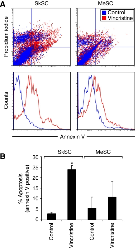
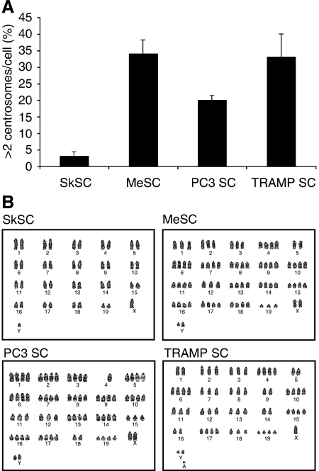
Alterations in the tumour suppressor p53 have been reported in tumour-associated stromal cells; however, the consequence of these alterations has not been elucidated. We investigated p53 status and responses to p53-activating drugs using tumour-associated stromal cells from A375 melanoma and PC3 prostate carcinoma xenografts, and a spontaneous prostate tumour model (TRAMP). p53 accumulation after treatment with different p53-activating drugs was diminished in tumour-associated stromal cells compared to normal stromal cells. Tumour-associated stromal cells were also less sensitive to p53-activating drugs - this effect could be reproduced in normal stromal cells by p53 knockdown. Unlike normal stromal cells, tumour stromal cells failed to arrest in G(2) after etoposide treatment, failed to upregulate p53-inducible genes, and failed to undergo apoptosis after treatment with vincristine. The lower levels of p53 in tumour stromal cells accompanied abnormal karyotypes and multiple centrosomes. Impaired p53 function in tumour stroma might be related to genomic instability and could enable stromal cell survival in the destabilising tumour microenvironment.


Figures

Figure 1
Figure 1
Stromal cell characterisation. (A) Morphology of SC from normal mouse skin or subcutaneous xenografts of A375 melanoma (a) and comparison of their growth properties in culture (b). (B) By immunofluorescence, SC stained uniformly positive for caldesmon, CD105, and FSP-1. α-SMA was expressed only in the tumour SC, indicative of a myofibroblast-like phenotype.
Figure 2
Figure 2
p53 function in tumour stromal cells. (A) Western blotting of whole-cell lysates from UV-treated SkSC and MeSC. Cells were treated with 100 mJ cm−2 for 5 min and lysates were prepared 6 h later. (B) Western blotting of whole-cell lysates from 8 h etoposide-treated (a), 1 nM vincristine-treated (b), or 10 μg ml−1 TNP-470-treated cells (c). The same blots were stripped and re-probed with rabbit polyclonal pSER15 p53 antibodies and then mouse monoclonal β-actin antibodies. (C) Dose–response curves for etoposide (a), vincristine (b), and TNP-470 (c). Cells were plated in triplicate and treated with each drug for 72 h before dispersing in trypsin and counting. *Results are statistically significant (P<0.05) by student's t-test.
Figure 3
Figure 3
p53 knockdown in SkSC decreases sensitivity to etoposide and vincristine. (A) p53 knockdown was complete at the RNA (a) and protein levels (b) using siRNA. (B) p53 knockdown resulted in multiple less-uniform centrosomes (a), and about 30% of the cells had an abnormal >2 pericentrin signals per cell (b). (C) Cell numbers determined in p53 knockdown SkSC after a 72 h treatment with etoposide (a), vincristine (b), and TNP-470 (c). Cells were plated in triplicate and treated with each drug for 72 h before dispersing in trypsin and counting. *Results are statistically significant (P<0.05) by student's t-test.
Figure 4
Figure 4
p53 localises to the nucleus in SkSC and MeSC, but the total cellular p53 pool is reduced. (A) p53 immunofluorescence in SkSC and MeSC treated with etoposide (10 μM, 8 h). Note the predominant nuclear staining for p53 in both the cell types, although the p53 signal in MeSC is diminished. (B) The luminosity for the nuclear p53 signal in treated and untreated cells was measured in 10 cells from three random fields and plotted. (C) Western blots of purified nuclear and cytoplasmic fractions from etoposide-treated cells (10 μM, 8 h). As loading controls, blots were stripped and re-probed with γ-tubulin (cytoplasm) and lamin A/C (nucleus). (D) Vincristine-treated cells (1 nM, 24 h) were methanol-fixed, and the nuclei stained with DAPI. The circled cells are single cells, and the arrows point to endoduplicated nuclei (a). Cells in 10 random fields were counted, and the average number of cells with multiple nuclei (>3 per cell) was plotted (b).
Figure 5
Figure 5
MeSC fail to arrest in G2 after etoposide treatment. (A) Cells treated with 1 μM etoposide were analysed by FACS 24 h later. Note the larger G2 peak on the histogram in etoposide-treated SkSC but not in MeSC. (B) DNA histograms were analysed and the results from two experiments were averaged and are shown in the figure (numbers are per cent). (C) Semiquantitative RT–PCR analysis for p53-indicible genes in SC treated with 1 μM etoposide for 24 h.
Figure 6
Figure 6
MeSC fail to undergo apoptosis after vincristine treatment. (A) Cells treated with 1 nM vincristine for 24 h were double stained with PI and AV and analysed by FACS. An increase in PI+/AV+ and PI/AV+ cells was detected in vincristine-treated SkSC compared to MeSC. (B) The average numbers of early apoptotic AV+ cells from two experiments were plotted. *Results are statistically significant (P<0.05) by student's t-test.
Figure 7
Figure 7
p53 function is impaired in stromal cells from PC3 and TRAMP prostate tumours. (A) Both PC3SC and TRAMPSC had a fibroblast-like morphology (a) and uniformly expressed FSP-1 (b). (B) After treatment with etoposide (10 μM, 8 h) or vincristine (1 nM, 24 h), p53 accumulation was diminished in TRAMPSC and PC3SC compared to normal SkSC (a). Western blotting for pSER15 and pSER20 after vincristine (b) (1 nM, 24 h) or etoposide (8 h) treatment (c). Blots were striped and re-probed with p53 or β-actin antibodies. (C) Viabilities of TRAMPSC and PC3SC after treatment with etoposide (a) or vincristine (b). Cells were plated in triplicate and treated with each drug for 72 h before dispersing in trypsin and counting. Asterisk (*) indicates results are statistically significant (P<0.05) by Student's t-test when comparing SkSC vs PC3 SC and a dagger (†) when comparing SkSc vs TRAMP SC.
Figure 8
Figure 8
Tumour stromal cells have multiple centrosomes and aneuploid karyotypes. (A) Centrosomes were labeled with pericentrin antibodies and counted. One hundred cells were scored and averaged from three different fields. (B) Karyotypes for SkSC, MeSC, PC3SC, and TRAMPSC showing aneuploidy in all tumour SC, whereas SkSC were normal.

References

    1. Allinen M, Beroukhim R, Cai L, Brennan C, Lahti-Domenici J, Huang H, Porter D, Hu M, Chin L, Richardson A, Schnitt S, Sellers WR, Polyak K (2004) Molecular characterization of the tumor microenvironment in breast cancer. Cancer Cell 6: 17–32 - PubMed
    1. Appella E, Anderson CW (2001) Post-translational modifications and activation of p53 by genotoxic stresses. Eur J Biochem 268: 2764–2772 - PubMed
    1. Arbiser JL, Raab G, Rohan RM, Paul S, Hirschi K, Flynn E, Price ER, Fisher DE, Cohen C, Klagsbrun M (1999) Isolation of mouse stromal cells associated with a human tumor using differential diphtheria toxin sensitivity. Am J Pathol 155: 723–729 - PMC - PubMed
    1. Bennett RA, Izumi H, Fukasawa K (2004) Induction of centrosome amplification and chromosome instability in p53-null cells by transient exposure to subtoxic levels of S-phase-targeting anticancer drugs. Oncogene 23: 6823–6829 - PubMed
    1. Bhowmick NA, Neilson EG, Moses HL (2004) Stromal fibroblasts in cancer initiation and progression. Nature 432: 332–337 - PMC - PubMed

Publication types