. 2008 Sep;82(17):8605-18.
doi: 10.1128/JVI.00904-08. Epub 2008 Jul 2.

Comprehensive characterization of extracellular herpes simplex virus type 1 virions

Affiliations

Comprehensive characterization of extracellular herpes simplex virus type 1 virions

Sandra Loret et al. J Virol. 2008 Sep.

Abstract

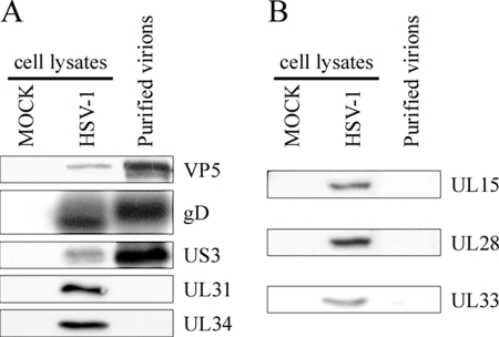
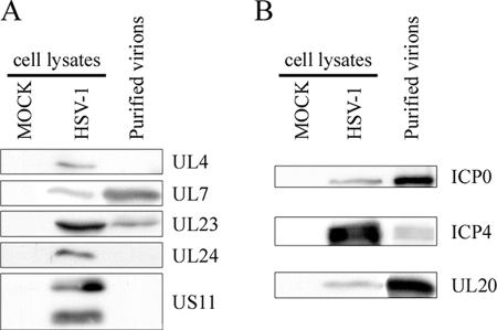
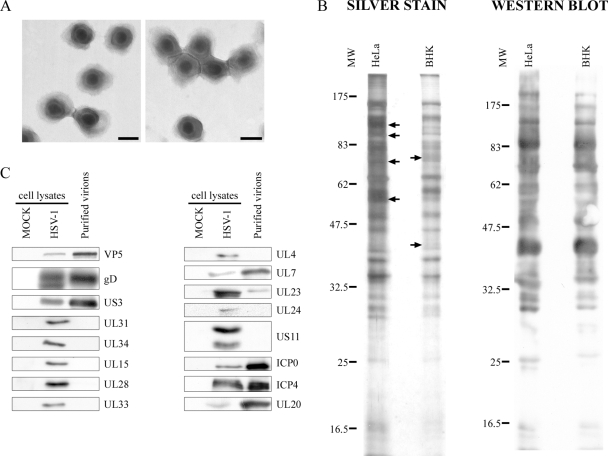
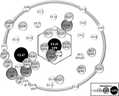
The herpes simplex virus type 1 (HSV-1) genome is contained in a capsid wrapped by a complex tegument layer and an external envelope. The poorly defined tegument plays a critical role throughout the viral life cycle, including delivery of capsids to the nucleus, viral gene expression, capsid egress, and acquisition of the viral envelope. Current data suggest tegumentation is a dynamic and sequential process that starts in the nucleus and continues in the cytoplasm. Over two dozen proteins are assumed to be or are known to ultimately be added to virions as tegument, but its precise composition is currently unknown. Moreover, a comprehensive analysis of all proteins found in HSV-1 virions is still lacking. To better understand the implication of the tegument and host proteins incorporated into the virions, highly purified mature extracellular viruses were analyzed by mass spectrometry. The method proved accurate (95%) and sensitive and hinted at 8 different viral capsid proteins, 13 viral glycoproteins, and 23 potential viral teguments. Interestingly, four novel virion components were identified (U(L)7, U(L)23, U(L)50, and U(L)55), and two teguments were confirmed (ICP0 and ICP4). In contrast, U(L)4, U(L)24, the U(L)31/U(L)34 complex, and the viral U(L)15/U(L)28/U(L)33 terminase were undetected, as was most of the viral replication machinery, with the notable exception of U(L)23. Surprisingly, the viral glycoproteins gJ, gK, gN, and U(L)43 were absent. Analyses of virions produced by two unrelated cell lines suggest their protein compositions are largely cell type independent. Finally, but not least, up to 49 distinct host proteins were identified in the virions.


Figures

FIG. 1.
FIG. 1.
Kinetics of infection. HeLa cells were mock treated or infected with HSV-1 and the cells (cell) and extracellular medium (sup) were collected at 9, 12, 16, and 24 hpi. Once cells were concentrated by centrifugation, they were analyzed by Western blotting with the Remus polyclonal antibody against HSV-1. “Mock” denotes an uninfected control that was analyzed at 24 hpi. The molecular weights (MW) of the protein markers are indicated (103) to the left.
FIG. 2.
FIG. 2.
Purification scheme. Schematic description of the different steps used to purify extracellular HSV-1 virions. See text and Materials and Methods for details.
FIG. 3.
FIG. 3.
Analysis of extracellular virions by EM. Purified virions from HeLa cells were negatively stained and analyzed by EM. Panels A to D show typical views of the sample. Note that the sample consisted exclusively of enveloped virions and was exempt of cellular debris and L particles. Bars represent 100 nm.
FIG. 4.
FIG. 4.
Analysis of purified virions by silver staining and Western blotting. (A) Five micrograms of unpurified (Extracellular medium) or purified virions from HeLa cells were loaded onto a 12% SDS-polyacrylamide gel and silver stained. As controls, equivalent amounts of total mock-treated and infected cell lysates were also loaded. Note the differences between the protein pattern of purified virions and that of control total cell lysates (dots at right). (B) The same samples were also analyzed by Western blotting with the Remus antibody, but in this case, 25 μg of control mock and infected total cell lysates was loaded. Viral enrichment of the purified virions was particularly evident compared to that of the unpurified sample. As before, the molecular weights (MW) of the protein markers are indicated at the left of the gels (103).
FIG. 5.
FIG. 5.
Western blotting analysis of the capsid. (A) Five micrograms of purified virions from HeLa cells was separated by SDS-PAGE, transferred to PVDF membranes, and probed with antibodies against the major VP5 capsid, a control glycoprotein D envelope protein (gD), the serine/threonine viral kinase (US3), and the UL31/UL34 complex. Fifteen micrograms of total mock-infected or infected cell lysates was also included as an antibody control. (B) As described above, except that the viral terminase components (UL15, UL28, UL33) were analyzed with specific antibodies.
FIG. 6.
FIG. 6.
Western blot analysis of the tegument. Five micrograms of purified virions from HeLa cells was separated by SDS-PAGE, transferred to PVDF membranes, and probed with antibodies against UL4, UL7, UL23, UL24, and US11 (A) or ICP0, ICP4 and UL20 (B). Fifteen micrograms of mock-infected or infected cell lysates was included as an antibody control.
FIG. 7.
FIG. 7.
Characterization of virions purified from BHK cells. (A) Virions were purified from BHK cells by the same procedure as that used for HeLa cells, negatively stained, and examined by EM. The viruses were all enveloped and exempt of cellular debris and L particles. Bars represent 100 nm. (B) Five micrograms of purified virions from HeLa and BHK cells was loaded onto a 12% SDS-polyacrylamide gel, and the overall protein composition was determined by silver staining (left panel). In contrast, viral enrichment was examined by Western blotting using a polyclonal HSV-1 antibody (right panel). Note the similar protein pattern (arrows indicate differences). (C) To more specifically probe differences between HeLa and BHK virions, 15 μg of mock-infected or infected cell lysates (antibody controls) and 5 μg of purified virions from BHK cells were separated by SDS-PAGE, transferred to PVDF membranes, and immunoblotted for various viral proteins as indicated. MW, molecular weight (103).
FIG. 8.
FIG. 8.
Schematic representation of mature extracellular virions. The viral composition of the capsid, tegument, and envelope is indicated. The diameter of each circle indicates the relative abundance of the proteins based on their NQPCT score (see Materials and Methods). Given the semiquantitative nature of this score, they are grouped into low abundance (<1% of total proteins), medium abundance (1 to 4%), high abundance (5 to 10%), and very high abundance (>10%). The potential cellular components identified in Table 3 have been omitted since they are in need of validation. However, all but one of them (profilin-1) falls within the first two lowest abundance brackets.

Similar articles

Cited by

References

    1. Adams, R., C. Cunningham, M. D. Davison, C. A. MacLean, and A. J. Davison. 1998. Characterization of the protein encoded by gene UL49A of herpes simplex virus type 1. J. Gen. Virol. 79813-823. - PubMed
    1. America, A. H., and J. H. Cordewener. 2008. Comparative LC-MS: a landscape of peaks and valleys. Proteomics 8731-749. - PubMed
    1. Anderson, L., and C. L. Hunter. 2006. Quantitative mass spectrometric multiple reaction monitoring assays for major plasma proteins. Mol. Cell. Proteomics 5573-588. - PubMed
    1. Baines, J., and C. Duffy. 2006. Nucleocapsid assembly and envelopment of herpes simplex virus, p. 175-204. In R. M. Sandri-Goldin (ed.), Alpha herpesviruses: molecular and cellular biology. Caister Academic Press, Norfolk, United Kingdom.
    1. Baines, J. D., A. H. Koyama, T. Huang, and B. Roizman. 1994. The UL21 gene products of herpes simplex virus 1 are dispensable for growth in cultured cells. J. Virol. 682929-2936. - PMC - PubMed

MeSH terms

LinkOut - more resources