. 2008 Sep;82(17):8687-94.
doi: 10.1128/JVI.00852-08. Epub 2008 Jul 2.

Vaccinia virus l1 protein is required for cell entry and membrane fusion

Affiliations

Vaccinia virus l1 protein is required for cell entry and membrane fusion

Himani Bisht et al. J Virol. 2008 Sep.

Abstract

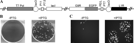
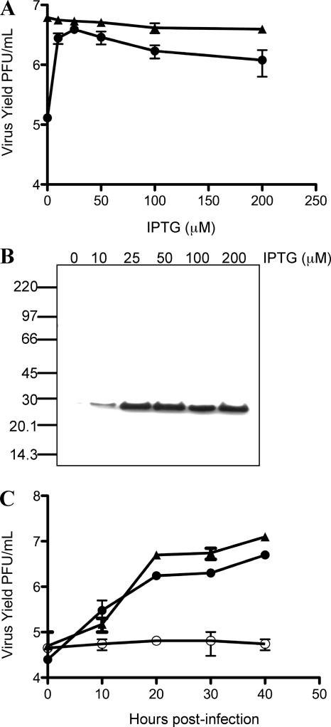
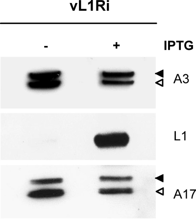
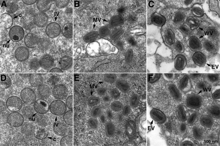
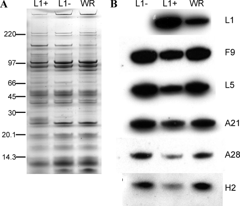
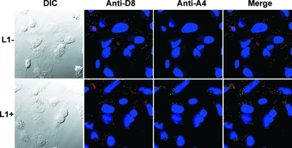
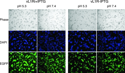
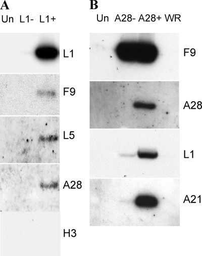
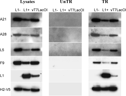
Genetic and biochemical studies have provided evidence for an entry/fusion complex (EFC) comprised of at least eight viral proteins (A16, A21, A28, G3, G9, H2, J5, and L5) that together with an associated protein (F9) participates in entry of vaccinia virus (VACV) into cells. The genes encoding these proteins are conserved in all poxviruses, are expressed late in infection, and are components of the mature virion membrane but are not required for viral morphogenesis. In addition, all but one component has intramolecular disulfides that are formed by the poxvirus cytoplasmic redox system. The L1 protein has each of the characteristics enumerated above except that it has been reported to be essential for virus assembly. To further investigate the role of L1, we constructed a recombinant VACV (vL1Ri) that inducibly expresses L1. In the absence of inducer, L1 synthesis was repressed and vL1Ri was unable to form plaques or produce infectious progeny. Unexpectedly, assembly and morphogenesis appeared normal and the noninfectious virus particles were indistinguishable from wild-type VACV as determined by transmission electron microscopy and analysis of the component polypeptides. Notably, the L1-deficient virions were able to attach to cells but the cores failed to penetrate into the cytoplasm. In addition, cells infected with vL1Ri in the absence of inducer did not form syncytia following brief low-pH treatment even though extracellular virus was produced. Coimmunoprecipitation experiments demonstrated that L1 interacted with the EFC and indirectly with F9, suggesting that L1 is an additional component of the viral entry apparatus.


Figures

FIG. 1.
FIG. 1.
Construction and characterization of an L1-inducible virus. (A) Diagram of vL1Ri. The relevant segment of the viral genome is shown. Abbreviations: T7 Pol, bacteriophage T7 RNA polymerase open reading frame; lacO, E. coli lac operator; lacI, E. coli lac repressor open reading frame; P11, VACV late promoter; PE/L and P7.5, VACV early/late promoters; PT7, T7 promoter; EGFP, EGFP open reading frame. (B) Plaque formation by vL1Ri in the absence and presence of IPTG. BS-C-1 cells were infected with vL1Ri in the absence (−) or presence (+) of 50 μM IPTG. After 48 h, monolayers were fixed and stained with crystal violet. (C) Cells were infected as for panel B except that at 24 h the plate was examined with a fluorescence microscope for EGFP expression.
FIG. 2.
FIG. 2.
Replication of vL1Ri. (A) Effect of IPTG concentrations on virus yield. BS-C-1 cells were infected with 5 PFU per cell of vT7lacOI (triangles) or vL1Ri (circles) in the presence of 0 to 200 μM IPTG. At 24 h postinfection, virus yields were determined by plaque assay on BS-C-1 cells in the presence of 50 μM IPTG. (B) Expression of L1. At 24 h, in the presence of indicated concentrations of IPTG, L1 was measured by Western blotting with anti-L1 MAb followed by horseradish peroxidase-linked secondary antibody. The masses in kDa of mobility marker proteins are shown on the left. (C) One-step growth curve. BS-C-1 cells were infected with 5 PFU per cell of vT7lacOI (triangles) or vL1Ri in the presence (solid circles) or absence (open circles) of IPTG. Cells were harvested at the indicated times, and the virus titers were determined by plaque assay as described above.
FIG. 3.
FIG. 3.
Synthesis and processing of viral proteins. BS-C-1 cells were infected with 5 PFU of vL1Ri per cell in the absence or presence of 50 μM IPTG. At 24 h postinfection, cells were harvested, lysed, and subjected to SDS-PAGE followed by Western blotting with anti-A3, anti-L1, or anti-A17 antibody followed by horseradish peroxidase-linked secondary antibody. Solid and open arrowheads point to precursor and product polypeptides, respectively.
FIG. 4.
FIG. 4.
Transmission electron microscopy of the cells infected with vL1Ri in the absence of IPTG. BS-C-1 cells were infected with 5 PFU per cell of vL1Ri in the absence of IPTG. At 12 h (A to C) or 20 h (D to F) the cells were fixed, embedded, sectioned, and examined by transmission electron microscopy. Abbreviations: c, crescent; IV, immature virion; nu, nucleoid within an immature virion; WV, wrapped virion; EV, extracellular virion. Bars, 100 nm.
FIG. 5.
FIG. 5.
Polypeptide composition of purified virions. Virions were purified through two sucrose cushions and a gradient from lysates of RK13 cells infected with VACV or vL1Ri in the presence and absence of IPTG. (A) Equal amounts of purified L1+, L1, and VACV WR virions were analyzed by SDS-PAGE and silver staining. Numbers at left are molecular masses in kilodaltons. (B) Following SDS-PAGE, Western blotting with MAb 7D11 to L1 and polyclonal antibodies to F9, L5, A21, A28, and H2 was followed by horseradish peroxidase-linked secondary antibody.
FIG. 6.
FIG. 6.
Cell binding of virions and entry of cores. HeLa cells were infected with 5 PFU per cell of L1+ or the equivalent numbers of L1 virions in the presence of cycloheximide for 1 h at 4°C and then for 2 h at 37°C. The cells were then fixed, permeabilized, stained red with a mouse MAb to the D8 MV membrane protein and Alexa Fluor 594-conjugated goat anti-mouse secondary antibody, and stained green with a rabbit antibody to the A4 core protein and Alexa Fluor 498-conjugated goat anti-rabbit secondary antibody. DNA was stained with DAPI, and the cells were examined by confocal microscopy. Dark-field (DIC [differential interference contrast]), anti-D8, anti-L1, and a merge of anti-D8 and anti-L1 images are labeled.
FIG. 7.
FIG. 7.
Low pH triggered cell-cell fusion. BS-C-1 monolayers were infected with 5 PFU per cell of vL1Ri in the presence (+) or absence (−) of IPTG. At 18 h, the cells were treated for 3 min with pH 5.3 or pH 7.4 buffer before replacement with normal medium. After 3 h, the cells were fixed, permeabilized, stained with DAPI, and visualized by phase-contrast and fluorescence microscopy.
FIG. 8.
FIG. 8.
Interaction of L1 with the EFC. (A) BS-C-1 cells were uninfected (Un) or infected with vL1Ri in the absence (L1−) or presence (L1+) of IPTG for 24 h. The lysates were incubated overnight with MAb 7D11 (anti-L1), and the antigen-antibody complexes were immobilized on protein G Sepharose. The bound proteins were then resolved by SDS-PAGE followed by Western blotting with polyclonal antibodies to L1, F9, L5, A28, and H3 and secondary antibody conjugated to horseradish peroxidase. (B) BS-C-1 cells were infected with vA28i/F9TAP in the absence (A28−) or presence (A28+) of IPTG or VACV WR (WR). The lysates were incubated overnight with streptavidin beads, and the bound proteins were eluted with biotin and analyzed by Western blotting using polyclonal antibodies to F9, A28, L1, and A21. Un, uninfected cells (control).
FIG. 9.
FIG. 9.
L1 was not required for assembly of the EFC. BS-C-1 cells were infected with vL1Ri in the absence (L1−) or presence (L1+) of IPTG or with control vT7LacOI and either untransfected (UnTR) or transfected (TR) with a plasmid expressing H2 with a C-terminal V5 tag controlled by its native promoter. Approximately 5% of soluble lysates were analyzed directly by SDS-PAGE followed by Western blotting with antibodies to A21, A28, L5, F9, L1, and V5. The Western blot of the lysate from transfected cells is shown on the left. The Western blot of the lysate from the untransfected cells was identical and is not shown. The remaining portions of the cell lysates from the untransfected (middle panel) and transfected (right panel) cells were incubated with immobilized anti-V5 antibody, and bound proteins were analyzed by Western blotting.

Similar articles

Cited by

References

    1. Aldaz-Carroll, L., J. C. Whitbeck, M. Ponce de Leon, H. Lou, L. K. Pannell, J. Lebowitz, C. Fogg, C. White, B. Moss, G. H. Cohen, and R. J. Eisenberg. 2005. Physical and immunological characterization of a recombinant secreted form of the membrane protein encoded by the vaccinia virus L1R gene. Virology 34159-71. - PubMed
    1. Ansarah-Sobrinho, C., and B. Moss. 2004. Role of the I7 protein in proteolytic processing of vaccinia virus membrane and core components. J. Virol. 786335-6343. - PMC - PubMed
    1. Appleyard, G., A. J. Hapel, and E. A. Boulter. 1971. An antigenic difference between intracellular and extracellular rabbitpox virus. J. Gen. Virol. 139-17. - PubMed
    1. Betakova, T., E. J. Wolffe, and B. Moss. 1999. Membrane topology of the vaccinia virus A17L envelope protein. Virology 261347-356. - PubMed
    1. Blasco, R., and B. Moss. 1991. Extracellular vaccinia virus formation and cell-to-cell virus transmission are prevented by deletion of the gene encoding the 37,000-dalton outer envelope protein. J. Virol. 655910-5920. - PMC - PubMed

Publication types

MeSH terms

LinkOut - more resources