. 2008 Oct;19(10):1879-90.
doi: 10.1681/ASN.2007101087. Epub 2008 Jul 2.

Establishment of conditionally immortalized mouse glomerular parietal epithelial cells in culture

Affiliations

Establishment of conditionally immortalized mouse glomerular parietal epithelial cells in culture

Takamoto Ohse et al. J Am Soc Nephrol. 2008 Oct.

Abstract

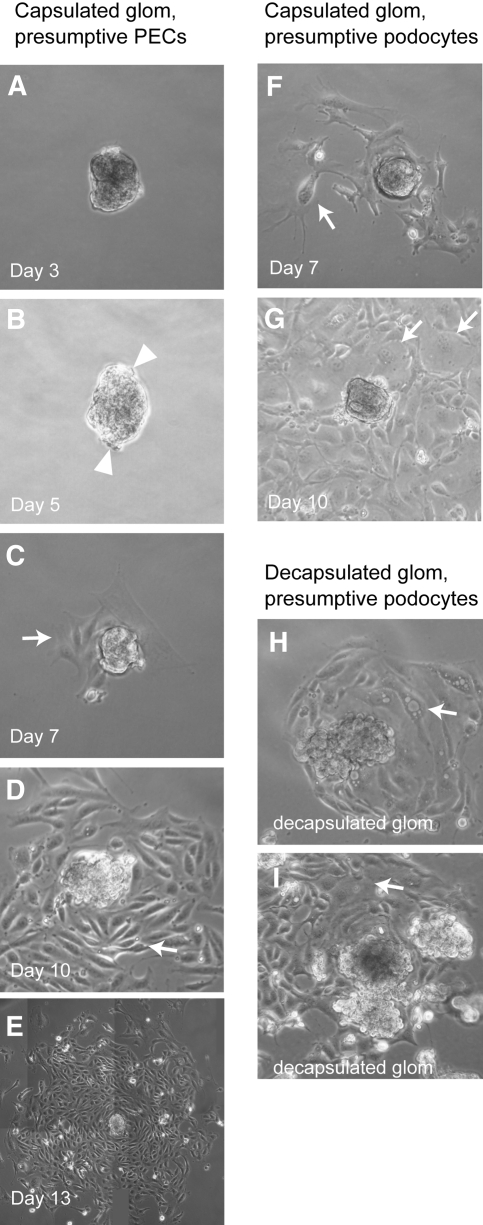
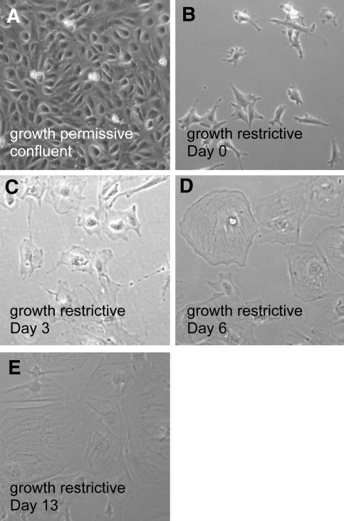
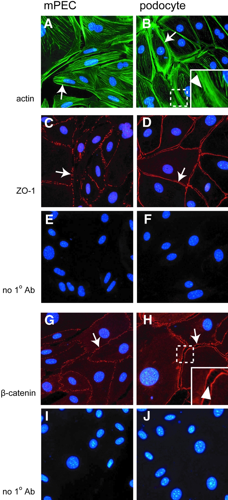
Parietal epithelial cells (PEC) are major constituents of crescents in crescentic glomerulonephritis. The purpose of these studies was to establish an immortalized PEC cell line with similar characteristics to PEC in vivo for use in future mechanistic studies. Glomeruli were isolated from H-2Kb tsA58 transgenic mice (ImmortoMouse) by standard differential sieving, and several candidate PEC cell lines were obtained by subcloning outgrowths of cells from capsulated glomeruli. One clone, designated mouse PEC (mPEC), was extensively characterized. mPEC exhibited a compact cell body with typical epithelial morphology when grown in permissive conditions, but the cell shape changed to polygonal after 14 d in growth-restrictive conditions. mPEC but not podocytes used as a negative control expressed claudin-1, claudin-2, and protein gene product 9.5, which are proteins specific to PEC in vivo, and did not express the podocyte-specific proteins synaptopodin and nephrin. The junctional proteins zonula occludens-1 and beta-catenin stained positively in both mPEC and podocytes, but the staining pattern at cell-cell contacts was intermittent in mPEC and linear in podocytes. Finally, mPEC had thin bundled cortical F-actin filaments and no F-actin projections compared with podocytes, which exhibited thick bundled cortical F-actin filaments and interdigitating F-actin projections at cell-cell contacts. We conclude that immortalized mPEC in culture exhibit specific features of PEC in vivo and that these cells are distinct from podocytes, despite having the same mesenchymal origin. This mPEC line will assist in future mechanistic studies of PEC and enhance our understanding of glomerular injury.


Figures

Figure 1.
Figure 1.
Protocol for isolating mPEC in culture. See text for details.
Figure 2.
Figure 2.
Cell outgrowths from capsulated glomeruli. Adherent isolated capsulated glomeruli were grown in culture under growth-permissive conditions and monitored daily by inverted microscopy. (A) No cellular outgrowths were observed in this attached glomerulus after 3 d in culture. (B) The first evidence of cells emerging from the glomerulus was after 5 d in culture (examples represented by arrow). (C) The number of cells increased by day 7 (examples represented by arrow). (D) By day 10, cells with compact bodies were abundant and surrounded the “parent” glomerulus. (E) By day 13, this merged image illustrates that several of the parent capsulated glomeruli were completely surrounded by a large colony of cells, all uniform in shape and size. These cells were used for dilution cloning to propagate putative mPEC. (F and G) A minority of capsulated glomeruli, such as the one represented, had cell outgrowths characterized by larger size, with distinct processes emerging form the cell body. The morphology of these cells was consistent with podocytes, not PEC. (H and I) For a comparison, representative images of decapsulated glomeruli with cellular outgrowths are shown. Arrows indicates the arborized putative podocyte.
Figure 3.
Figure 3.
Differentiation of mPEC. (A) After dilutional cloning, putative mPEC were initially cultured under growth-permissive conditions to enhance cell proliferation and thus expansion of the cell colony. The figure shows cells with small bodies in a confluent monolayer. For induction of cell differentiation and quiescence, similar to the in vivo phenotype, cells were replated and grown under growth-restrictive conditions, and the results are shown in B through E. (B) mPEC show small cell bodies and had small processes. (C) By day 3 of growth-restrictive condition, mPEC started to spread out, with an increase in cell size. (D) At day 6, mPEC spread out further, and the edges of cells were polygonal. (E) At day 13, mPEC had spread out even more and were uniformly polygonal.
Figure 4.
Figure 4.
PEC proliferation decreased in growth-restrictive conditions. PEC were grown in growth-permissive (GP; 33°C + IFN) and growth-restrictive (GR; 37°C) conditions. Immunofluorescence for Ki-67, a marker of cell proliferation, and DAPI as a cell marker were performed. (A and B) GP cells at day 4 showed positive Ki-67 staining in the majority of cells. (C and D) GR PEC at day 14 revealed minimal Ki-67 staining. (A′ and B′) No primary antibody revealed lack of background staining. Not shown here are similar results obtained with PCNA. (E) MTT assay (quantification of cell viability) demonstrated lower cellular activity in GR cells than GP PEC. These data demonstrate that in vitro PEC proliferate in GP conditions whereas they lack proliferation in GR conditions.
Figure 5.
Figure 5.
Immunostaining for PEC- and podocyte-specific proteins in normal adult mouse kidneys. To ensure that the antibodies used in culture stained specific cells in vivo, we performed immunostaining on normal adult mouse kidney sections. Three PEC markers (claudin-1, claudin-2, and PGP9.5) and two podocyte markers (nephrin and synaptopodin) were examined. Examples of positive staining are illustrated by arrows. (A through F) claudin-1 (A and B) and claudin-2 (C through F) showed a cytoplasmic staining pattern in PEC along with Bowman's basement membrane. (G through J) Cytoplasmic staining for PGP9.5 was confined to PEC along Bowman's basement membrane. (K through N) Nephrin (K and L) and synaptopodin (M and N) staining was abundant and restricted to podocytes. (O through R) Double staining of claudin-1 and synaptopodin is shown. For ruling out the possibility that signals are produced by nonspecific reaction, primary antibody–omitted staining is shown (O and P), in addition to no primary antibody control (R). These results show that PEC and podocytes express unique proteins in normal mice in vivo, which can be used to distinguish specific glomerular epithelial cells in vivo and in vitro.
Figure 6.
Figure 6.
Specific indirect immunofluorescence immunostaining characterizes and distinguishes mPEC from podocytes in culture. (A through J) Putative mPEC and previously characterized podocytes were grown separately under different cell culture conditions and stained with antibodies that specifically stained either PEC or podocyte in vivo. Examples of positive staining are illustrated by arrows. (A through D) Claudin-1 staining was abundant in mPEC along the cell border and in the cytoplasm in both growth-permissive and growth-restrictive conditions; this was absent in podocytes. (E through H) Claudin-2 staining was detected in mPEC along the cell membrane only in growth-restrictive conditions; in contrast, claudin-2 staining was not detected in podocytes. (I through L) PGP9.5 was restricted to the cell membrane of mPEC in growth-restrictive conditions. In contrast, staining was not detected in podocytes for PGP9.5. These results demonstrate that the cells isolated in culture were indeed mPEC. mPEC did not show the positive staining for nephrin (M) or synaptopodin (O), whereas podocytes showed positive linear staining of nephrin at cell borders (N, arrow) and positive linear staining of synaptopodin in cytoplasm and cell border (P, arrow). These results demonstrate that the mPEC grown in culture are distinct from podocytes.
Figure 7.
Figure 7.
Western blot analysis for mPEC- and podocyte-specific proteins. For ensuring antibody and cell specificity, protein was extracted from mPEC and podocytes for Western blot analysis. Glyceraldehyde-3-phosphate dehydrogenase (GAPDH) was used as a loading control for all proteins except nephrin, for which Na-K-ATPase was used. The molecular weight (kD) is shown on the left of each Western blot. mPEC but not podocytes expressed PGP9.5. The abundance of claudin-1 and claudin-2 was substantially greater in mPEC than in podocytes. In contrast, synaptopodin was detected only in podocytes. There was a very slight band detected for nephrin in mPEC. In contrast, nephrin was abundant in podocytes. These results validate the staining results and provide further proof of the establishment of an mPEC-specific cell line in culture.
Figure 8.
Figure 8.
Characterizing and distinguishing cytoskeletal and junctional proteins in cultured mPEC and podocytes. Staining with specific antibodies was used to delineate the cytoskeletal architecture of mPEC and to determine the possibility that mPEC express junctional proteins distinct from podocytes. Examples are illustrated with arrows, and magnified insets highlight the pattern of staining. (A) F-actin staining was abundant in the cytoplasm of mPEC in a linear distribution. (B) Podocytes are characterized by thick bundled cortical actin and thin bridging structures between adjacent cells (arrowhead). (C) ZO-1 staining was readily detected at the cell border of mPEC in a punctuated distribution. (D) ZO-1 staining was abundant at the cell membrane of podocytes in a continuous and linear pattern. (G) β-Catenin staining was intermittent and restricted to the cell border in mPEC. (H) Positive staining was seen in podocyte with a mixture of linear and intermittent patterns at cell borders (arrow) and with thin bridging signals between adjacent cells (arrowhead).

Similar articles

Cited by

References

    1. Shimizu M, Kondo S, Urushihara M, Takamatsu M, Kanemoto K, Nagata M, Kagami S: Role of integrin-linked kinase in epithelial-mesenchymal transition in crescent formation of experimental glomerulonephritis. Nephrol Dial Transplant 21: 2380–2390, 2006 - PubMed
    1. Fujigaki Y, Sun DF, Fujimoto T, Suzuki T, Goto T, Yonemura K, Morioka T, Yaoita E, Hishida A: Mechanisms and kinetics of Bowman's epithelial-myofibroblast transdifferentiation in the formation of glomerular crescents. Nephron 92: 203–212, 2002 - PubMed
    1. Kanemoto K, Usui J, Tomari S, Yokoi H, Mukoyama M, Aten J, Weening JJ, Nagata M: Connective tissue growth factor participates in scar formation of crescentic glomerulonephritis. Lab Invest 83: 1615–1625, 2003 - PubMed
    1. Sagrinati C, Netti GS, Mazzinghi B, Lazzeri E, Liotta F, Frosali F, Ronconi E, Meini C, Gacci M, Squecco R, Carini M, Gesualdo L, Francini F, Maggi E, Annunziato F, Lasagni L, Serio M, Romagnani S, Romagnani P: Isolation and characterization of multipotent progenitor cells from the Bowman's capsule of adult human kidneys. J Am Soc Nephrol 17: 2443–2456, 2006 - PubMed
    1. Kiuchi-Saishin Y, Gotoh S, Furuse M, Takasuga A, Tano Y, Tsukita S: Differential expression patterns of claudins, tight junction membrane proteins, in mouse nephron segments. J Am Soc Nephrol 13: 875–886, 2002 - PubMed

Publication types