. 2008 Jul 2;28(27):6787-93.
doi: 10.1523/JNEUROSCI.2377-07.2008.

Immunotherapy reduces vascular amyloid-beta in PDAPP mice

Affiliations

Immunotherapy reduces vascular amyloid-beta in PDAPP mice

Sally Schroeter et al. J Neurosci. .

Abstract

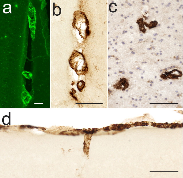
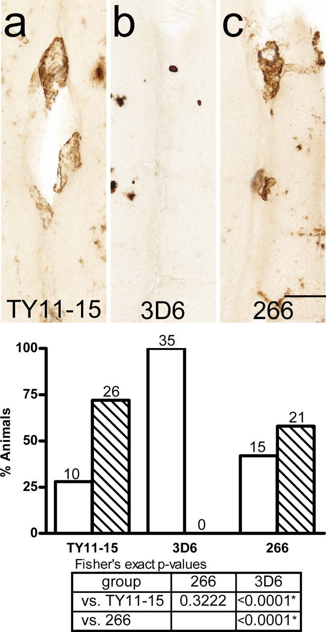
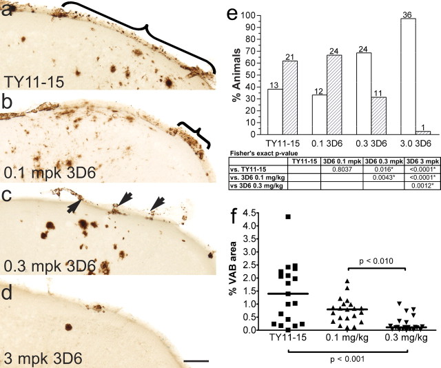
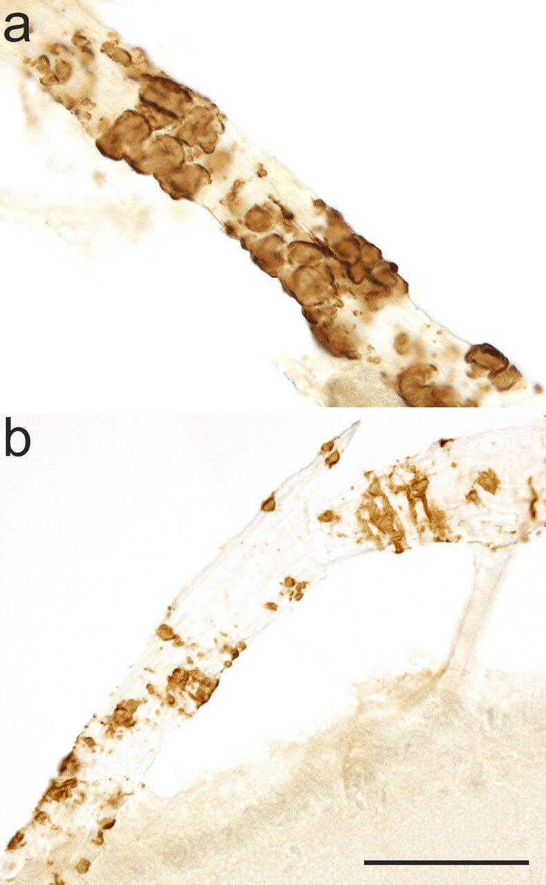
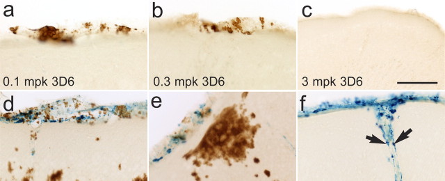
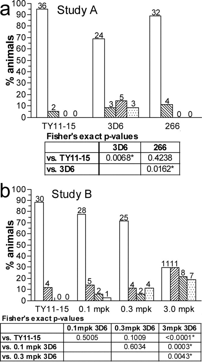
In addition to parenchymal amyloid-beta (Abeta) plaques, Alzheimer's disease (AD) is characterized by Abeta in the cerebral vasculature [cerebral amyloid angiopathy (CAA)] in the majority of patients. Recent studies investigating vascular Abeta (VAbeta) in amyloid precursor protein transgenic mice have suggested that passive immunization with anti-Abeta antibodies may clear parenchymal amyloid but increase VAbeta and the incidence of microhemorrhage. However, the influences of antibody specificity and exposure levels on VAbeta and microhemorrhage rates have not been well established, nor has any clear causal relationship been identified. This report examines the effects of chronic, passive immunization on VAbeta and microhemorrhage in PDAPP mice by comparing antibodies with different Abeta epitopes (3D6, Abeta(1-5); 266, Abeta(16-23)) and performing a 3D6 dose-response study. VAbeta and microhemorrhage were assessed using concomitant Abeta immunohistochemistry and hemosiderin detection. 3D6 prevented or cleared VAbeta in a dose-dependent manner, whereas 266 was without effect. Essentially complete absence of VAbeta was observed at the highest 3D6 dose, whereas altered morphology suggestive of ongoing clearance was seen at lower doses. The incidence of microhemorrhage was increased in the high-dose 3D6 group and limited to focal, perivascular sites. These colocalized with Abeta deposits having altered morphology and apparent clearance in the lower-dose 3D6 group. Our results suggest that passive immunization can reduce VAbeta levels, and modulating antibody dose can significantly mitigate the incidence of microhemorrhage while still preventing or reducing VAbeta. These observations raise the possibility that Abeta immunotherapy can potentially slow or halt the course of CAA development in AD that is implicated in vascular dysfunction.


Figures

Figure 1.
Figure 1.
Meningeal foci of VAβ are found in the PDAPP mouse model and appear similar to human VAβ deposits. a, Thioflavin S staining reveals Aβ in midline vessels of an 18-month-old PDAPP mouse. b, 3D6 antibody detection reveals Aβ in a similar pattern. c, 3D6 immunohistochemistry similarly labels VAβ in a human AD section. d, The leptomeninges and superficial parenchymal vessels are also major sites of VAβ in the PDAPP mouse. Scale bars, 100 μm.
Figure 2.
Figure 2.
Study A: 3D6 (Aβ1–5), but not 266 (Aβ16–23), clears/prevents VAβ at a dose of 3 mg/kg. Representative observations are shown using the midline vessels as examples. a, VAβ in midline vessels in control-treated animals. b, VAβ in 3 mg/kg 3D6-treated animals is cleared/prevented. c, VAβ in 3 mg/kg 266-treated animals is similar to the control. The graph shows the percentage of animals in each group with none or little VAβ (white bars) and moderate VAβ (hatched bars). Statistical analyses show significant VAβ clearance/prevention by 3D6 compared with the control and 266 groups and no difference in clearance/prevention between the TY11–15 control and 266 groups. FET p values <0.025, indicated by asterisks in the table, are statistically significant using the Hochberg method of multiple comparisons. Scale bar, 50 μm.
Figure 3.
Figure 3.
Study B: VAβ is cleared at 3 mg/kg 3D6 and partially cleared at lower doses of 3D6. a, VAβ is abundant in leptomeningeal vessels in control-treated animals. b, VAβ in the 0.1 mg/kg group. c, VAβ in the 0.3 mg/kg group is partially cleared. d, VAβ in the 3 mg/kg group is cleared. e, Percentage of animals in each group with none or little VAβ (white bars) and moderate VAβ (hatched bars). Statistical analyses show significant VAβ clearance/prevention in the 0.3 and 3 mg/kg 3D6-treated groups compared with the other groups. FET p values <0.025, indicated by asterisks in the table, are statistically significant using the Hochberg method of multiple comparisons. f, Quantitation of VAβ by area shows the same result. Brackets and arrows indicate VAβ. Scale bar, 100 μm.
Figure 4.
Figure 4.
Morphology of VAβ during clearance. a, Rounded masses and bands of intact Aβ encompass an unaffected leptomeningeal vessel. b, VAβ has a patchy, eroded appearance during partial clearance. This morphology is not seen in untreated mice. Both images are from 0.1 mg/kg 3D6-treated animals. Scale bar, 50 μm.
Figure 5.
Figure 5.
VAβ clearance results in microhemorrhage in a subset of vessels. a–c, Partial (a, b) and complete (c) clearance or prevention of VAβ is seen at all doses of 3D6 with no evidence of microhemorrhage in most animals. d–f, In a subset of mice, microhemorrhage was seen at some sites of partial clearance at lower doses (d, e) and at sites of complete clearance at 3 mg/kg 3D6 (f). Arrows indicate phagocytes containing hemosiderin but not amyloid. Scale bar, 100 μm.
Figure 6.
Figure 6.
The incidence of microhemorrhage is mitigated by dose. a, Study A: the mean hemosiderin rating of the 3D6 group at 3 mg/kg was significantly higher than the TY11–15 group, and no significant difference was seen between the 266 and TY11–15 groups. b, Study B: the two lower-dose groups did not differ from the control, and the mean hemosiderin rating was significantly higher in the 3D6 3 mg/kg group compared with the TY11–15 and the 0.3 and 0.1 mg/kg 3D6 groups. Hemosiderin ratings 0–3 are represented by the white and hatched bars from left to right in each group. For studies A and B, all FET p values <0.025 and <0.0125, respectively, are statistically significant using the Hochberg method of multiple comparisons.

References

    1. Bard F, Cannon C, Barbour R, Burke RL, Games D, Grajeda H, Guido T, Hu K, Huang J, Johnson-Wood K, Khan K, Kholodenko D, Lee M, Lieberburg I, Motter R, Nguyen M, Soriano F, Vasquez N, Weiss K, Welch B, et al. Peripherally administered antibodies against amyloid beta-peptide enter the central nervous system and reduce pathology in a mouse model of Alzheimer disease. Nat Med. 2000;6:916–919. - PubMed
    1. Bard F, Barbour R, Cannon C, Carretto R, Fox M, Games D, Guido T, Hoenow K, Hu K, Johnson-Wood K, Khan K, Kholodenko D, Lee C, Lee M, Motter R, Nguyen M, Reed A, Schenk D, Tang P, Vasquez N, et al. Epitope and isotype specificities of antibodies to beta-amyloid peptide for protection against Alzheimer's disease-like neuropathology. Proc Natl Acad Sci U S A. 2003;100:2023–2028. - PMC - PubMed
    1. Boche D, DeBeer S, Cox A, Wilkinson D, Holmes C, Neal J, Love S, Esiri M, Bridges L, Weller R, Nicoll J. Evidence of a transient increase in cerebral amyloid angiopathy after Abeta42 immunization in human Alzheimer's disease. Neurodegenerative Dis. 2007;4(Suppl 1):273.
    1. Burbach GJ, Vlachos A, Ghebremedhin E, Del Turco D, Coomaraswamy J, Staufenbiel M, Jucker M, Deller T. Vessel ultrastructure in APP23 transgenic mice after passive anti-Abeta immunotherapy and subsequent intracerebral hemorrhage. Neurobiol Aging. 2007;28:202–212. - PubMed
    1. Buttini M, Masliah E, Barbour R, Grajeda H, Motter R, Johnson-Wood K, Khan K, Seubert P, Freedman S, Schenk D, Games D. Beta-amyloid immunotherapy prevents synaptic degeneration in a mouse model of Alzheimer's disease. J Neurosci. 2005;25:9096–9101. - PMC - PubMed

MeSH terms