. 2008 Jul 2;28(27):6807-17.
doi: 10.1523/JNEUROSCI.4206-07.2008.

A retinal circuit that computes object motion

Affiliations

A retinal circuit that computes object motion

Stephen A Baccus et al. J Neurosci. .

Abstract

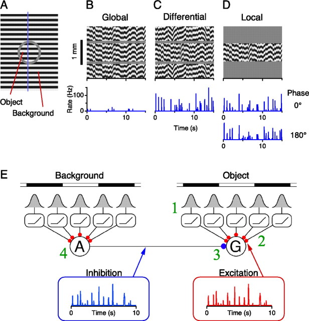
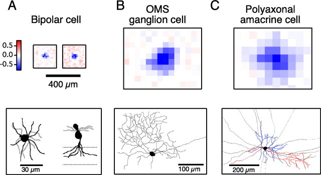
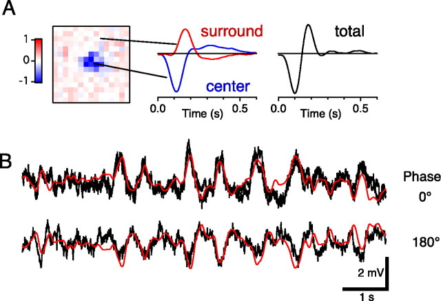
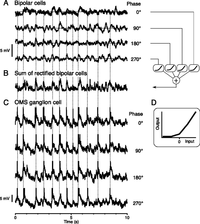
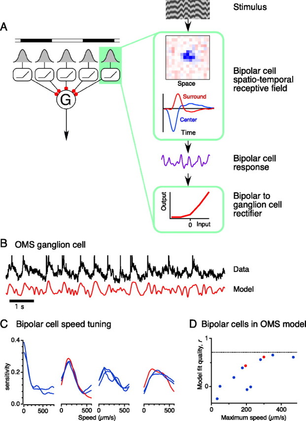
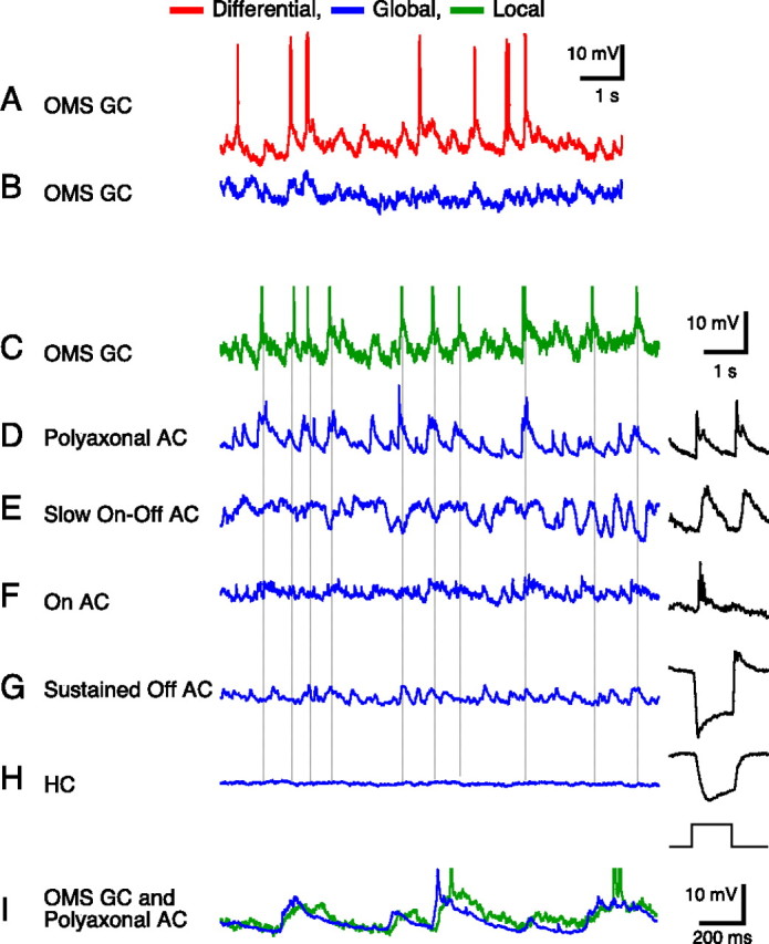
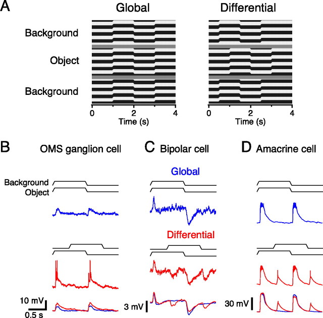
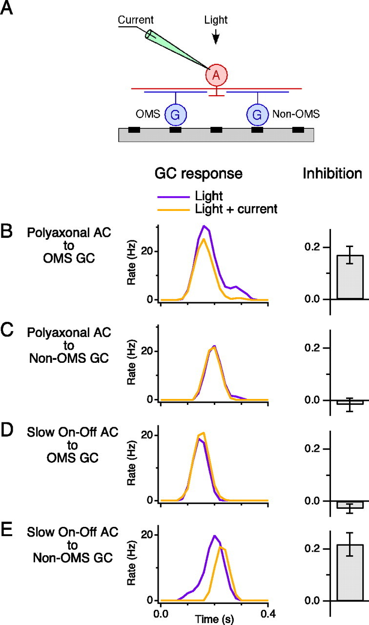
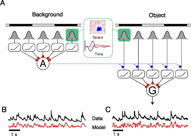
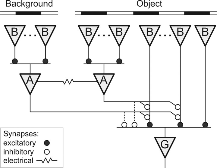
Certain ganglion cells in the retina respond sensitively to differential motion between the receptive field center and surround, as produced by an object moving over the background, but are strongly suppressed by global image motion, as produced by the observer's head or eye movements. We investigated the circuit basis for this object motion sensitive (OMS) response by recording intracellularly from all classes of retinal interneurons while simultaneously recording the spiking output of many ganglion cells. Fast, transient bipolar cells respond linearly to motion in the receptive field center. The synaptic output from their terminals is rectified and then pooled by the OMS ganglion cell. A type of polyaxonal amacrine cell is driven by motion in the surround, again via pooling of rectified inputs, but from a different set of bipolar cell terminals. By direct intracellular current injection, we found that these polyaxonal amacrine cells selectively suppress the synaptic input of OMS ganglion cells. A quantitative model of these circuit elements and their interactions explains how an important visual computation is accomplished by retinal neurons and synapses.


Figures

Figure 1.
Figure 1.
Stimuli and working model for studying the OMS circuit. A, Diagram of object and background regions in the stimulus display. B–D, First row, Space–time plot of a vertical cross section through the center of the stimulus (line in A), showing trajectories for global motion, differential motion, and local motion. Second row, Firing rate of a sample OMS ganglion cell in response to 10 repeats of each stimulus sequence. Third row (D), Response to a local motion stimulus having the same trajectory but reversed grating phase (180°), with black and white bars exchanged relative to 0° phase. E, Working model of the OMS circuit. An OMS ganglion cell (G) receives excitatory input in the object region from multiple small subunits. Each subunit applies a linear spatiotemporal filter to the stimulus in its receptive field. The result is then rectified and summed with the output of other subunits. An inhibitory amacrine cell (A) in the background region receives input from a similar set of rectified subunits. The output of the amacrine cell then inhibits the ganglion cell. Both inhibition and excitation are temporally sparse. Traces show the excitatory and inhibitory components of the model's response to global motion. Numbers identify the key circuit elements to be identified, as listed in the text.
Figure 2.
Figure 2.
Receptive fields and morphology of relevant neurons. A, Top, Spatial profile of the receptive field for two OFF bipolar cells; the same scale bar applies to receptive fields in B and C. Bottom, Tracing of an OFF bipolar cell in tangential and radial views. Dotted lines indicate borders of the inner plexiform layer. The axon terminals ramify in the outer (OFF) sublamina. B, Top, Receptive field of an OMS ganglion cell. Bottom, Tracing in tangential view. C, Top, Receptive field of a polyaxonal amacrine cell. Bottom, Tracing in tangential view showing ON dendrites (red), OFF dendrites (blue), and axons (black). Dashed lines indicate missing image information. The axons were followed beyond the pictured region and extended >3 mm from the soma. Receptive fields and tracings are from different cells.
Figure 3.
Figure 3.
Bipolar cells encode the spatial pattern. A, Bipolar cell spatiotemporal receptive field. Left, Spatial profile of a bipolar cell receptive field with OFF center and ON surround. Right, Temporal profile of the receptive field, normalized to the peak sensitivity, and summed over all pixels (total) or only pixels in the center or the surround. B, Bipolar cell response to a jittering local motion stimulus. Top, Traces from two identical presentations of the same trajectory. Bottom, The same trajectory with the grating reversed (180° spatial phase shift). Red line shows a prediction of the bipolar cell response generated from Equation 2, using only the measured spatiotemporal receptive field and the stimulus trajectory.
Figure 4.
Figure 4.
The transformation from bipolar cells to OMS ganglion cell involves rectification and summation. A, Intracellular recordings from a single bipolar cell responding to a jittering local motion stimulus, with the grating positioned at four different phases. The top plot includes two traces for identical stimuli to illustrate reproducibility of the response. B, The measured bipolar cell responses were each rectified (see D) and then summed to yield a prediction for the ganglion cell response. C, Measured response of an OMS ganglion cell to four phases of a jittering local motion stimulus. D, The nonlinear function used to rectify the bipolar output. This shape was chosen to optimize the fit between the trace in B and the traces in C, ignoring the action potentials. Vertical lines indicate action potentials of the ganglion cell.
Figure 5.
Figure 5.
Model of the OMS ganglion cell excitatory input. A, Detailed structure of a subunit based on bipolar cell measurements. The stimulus trajectory of the object region during local motion is filtered by the bipolar cell spatiotemporal receptive field, yielding the bipolar cell response. Each bipolar response is then passed through the nonlinear transfer function from Figure 4D and summed, yielding the ganglion cell membrane potential. B, Comparison of ganglion cell membrane potential and model output. C, Speed tuning curves of bipolar cells to a moving 184 μm period grating (see Materials and Methods). Each trace shows the normalized sensitivity from a different bipolar cell, grouped into four types of tuning profile. Cells with faster responses are at right. D, Each bipolar cell receptive field was used in a separate model of the OMS response (A). The quality of the model fit was measured by the correlation coefficient between predicted and observed OMS ganglion cell response (B). The high speed cutoff of the bipolar cell was measured by the speed at which the sensitivity falls to 90% of the peak value (C). Here, the model quality is plotted against the high speed cutoff. The dotted line indicates the correlation coefficient calculated between ganglion cell responses to repeated jittering stimuli.
Figure 6.
Figure 6.
Signals of inhibitory interneurons during the OMS response. A, B, Intracellular recording from an OMS ganglion cell responding to differential motion or global motion. C–H, Response of an OMS ganglion cell and a panel of different inhibitory interneurons to the same motion trajectory: local motion for the OMS ganglion cell, and global motion for the inhibitory interneurons (different trajectory from panels A and B). Vertical lines indicate action potentials of the OMS ganglion cell. Right, Response of the same neuron to a periodic stimulus, used to characterize the cell type: a contrast-reversing grating in D and E, and a uniform field flash in F–H. I, Expanded time scale comparing the polyaxonal amacrine cell response to global motion with the OMS ganglion cell response to local motion. AC, Amacrine cell; GC, ganglion cell; HC, horizontal cell.
Figure 7.
Figure 7.
Convergence of signals from object and background. Periodic jitter stimuli were composed of an object and background grating shifting back and forth 40 μm every 1 s. A, Space–time plot as in Figure 1B. The two gratings were shifted in synchrony for global motion and in alternation for differential motion. B–D, Membrane potential responses to periodic jitter. First row, Global motion. Second row, Differential motion. Third row, Average of 15 responses under each condition. B, OMS ganglion cell. The third row shows the average of the subthreshold potential after spikes were removed. C, Fast, transient OFF-type bipolar cell. D, Polyaxonal amacrine cell. All these neurons had a receptive field center in the object region.
Figure 8.
Figure 8.
Polyaxonal amacrine cells selectively suppress OMS ganglion cells. A, Schematic diagram of experiment. Contrast reversal of a grating was used to visually stimulate the retina either alone or synchronous with a depolarizing current pulse (500 pA, 0.5 s) delivered intracellularly to an amacrine cell. Spiking activity from OMS and non-OMS ganglion cells (G) was recorded with a multielectrode array. Responses were analyzed from ganglion cells within 200 μm of the amacrine cell (A). B, C, Results from current injection into a polyaxonal amacrine cell (AC). B, Left, Average firing rate over 30 trials of an OMS ganglion cell (GC) responding to the visual stimulus with and without the current pulse. Trials for the two conditions were interleaved. Right, Fractional inhibition of ganglion cell firing by the current pulse (mean ± SEM, 10 GCs, 3 ACs). C, Left, Same as B for a non-OMS OFF-type ganglion cell. Right, Fractional inhibition of non-OMS ganglion cells (13 GCs, 3 ACs). D, E, Results from current injection into a slow ON–OFF amacrine cell, presented as in B and C. D, Left, OMS ganglion cell light response with and without the current pulse. Right, Fractional inhibition of OMS ganglion cells (10 GCs, 3 ACs). E, Left, Same as D for a non-OMS OFF-type ganglion cell. Right, Fractional inhibition of non-OMS ganglion cells (10 GCs, 3 ACs).
Figure 9.
Figure 9.
Model of object motion sensitivity. A, Amacrine and ganglion cells each sum the rectified output of many linear bipolar cells. Amacrine inhibition is delivered to the output of the bipolar cell before rectification, for example, at the bipolar cell presynaptic terminal. The output of the amacrine cell scales the bipolar output by a factor ranging between ∼0.5 and 1 (see Materials and Methods). B, Comparison of actual and model amacrine cell membrane potential response to background motion. C, Comparison of actual and model ganglion cell membrane potential response to differential motion. The motion trajectory is different from that in B.
Figure 10.
Figure 10.
A circuit for object motion sensitivity. Proposed neural circuitry underlying the OMS response, linking bipolar cells (B), polyaxonal amacrine cells (A), and OMS ganglion cells (G). The bipolar cells have an OFF-type transient response, and their synapses are rectifying. The polyaxonal amacrine cells have a restricted dendritic field that pools excitation from many bipolars and are probably coupled electrically to more distant amacrines in the population. The OMS ganglion cell also pools over many bipolars, but their terminals are inhibited by the polyaxonal amacrine cells. Direct, shunting inhibition of the OMS ganglion cell may also exist. Note that the bipolar cell synapses onto amacrines do not receive presynaptic inhibition.

Similar articles

Cited by

References

    1. Baccus SA, Meister M. Fast and slow contrast adaptation in retinal circuitry. Neuron. 2002;36:909–919. - PubMed
    1. Born RT, Tootell RB. Segregation of global and local motion processing in primate middle temporal visual area. Nature. 1992;357:497–499. - PubMed
    1. Burkhardt DA, Fahey PK. Contrast enhancement and distributed encoding by bipolar cells in the retina. J Neurophysiol. 1998;80:1070–1081. - PubMed
    1. Cook PB, McReynolds JS. Lateral inhibition in the inner retina is important for spatial tuning of ganglion cells. Nat Neurosci. 1998;1:714–719. - PubMed
    1. Costa LF, Velte TJ. Automatic characterization and classification of ganglion cells from the salamander retina. J Comp Neurol. 1999;404:33–51. - PubMed

Publication types

MeSH terms

LinkOut - more resources