. 2008 Jul 2;28(27):6836-47.
doi: 10.1523/JNEUROSCI.5372-07.2008.

Regenerative growth of corticospinal tract axons via the ventral column after spinal cord injury in mice

Affiliations

Regenerative growth of corticospinal tract axons via the ventral column after spinal cord injury in mice

Oswald Steward et al. J Neurosci. .

Abstract

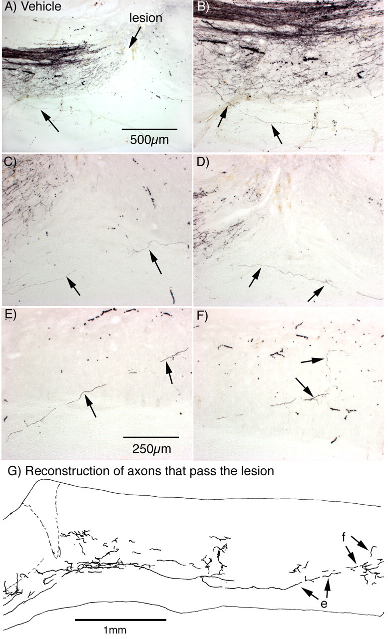
Studies that have assessed regeneration of corticospinal tract (CST) axons in mice after genetic modifications or other treatments have tacitly assumed that there is little if any regeneration of CST axons in normal mice in the absence of some intervention. Here, we document a previously unrecognized capability for regenerative growth of CST axons in normal mice that involves growth past the lesion via the ventral column. Mice received dorsal hemisection injuries at thoracic level 6-7, which completely transect descending CST axons in the dorsal and dorsolateral column. Corticospinal projections were traced by injecting biotinylated dextran amine (BDA) into the sensorimotor cortex of one hemisphere either at the time of the injury or 4 weeks after injury, and mice were killed at 20-23 or 46 d after injury. At 20-23 d after injury, BDA-labeled CST axons did not extend past the lesion except in one animal. By 46 d after injury, however, a novel population of BDA-labeled CST axons could be seen extending from the gray matter rostral to the injury into the ventral column, past the lesion, and then back into the gray matter caudal to the injury in which they formed elaborate terminal arbors. The number of axons with this highly unusual trajectory was small ( approximately 1% of the total number of labeled CST axons rostral to the injury). The BDA-labeled axons in the ventral column were on the same side as the main tract and thus are not spared ventral CST axons (which would be contralateral to the main tract). These results indicate that normal mice have a capacity for CST regeneration that has not been appreciated previously, which has important implications in studying the effect of genetic or pharmacological manipulations on CST regeneration in mice.


Figures

Figure 1.
Figure 1.
Distribution of CST axons in mice killed 20 d after a dorsal hemisection: cross sections (animal 071405C). A, Cross section taken at a high thoracic level rostral to the injury. BDA-labeled axons are prominent in the dCST and dlCST, which can be more clearly seen in C on the left side of the spinal cord, and a dense plexus of BDA-labeled axon arbors are present in the gray matter. B, Cross section taken from a high lumbar level caudal to the injury. Note the complete absence of labeled axons in the dorsal column (dc) and ventral column (vc). C, Higher-power view of the section illustrated in A. Note that, in this mouse, there are an unusually large number of BDA-labeled axons in the dorsal and dorsolateral CST ipsilateral to the injection (right side). D, Higher-power view of the dorsal column and ventral column from the section illustrated in B. E, Higher-power view of axons extending into the ventral column from the gray matter (arrows). F, Injection site in the sensorimotor cortex. Note that the injection did not penetrate the cerebral ventricle.
Figure 2.
Figure 2.
Distribution of CST axons in mice killed 20 d after a dorsal hemisection: sagittal sections (animal 071405C). A, Schematic illustration of a dorsal hemisection. B, C, Illustration of two sagittal sections from the block containing the lesion site from the mouse illustrated in Figure 1. B, Sagittal section through the main CST in the dorsal column; C, sagittal section through the gray matter lateral to the main tract. The unlabeled arrow in B indicates an axon that extends down into the ventral column. The schematic illustration is modified from Zheng et al. (2006).
Figure 3.
Figure 3.
CST axons in the dlCST in mice killed 20 d after a dorsal hemisection (animal 071405C). The panels illustrate sagittal sections through the dlCST on each side. A, The dlCST on the left side. B, The dlCST on the right side. The BDA injection was made into the right sensorimotor cortex.
Figure 4.
Figure 4.
Extension of CST axons past the lesion via the ventral column in mice killed 46 d after a dorsal hemisection: sagittal sections (animal 060106I). A, Sagittal section through the dCST in the dorsal column. Note axon extending down into the ventral column (arrow). B, Higher-magnification view of the section shown in A. Arrows indicate the axon extending from the gray matter into the ventral column. C, D, High-magnification views of BDA-labeled axons in the ventral column just below the lesion site. E, Axon reentering the gray matter from the ventral column caudal to the lesion. F, Complex terminal arbor in the gray matter. G, Drawing of labeled axons from serial sagittal sections. e and f indicate the labeled axons shown in E and F.
Figure 5.
Figure 5.
Cross sections through the segments rostral and caudal to the block containing the lesion site ∼5 mm rostral and caudal to the lesion, respectively (animal 060106I). A, C, Distribution of BDA-labeled CST axons rostral to the injury. C is a higher-magnification view of the ventral part of A. Note absence of BDA-labeled axons in the ventral column (vc). B, D, Cross section taken caudal to the injury indicates the complete absence of BDA-labeled axons in the dCST, dlCST, and ventral column. D is a higher-magnification view of the ventral part of B. Note two small labeled axon segments in the gray matter in B (shown at higher magnification in F). These are branches of arbors at the caudal end of the block containing the lesion site. E, BDA-labeled arbor at the very caudal end of the sagittal section of the block containing the lesion site. The labeled axon continues off the end of the section (arrow) and continues as the axon labeled 1 in F. cc, Central canal.
Figure 6.
Figure 6.
CST axons that extend past the lesion via the ventral column originate as collaterals from the gray matter in rostral segments (animal 060106L). A–C illustrate three adjacent sagittal sections taken near the midline (identified as the center of the central canal). The panels illustrate a region ∼1–3 mm rostral to the lesion (which is just to the right out of the frame of the photomicrograph). Arrows indicate axons that extend ventrally into the ventral column from the gray matter. cc, Central canal.
Figure 7.
Figure 7.
Composite reconstruction of CST axons extending past the lesion via the ventral column (animal 060106L). The panels illustrate composite reconstructions of an axon that emerges from the ventral gray matter ∼1 mm rostral to the injury (A, arrows) and extends along the ventral column bypassing the lesion (B). The images were constructed by taking images of segments of the axon from four adjacent sections and superimposing them on a single image. A second, smaller axon is partially reconstructed in B (below the main axon indicated by arrows). Note the irregular diameter and tortuous course of the axons as they pass the lesion.
Figure 8.
Figure 8.
Axons that extend into the ventral column in a mouse killed 20 d after a dorsal hemisection: sagittal sections (animal 071405C). The panels illustrate two adjacent sagittal sections taken through the main labeled tract in the dorsal column. These are from the same mouse illustrated in Figure 1. Arrows indicate axons that extend from the gray matter into the ventral column.
Figure 9.
Figure 9.
Axons that extend into the ventral column in a mouse killed 23 d after a dorsal hemisection: sagittal sections (animal 121405L). The panel illustrates a composite reconstruction of an axon (axon #1) that extends from a position very near the cut axons in the dCST and extends ventrally into the ventral column. The images were constructed by taking images of segments of the axon from four adjacent sections and superimposing them on a single image. This axon ended in the small swelling indicated by the arrow and label (axon #1). A segment of another axon can be seen in the gray matter caudal to the lesion (axon #2), the course of which is described in Results.

Similar articles

Cited by

References

    1. Bareyre FM, Kerschensteiner M, Raineteau O, Mettenleiter TC, Weinmann O, Schwab ME. The injured spinal cord spontaneously forms a new intraspinal circuit in adult rats. Nat Neurosci. 2004;7:269–277. - PubMed
    1. Bareyre FM, Kerschensteiner M, Misgeld T, Sanes JR. Transgenic labeling of the corticospinal tract for monitoring axonal responses to spinal cord injury. Nat Med. 2005;11:1355–1360. - PubMed
    1. Brösamle C, Schwab ME. Ipsilateral, ventral corticospinal tract of the adult rat: ultrastructure, myelination and synaptic connections. J Neurocytol. 2000;29:499–507. - PubMed
    1. Brösamle C, Huber AB, Fiedler M, Skerra A, Schwab ME. Regeneration of lesioned corticospinal tract fibers in the adult rat induced by a recombinant, humanized IN-1 antibody fragment. J Neurosci. 2000;20:8061–8068. - PMC - PubMed
    1. Bulsara KR, Iskandar BJ, Villavicencio AT, Skene JH. A new millenium for spinal cord regeneration: growth-associated genes. Spine. 2002;27:1946–1949. - PubMed

Publication types

MeSH terms