. 2008 Jul 2;28(27):6904-13.
doi: 10.1523/JNEUROSCI.1901-08.2008.

Postnatal inflammation increases seizure susceptibility in adult rats

Affiliations

Postnatal inflammation increases seizure susceptibility in adult rats

Michael A Galic et al. J Neurosci. .

Abstract

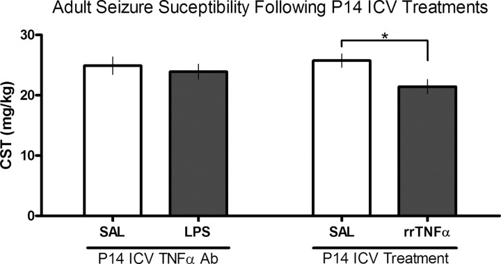
There are critical postnatal periods during which even subtle interventions can have long-lasting effects on adult physiology. We asked whether an immune challenge during early postnatal development can alter neuronal excitability and seizure susceptibility in adults. Postnatal day 14 (P14) male Sprague Dawley rats were injected with the bacterial endotoxin lipopolysaccharide (LPS), and control animals received sterile saline. Three weeks later, extracellular recordings from hippocampal slices revealed enhanced field EPSP slopes after Schaffer collateral stimulation and increased epileptiform burst-firing activity in CA1 after 4-aminopyridine application. Six to 8 weeks after postnatal LPS injection, seizure susceptibility was assessed in response to lithium-pilocarpine, kainic acid, and pentylenetetrazol. Rats treated with LPS showed significantly greater adult seizure susceptibility to all convulsants, as well as increased cytokine release and enhanced neuronal degeneration within the hippocampus after limbic seizures. These persistent increases in seizure susceptibility occurred only when LPS was given during a critical postnatal period (P7 and P14) and not before (P1) or after (P20). This early effect of LPS on adult seizures was blocked by concurrent intracerebroventricular administration of a tumor necrosis factor alpha (TNFalpha) antibody and mimicked by intracerebroventricular injection of rat recombinant TNFalpha. Postnatal LPS injection did not result in permanent changes in microglial (Iba1) activity or hippocampal cytokine [IL-1beta (interleukin-1beta) and TNFalpha] levels, but caused a slight increase in astrocyte (GFAP) numbers. These novel results indicate that a single LPS injection during a critical postnatal period causes a long-lasting increase in seizure susceptibility that is strongly dependent on TNFalpha.


Figures

Figure 1.
Figure 1.
In vitro hippocampal slice electrophysiological recordings from 5-week-old rats treated postnatally with either SAL or LPS (100 μg/kg). A, CA1 recordings of fEPSP slopes to stimulation of afferent fibers in the Schaffer collateral pathway from rats treated with either SAL or LPS at P14 showed significantly (p < 0.05) greater slopes (in microvolts per millisecond) at 75 and 100% maximum stimulation in the LPS-treated group compared with controls (n = 9–12/group). B, Representative fEPSP traces from the two treatment conditions. C, The number of epileptiform bursts per minute from the pyramidal cell layer of CA1 during bath application of 4-AP in aCSF from SAL- or LPS-treated rats (n = 8–14/group). There were significantly (p < 0.05) more burst-firing episodes in the LPS-treated group compared with controls. D, Representative traces of 4-AP-induced epileptiform activity from the two treatment conditions. An asterisk denotes LPS-treated groups that differ significantly (p < 0.05) from controls. Error bars indicate SEM.
Figure 2.
Figure 2.
Adult seizure susceptibility to LI-PILO and PTZ after postnatal treatments of either SAL or LPS. A, Latency in minutes to SOT in adult male rats that received LI-PILO after a P14 injection of either SAL (n = 6), or LPS at 25, 100, or 250 μg/kg (n = 4–5/group). All rats that received LPS regardless of the dosage showed significantly (p < 0.05) faster SOTs compared with controls. Error bars indicate SEM. B, Adult seizure susceptibility to PTZ after postnatal treatments of either SAL or LPS (100 μg/kg) on either P1, P7, P14, or P20 (n = 5–6/group). Data are presented as means ± SEM for PTZ-induced clonic seizure threshold (CST) in mg/kg. Rats that received LPS on P7 and P14, but not on P1 or P20, showed significantly (p < 0.05) lower CSTs compared with controls. An asterisk denotes LPS-treated groups that differ significantly (p < 0.05) from controls.
Figure 3.
Figure 3.
Adult seizure susceptibility to KA after postnatal treatments of either SAL or LPS (100 μg/kg). A, Latency in minutes to SOT in adult rats that received KA after a P14 injection of SAL or LPS (n = 6–7/group). Rats that received LPS at P14 showed significantly (p < 0.05) faster SOTs compared with controls. B, Levels of IL-1β and TNFα (in picograms per milligram of protein) within the hippocampus (HPC) 6 h after seizure onset in adult animals that received KA after a postnatal injection of either SAL or LPS (n = 5–7/group). C, Total number of Fluoro-Jade-staining neurons within the DG, CA3, and CA1 hippocampus of adult rats that received KA after a postnatal injection of either SAL or LPS (n = 3–4/group). Significantly (p < 0.05) more degenerating neurons were found in the CA3 and CA1 region of the hippocampus 24 h after KA in LPS-treated rats compared with controls. D, Photomicrograph of degenerating neurons (arrow) stained with Fluoro-Jade from the CA1 hippocampus of a postnatally LPS-treated rat after adult KA. Scale bar, 25 μm. An asterisk denotes LPS-treated groups that differ significantly (p < 0.05) from controls. Error bars indicate SEM.
Figure 4.
Figure 4.
Adult seizure susceptibility to PTZ after P14 treatments of either SAL or LPS (100 μg/kg) and intracerebroventricular (ICV) injections of TNFα antibody (Ab) (50 μg) (n = 9–11/group) or intracerebroventricular injection of SAL or rrTNFα (2 μg) (n = 5–7/group). Data are presented as means ± SEMs for the clonic seizure threshold (CST) in milligrams per kilogram for adult rats that received PTZ. Rats that received LPS and the TNFα Ab showed no difference in CST compared with SAL controls suggesting that this drug was capable of blocking the LPS-induced facilitation of seizure susceptibility. Moreover, P14 intracerebroventricular rrTNFα alone can reduce adult seizure threshold similar to LPS. The asterisk denotes that the rrTNFα-treated group differed significantly (p < 0.05) from controls.
Figure 5.
Figure 5.
Acute cytokine (IL-1β and TNFα) and microglia (Iba1 immunohistochemistry) data from P14 SAL- or LPS (100 μg/kg)-treated rats (n = 6/group). A, IL-1β and TNFα concentrations in the plasma (in picograms per milliliter) of P14-treated rats 2 h after injection. Both cytokines are significantly (p < 0.05) increased in LPS-treated rats compared with controls. Values lower than the detection limit of the ELISA kit are labeled as below detectable levels (BDL). B, IL-1β and TNFα concentrations (in picograms per milligram of protein) in the hippocampus (HPC) of P14-treated rats 6 h after injection. Both cytokines are significantly (p < 0.05) increased in LPS-treated rats compared with controls. C, Total number of microglia (Iba1-positive) cells (left axis) and the percentage activated microglia count (right axis) of P14 SAL- or LPS-treated rats 6 h after injection. There is no difference in the number of Iba1-positive cells between LPS and control tissues; however, there are significantly (p < 0.05) greater numbers of activated microglia within the CA1 HPC of LPS-treated rats 6 h after injection. D, Photomicrograph of activated microglia cells (arrows) from the CA1 HPC of P14 LPS-treated rats. Scale bar, 20 μm. An asterisk denotes LPS-treated groups that differ significantly (p < 0.05) from controls. Error bars indicate SEM.
Figure 6.
Figure 6.
Adult cytokine (IL-1β and TNFα), microglia (Iba1), and GFAP immunohistochemistry data from rats treated with SAL or LPS (100 μg/kg) on P14 (n = 4–6/group). A, IL-1β and TNFα concentrations in adult hippocampus (HPC) of rats treated at P14 with either SAL or LPS. Both cytokines are not significantly different between SAL- and LPS-treated groups. B, Total number of microglia (Iba1-positive) cells (left axis) and the percentage activated microglia count (right axis) of adult rats treated at P14 with SAL or LPS. There is no difference in the number of Iba1-positive cells or percentage activated between LPS and control tissues. C, Total number of GFAP-immunopositive cells within the DG, CA3, and CA1 regions of the HPC of adult animals treated at P14 with either SAL or LPS. There was a significant (p < 0.05) increase in the number of GFAP-positive cells in the CA1 HPC of rats treated at P14 with LPS compared with controls. D, Photomicrographs of GFAP-positive cells in the CA1 HPC of adult SAL- and LPS-treated rats. Scale bar, 150 μm. An asterisk denotes LPS-treated groups that differ significantly (p < 0.05) from controls. Error bars indicate SEM.

Similar articles

Cited by

References

    1. Agrawal AK, Shapiro BH. Neonatal phenobarbital imprints overexpression of cytochromes P450 with associated increase in tumorigenesis and reduced life span. FASEB J. 2005;19:470–472. - PubMed
    1. Allan SM, Rothwell NJ. Cytokines and acute neurodegeneration. Nat Rev Neurosci. 2001;2:734–744. - PubMed
    1. Allan SM, Tyrrell PJ, Rothwell NJ. Interleukin-1 and neuronal injury. Nat Rev Immunol. 2005;5:629–640. - PubMed
    1. Annegers JF, Hauser WA, Beghi E, Nicolosi A, Kurland LT. The risk of unprovoked seizures after encephalitis and meningitis. Neurology. 1988;38:1407–1410. - PubMed
    1. Avishai-Eliner S, Brunson KL, Sandman CA, Baram TZ. Stressed-out, or in (utero)? Trends Neurosci. 2002;25:518–524. - PMC - PubMed

Publication types

MeSH terms

Grants and funding