. 2008 Jul 2;28(27):6938-49.
doi: 10.1523/JNEUROSCI.0542-08.2008.

Transcranial magnetic stimulation over posterior parietal cortex disrupts transsaccadic memory of multiple objects

Affiliations

Transcranial magnetic stimulation over posterior parietal cortex disrupts transsaccadic memory of multiple objects

Steven L Prime et al. J Neurosci. .

Abstract

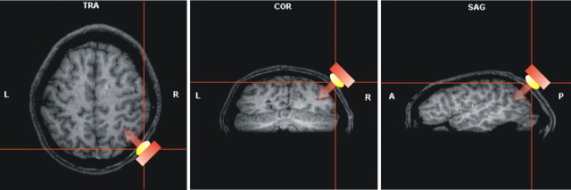
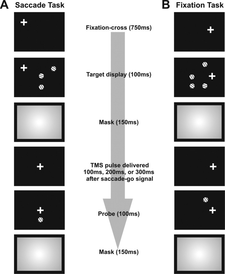
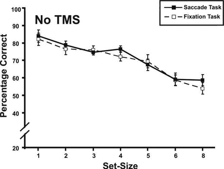
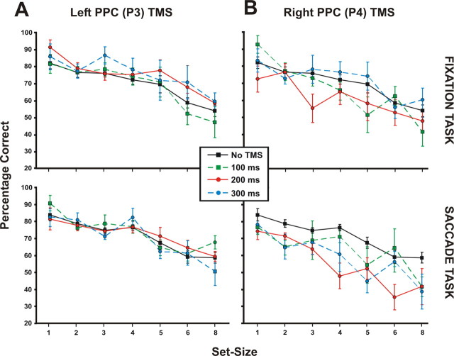
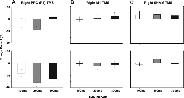
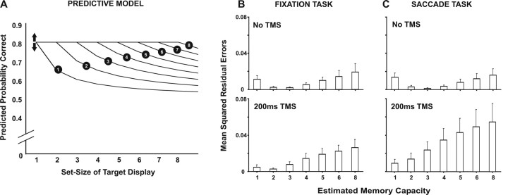
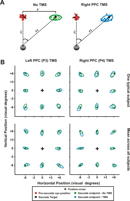
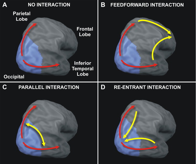
The posterior parietal cortex (PPC) plays a role in spatial updating of goals for eye and arm movements across saccades, but less is known about its role in updating perceptual memory. We reported previously that transsaccadic memory has a capacity for storing the orientations of three to four Gabor patches either within a single fixation (fixation task) or between separate fixations (saccade task). Here, we tested the role of the PPC in transsaccadic memory in eight subjects by simultaneously applying single-pulse transcranial magnetic stimulation (TMS) over the right and left PPC, over several control sites, and comparing these to behavioral controls with no TMS. In TMS trials, we randomly delivered pulses at one of three different time intervals around the time of the saccade, or at an equivalent time in the fixation task. Controls confirmed that subjects could normally retain at least three visual features. TMS over the left PPC and a control site had no significant effect on this performance. However, TMS over the right PPC disrupted memory performance in both tasks. This TMS-induced effect was most disruptive in the saccade task, in particular when stimulation coincided more closely with saccade timing. Here, the capacity to compare presaccadic and postsaccadic features was reduced to one object, as expected if the spatial aspect of memory was disrupted. This finding suggests that right PPC plays a role in the spatial processing involved in transsaccadic memory of visual features. We propose that this process uses saccade-related feedback signals similar to those observed in spatial updating.


Figures

Figure 1.
Figure 1.
Location of individual parietal TMS sites for one representative subject. The stimulation site for the right posterior parietal cortex is shown with the position of high-intensity signal markers placed on the subject's skull (P4). Red bars indicate the position of the TMS coil. The coil was placed tangential to the skull with the handle pointing backward parallel to the midline. This produces a current flowing in a posterior–anterior direction in the underlying brain areas. Stimulation sites were verified a posteriori using the MRI of the individual subject. The anatomical site of stimulation for the right PPC (shown here) is indicated by the line intersection in the transverse (TRA), coronal (COR), and sagittal (SAG) sections of T1-weighted MRI.
Figure 2.
Figure 2.
General experimental paradigm for our study. The rectangles of each panel show the temporal order during a typical trial for presentation of fixation crosses and the visual stimuli. A, The saccade task. Subjects fixated on the fixation cross while the target display was briefly presented (100 ms) containing either a lone target or a target accompanied by a random number of distracters (i.e., total set size of target + distracters was 1–6 or 8). After the mask (150 ms), subjects moved their eyes to the new location of fixation cross. In TMS trials, TMS pulses were time locked to the onset of the second fixation cross. Pulses were delivered either 100, 200, or 300 ms after the second fixation cross was presented. After the saccade, a probe was presented (100 ms) at the same location as the target. Subjects were required to indicate how the probe's orientation differed relative to the target's orientation. B, The fixation task is the same as the saccade task except that subjects were required to maintain eye fixation through target display and probe presentations. The fixation cross remained fixed in the same position throughout the trial. Again, TMS pulses were delivered at one of the three time intervals relative to the onset of the second fixation cross in TMS trials.
Figure 3.
Figure 3.
Results of the baseline No TMS trials. This figure shows the mean percentage correct responses across all subjects (n = 8) in both the saccade and fixation tasks for different set sizes. Saccade task performance is represented by the solid curve with the closed squares. Fixation task performance is represented by the dashed curve with the open squares. Performance in these tasks was statistically the same according to a goodness-of-fit analysis. These data replicated our previous findings (Prime et al., 2007b). Error bars represent 1 SE.
Figure 4.
Figure 4.
A, B, Main results of left PPC TMS (A) and right PPC TMS (B) conditions for both the fixation task (top) and saccade task (bottom). These data are shown as mean percentage correct responses across all subjects (n = 8) against different set sizes. Each colored data curve represents the different TMS time intervals in the TMS trials. The green curve represents the TMS data when TMS was delivered at the 100 ms time interval. Similarly, the red curve represents the 200 ms time interval, and the blue curve represents the 300 ms time interval. As a comparison, we replotted the baseline No TMS data curves from Fig. 3 for each task (black curves). Separate goodness-of-fit tests comparing each TMS data curve to their respective baseline No TMS data show that TMS only had an effect in the right PPC condition: fixation task performance was only disrupted when stimulation occurred at the 200 ms time interval. Saccade task performance was disrupted for all three TMS time intervals (100, 200, and 300 ms). Error bars represent 1 SE.
Figure 5.
Figure 5.
Magnitude of TMS effect. To determine the magnitude of the TMS effect we subtracted the TMS data curves of Fig. 4 from their respective baseline No TMS data from Fig. 3. A–C, The change in mean percentage correct is shown for the right PPC (A), right M1 TMS (B), and right sham TMS (C). The top panels represent the change in the fixation task and the bottom panels represent the change in the saccade task. Positive numbers reflect a greater percentage correct and negative numbers reflect a lesser percentage correct compared with baseline. The line at zero represents no change from baseline. Consistent with Fig. 4, right PPC TMS in the fixation task only increased errors when TMS was delivered at the 200 ms time interval. Also, saccade task performance during right PPC TMS was less accurate for all three TMS time intervals. As shown in the other TMS conditions, no disruption was found for the right M1 (B), right sham (C), and left sham (data not shown).
Figure 6.
Figure 6.
Estimating memory capacity during right PPC stimulation. A, A simple predictive model where each curve predicts the probability of correct response as a function of set size for different theoretical capacities of transsaccadic memory, indicated by the numbers above each curve. To determine which predictive curve provided the best fit, we computed the MSR errors between each of these curves and both the No TMS data and the 200 ms right parietal TMS data (where we saw the largest effect) from Figure 4B. The arrows shown in the predictive model indicate that the intercept of the predictive curves was adjusted to take into account the different intercepts (i.e., percentage correct obtained at one item set size) of each data curve. B, C, MSR errors for both fixation and saccade tasks, respectively, in the No TMS condition (top panels) and 200 ms right PPC TMS condition (bottom panels). The bar graphs represent the average MSR errors across all subjects after calculating the MSR errors for each subject individually. The least average MSR error indicates the best fit to a theoretical memory capacity according to our predictive model. First, during the No TMS condition, we found the average MSR errors of both the fixation (B, top) and saccade (C, top) tasks show the best fit to our predictive model estimating a memory capacity of three items, replicating our previous findings (Prime et al., 2007b). During 200 ms right PPC stimulation, where we saw the largest TMS effect, memory capacity in the fixation task (B, bottom) was reduced to two items and reduced to one item in the saccade task (C, bottom).
Figure 7.
Figure 7.
Saccade accuracy during No TMS, left PPC TMS, and right PPC TMS. A, Saccade accuracy of one typical subject during trials involving 6° saccades in the baseline No TMS and right PPC TMS conditions. Red dots represent the presaccadic eye position shown at the black fixation cross. The green dots represent the saccade end points for saccades in the No TMS condition and the blue dots represent the saccade end points for saccades in the right PPC TMS condition. Both saccade end points are shown at the saccade target (i.e., second fixation cross) indicated by the black dot. The mean saccade error was determined by the average distance between the saccade end point and the saccade target. Saccade accuracy was calculated by fitting an ellipse around the saccade end points in both the baseline No TMS and right PPC TMS conditions, shown by the green and blue ellipses. B, Saccade accuracy for all saccade directions across different set sizes both for one typical subject (top panels) and across all subjects (bottom panels). We also included saccade accuracy data in the left PPC TMS condition (left column) along with saccade accuracy in the right PPC TMS condition (right column), shown by the blue ellipses. Saccade accuracy from the No TMS condition is represented by the green ellipses. Presaccadic eye position was normalized for the sake of simplifying the figure. As shown in B, no differences in saccade accuracy were found between the baseline No TMS and TMS conditions. We also found that saccade latency (data not shown) did not significantly differ between baseline No TMS and TMS conditions. We conclude that TMS effects found in our data cannot be attributed to disruptions to subjects' saccadic eye movements.
Figure 8.
Figure 8.
Different possible explanations of how visual streams may be involved in transsaccadic memory. A, The “no interaction” hypothesis suggests that transsaccadic memory does not rely on binding of visual information between the dorsal and ventral streams. B, Alternatively, integration of visual information for transsaccadic memory may occur by feedforward connections to frontal cortical regions. C, Another possibility is that visual information is integrated for transsaccadic memory by parallel connections between dorsal and ventral streams. D, Last, transsaccadic memory may result from interactions between the visual streams through re-entrant pathways to earlier visual areas.

Similar articles

Cited by

References

    1. Andersen RA, Essick GK, Siegel RM. Encoding of spatial location by posterior parietal neurons. Science. 1985;230:456–458. - PubMed
    1. Astafiev SV, Shulman GL, Stanley CM, Snyder AZ, Van Essen DC, Corbetta M. Functional organization of human intraparietal and frontal cortex for attending, looking, and pointing. J Neurosci. 2003;23:4689–4699. - PMC - PubMed
    1. Beck DM, Muggleton N, Walsh V, Lavie N. Right parietal cortex plays a critical role in change blindness. Cereb Cortex. 2006;16:712–717. - PubMed
    1. Beckers G, Zeki S. The consequences of inactivating areas V1 and V5 on visual motion perception. Brain. 1995;118:49–60. - PubMed
    1. Bledowski C, Cohen Kadosh K, Wibral M, Rahm B, Bittner RA, Hoechstetter K, Scherg M, Maurer K, Goebel R, Linden DE. Mental chronometry of working memory retrieval: a combined functional magnetic resonance imaging and event-related potentials approach. J Neurosci. 2006;26:821–829. - PMC - PubMed

Publication types

MeSH terms