. 2008 Jul 2;28(27):7013-23.
doi: 10.1523/JNEUROSCI.1673-08.2008.

Backpropagating action potentials trigger dendritic release of BDNF during spontaneous network activity

Affiliations

Backpropagating action potentials trigger dendritic release of BDNF during spontaneous network activity

Nicola Kuczewski et al. J Neurosci. .

Abstract

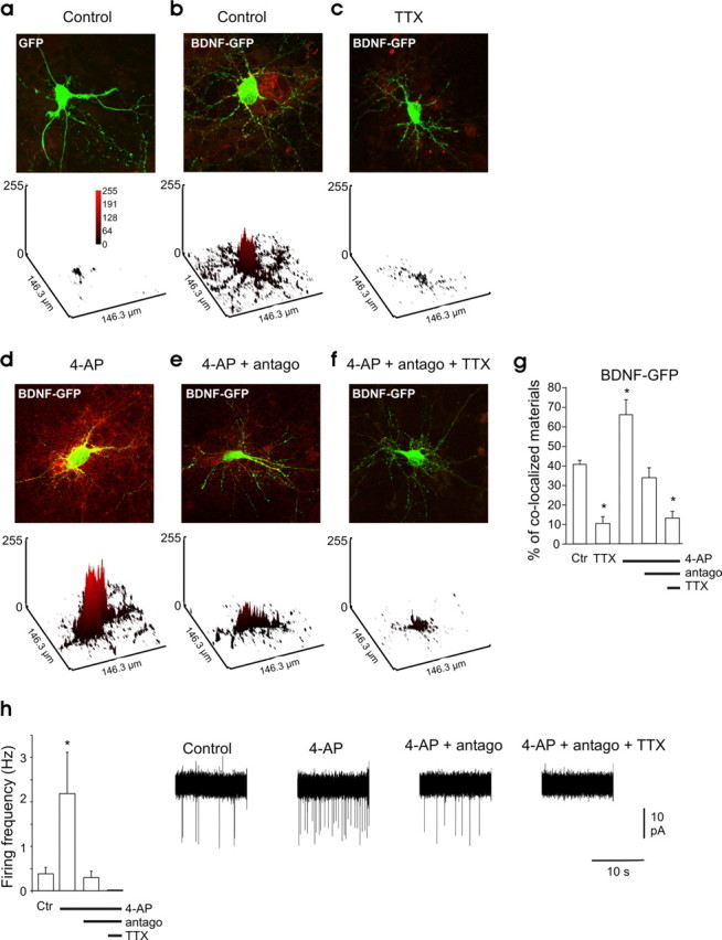
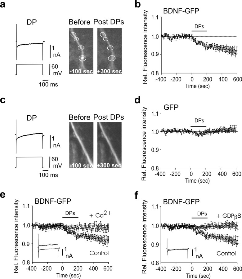
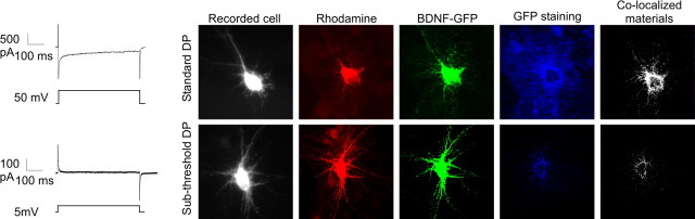
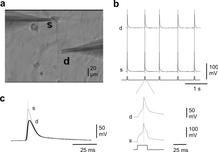
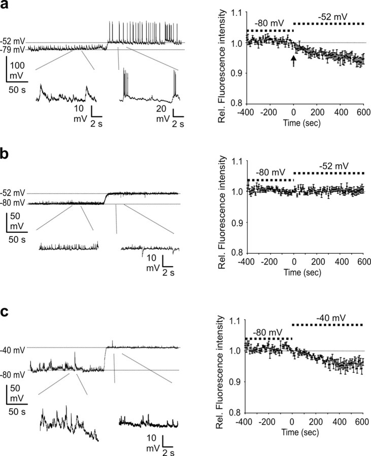
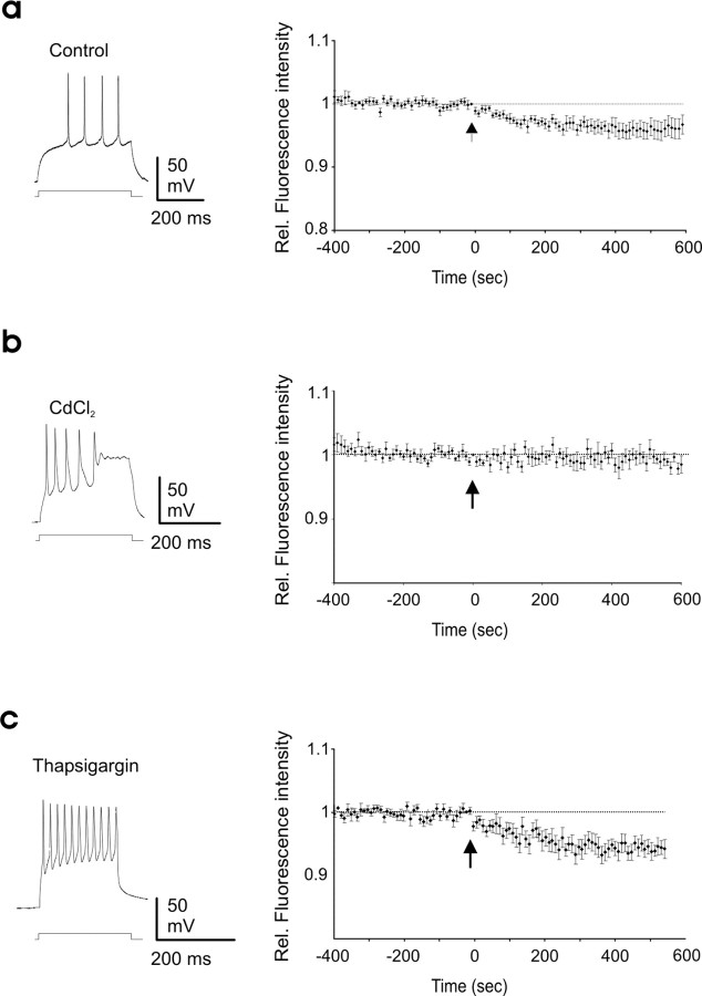
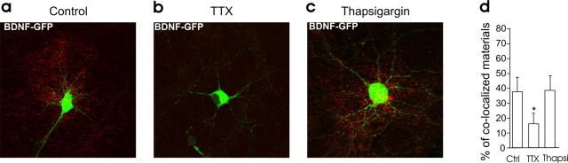
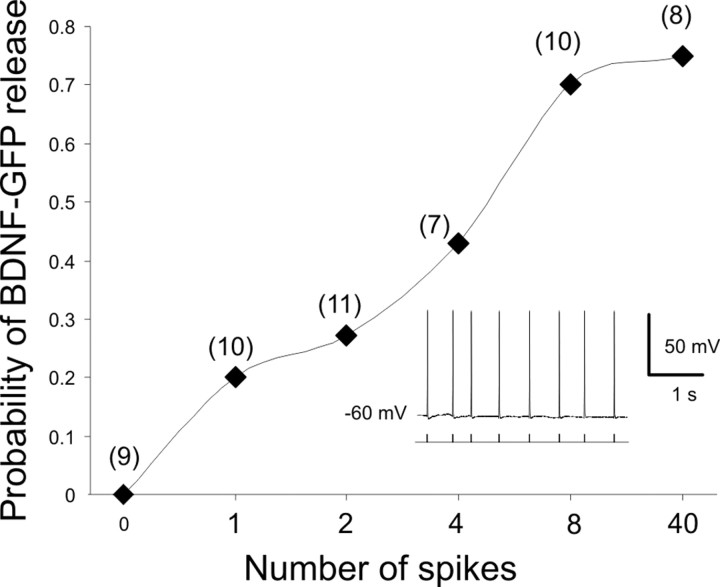
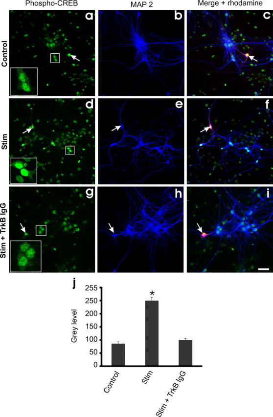
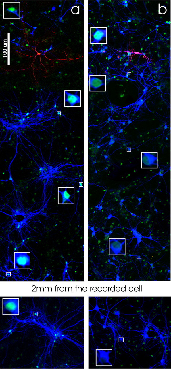
Brain-derived neurotrophic factor (BDNF) is a major regulator of activity-dependent synapse development and plasticity. Because BDNF is a secreted protein, it has been proposed that BDNF is released from target neurons in an activity-dependent manner. However, direct evidence for postsynaptic release of BDNF triggered by ongoing network activity is still lacking. Here we transfected cultures of dissociated hippocampal neurons with green fluorescent protein (GFP)-tagged BDNF and combined whole-cell recording, time-lapse fluorescent imaging, and immunostaining to monitor activity-dependent dendritic release of BDNF. We found that spontaneous backpropagating action potentials, but not synaptic activity alone, led to a Ca2+-dependent dendritic release of BDNF-GFP. Moreover, we provide evidence that endogenous BDNF released from a single neuron can phosphorylate CREB (cAMP response element-binding protein) in neighboring neurons, an important step of immediate early gene activation. Therefore, together, our results support the hypothesis that BDNF might act as a target-derived messenger of activity-dependent synaptic plasticity and development.


Figures

Figure 1.
Figure 1.
Spontaneous network activity triggers BDNF-GFP release. a–f, Overlapped images showing intracellular GFP fluorescence (green) and secreted BDNF-GFP detected using anti-GFP antibody (red) under nonpermeable conditions. a, Hippocampal neurons transfected with GFP alone. b–f, Neurons transfected with BDNF-GFP. b, Control condition. c, In the presence of TTX (1 μm). d, Stimulated with 4-AP (50 μm). e, Stimulated with 4-AP (50 μm) in the presence of NBQX (5 μm), d-APV (40 μm), and bicuculline (10 μm), indicated in the figure as “antago.” f, Stimulated with 4-AP (50 μm) in the presence of NBQX, d-APV, bicuculline, and TTX (1 μm). Top, Merged picture of both fluorescence channels of neurons transfected with the BDNF-GFP construct (green) and immunocytochemistry staining using an antibody against the GFP (red). Bottom, Quantification of the surface-bound BDNF-GFP on the transfected neuron (yellow signal); the plots are a three-dimensional representation of the mean gray level values. g, Quantitative analysis of surface-bound GFP comparing BDNF-GFP-transfected neurons in different ACSF conditions (n = 4 different cultures, 4 neurons per culture in each condition). h, Average firing rate of the neurons in the different conditions, measured in cell-attached configuration (n = 5 neurons in each condition). Right, Representative electrophysiological traces for each condition. The green signal produced by released BDNF-GFP is not visible in the images presented here because of the low laser intensity used to avoid saturation of the green signal in the transfected neurons. The red signal in the untransfected cell is attributable to the BDNF-GFP secreted by the transfected neurons that bound to membrane TrkB receptors of the neighboring cells. Antago in e, f, and h stands for NBQX (5 μm), APV (40 μm), and bicuculline (10 μm). Ctr, Control. In this and following figures, * indicates p < 0.05 compared with control condition.
Figure 2.
Figure 2.
Depolarizing steps trigger dendritic secretion of BDNF-GFP. a, Decrease of fluorescence intensity from BDNF-GFP granules localized in the dendrites was produced by 20 depolarizing steps of 50–60 mV depolarization, 500 ms long, given at 0.01 Hz. Note that fluorescence decreased within the white circle and did not increase in the surrounding area, indicating that the decrease in fluorescence intensity was not attributable to lateral movements in the x–y-axes. Left, Representative traces of a DP; note the inward Ca2+ current produced by the depolarization. Right, Example of dendritic BDNF-GFP granules (indicated by circles) before and after the DPs. b, Average time course of dendritic fluorescence change evoked by the DPs (n = 11). c, d, DP failed to produce significant variation of fluorescence in GFP-only-transfected neurons (n = 9). e, The effect of DPs on dendritic fluorescence was abolished by bath-applied Cd2+ (200 μm; n = 8); note the absence of Ca2+ current in response to DP (superimposed control and Cd2+ traces). f, Postsynaptic loading of GDP β-S (0.6 mm), a G-protein inhibitor that blocks granular secretion, prevented the DP-induced decrease of fluorescence in the dendrites of BDNF-GFP-transfected cells (n = 8); note that the Ca2+ current in response to DP was not affected (superimposed control and Cd2+ traces). All the experiments were performed in the presence of NBQX and APV. Rel., Relative.
Figure 3.
Figure 3.
Surface staining confirmed the BDNF-GFP secretion produced by the depolarizing steps. BDNF-GFP-expressing neurons received either 20 suprathreshold or 20 subthreshold DPs applied through the recording pipette. The intracellular solution contained rhodamine for post hoc identification. Surface staining of released BDNF-GFP was detected by immunocytochemical staining against GFP under nonpermeabilized conditions (blue staining).
Figure 4.
Figure 4.
Dendritic backpropagation of APs in cultured neurons. a, Example of paired somatic (s) and dendritic (d) whole-cell recording of a neuron in culture. b, APs elicited by somatic current injection backpropagate into the dendrite. c, Spontaneous AP generated in the soma backpropagates into the dendrite. Note the delay between somatic and dendritic AP.
Figure 5.
Figure 5.
Synaptically driven b-APs trigger dendritic BDNF secretion through membrane depolarization. a, Synaptically induced b-APs, occurring when neurons were held at −52 ± 1 mV, produced a dendritic BDNF-GFP secretion (n = 5); the arrow indicates the arrival of the first AP. Left, Representative trace. b, BDNF-GFP secretion did not occur when APs were prevented by QX314 in the recording pipette (n = 9). Left, Representative trace. c, Membrane depolarization to −40 mV, with QX314 in the pipette, produced a BDNF-GFP secretion even in the absence of APs (n = 6). Left, Representative trace. Rel., Relative.
Figure 6.
Figure 6.
VDCC activation, but not intracellular Ca2+ stores, is required for b-AP-induced BDNF secretion. a, BDNF-GFP secretion was induced by firing activity produced by somatic current injection. Left, Representative trace of step-induced firing activity (n = 17). b, BDNF-GFP release was prevented by bath application of the VDCC blocker CdCl (200 μm; n = 9). c, Thapsigargin (10 μm) did not prevent BDNF-GFP release elicited by b-APs (n = 8). Left, Representative traces of step-induced firing activity. Arrows indicate the arrival of the first action potential. In a–c, Vh = −70 mV. Rel., Relative.
Figure 7.
Figure 7.
Intracellular Ca2+ stores are not required for BDNF secretion produced by ongoing activity. a–c, Surface immunofluorescence staining on neuronal cultures in control condition (a) and incubated in TTX (b) or thapsigargin (10 μm; c) for 3 h. d, Quantitative analysis of surface-bound GFP (yellow signal/green signal) in the different conditions (n = 3 cultures, 4 neurons per cultures in each condition). Thapsi, Thapsigargin.
Figure 8.
Figure 8.
The probability of BDNF-GFP release increases with the number of APs. The probability to induce a dendritic BDNF-GFP secretion (fluorescence decrease >3% between 100 and 200 s after stimuli) is a function of the number of APs elicited by somatic current injection (at 3.96 ± 0.5 Hz). Inset, Representative trace of APs. In parentheses is the number of experiments.
Figure 9.
Figure 9.
Endogenous BDNF is released by firing activity. Shown is activation of pCREB in nontransfected neuronal cells after electrophysiological stimulation. Left and middle, Images of pCREB (left) and MAP2 (middle), in neurons neighboring the patched rhodamine-filled cell (arrows). Right, Merged pictures of the rhodamine-filled cell immunofluorescence (red) in the presence of MAP2 and pCREB immunostaining (blue) for the same field of view. Scale bar, 45 μm. a–c, Control condition; cell patched but not stimulated. d–f, Cell patched and stimulated to fire (40 spike at 10 Hz). g–i, Cell patched and stimulated to fire (40 spikes at 10 Hz) in the presence of the BDNF scavenger TrkB IgG. j, Histogram showing the pCREB level activation by measurement of fluorescence intensity (mean gray level values) (n = 3 cultures in each condition. The amount of pCREB was calculated through a distance of 250 μm from the recorded neuron. A total of 47, 56, and 49 neurons, respectively, were used for analysis of the intensity of pCREB in control, stimulated, and TrkB-IgG conditions). Electrophysiology was performed in ACSF supplemented with NBQX and APV. Stim, Stimulated.
Figure 10.
Figure 10.
Distribution of pCREB-positive neurons after single-cell stimulation. a, CREB phosphorylation produced by neuronal stimulation (40 spikes at 10 Hz) in current clamp is distributed in the culture and can be observed at a distance from the stimulated cell. b, CREB phosphorylation produced by neuronal stimulation (20 DP of 50 mV at 0.1 Hz) in voltage clamp in the presence of TTX is limited to the neurons surrounding the stimulated cell. Squares show nuclei at higher magnification.

References

    1. Aicardi G, Argilli E, Cappello S, Santi S, Riccio M, Thoenen H, Canossa M. Induction of long-term potentiation and depression is reflected by corresponding changes in secretion of endogenous brain-derived neurotrophic factor. Proc Natl Acad Sci U S A. 2004;101:15788–15792. - PMC - PubMed
    1. Balkowiec A, Katz DM. Cellular mechanisms regulating activity-dependent release of native brain-derived neurotrophic factor from hippocampal neurons. J Neurosci. 2002;22:10399–10407. - PMC - PubMed
    1. Bonhoeffer T. Neurotrophins and activity-dependent development of the neocortex. Curr Opin Neurobiol. 1996;6:119–126. - PubMed
    1. Bramham CR, Messaoudi E. BDNF function in adult synaptic plasticity: the synaptic consolidation hypothesis. Prog Neurobiol. 2005;76:99–125. - PubMed
    1. Brigadski T, Hartmann M, Lessmann V. Differential vesicular targeting and time course of synaptic secretion of the mammalian neurotrophins. J Neurosci. 2005;25:7601–7614. - PMC - PubMed

Publication types

MeSH terms

Substances

LinkOut - more resources