. 2008 Oct;17(10):1805-14.
doi: 10.1110/ps.036384.108. Epub 2008 Jul 2.

Differential ubiquitin binding of the UBA domains from human c-Cbl and Cbl-b: NMR structural and biochemical insights

Affiliations

Differential ubiquitin binding of the UBA domains from human c-Cbl and Cbl-b: NMR structural and biochemical insights

Zi-Ren Zhou et al. Protein Sci. 2008 Oct.

Abstract

The Cbl proteins, RING-type E3 ubiquitin ligases, are responsible for ubiquitinating the activated tyrosine kinases and targeting them for degradation. Both c-Cbl and Cbl-b have a UBA (ubiquitin-associated) domain at their C-terminal ends, and these two UBA domains share a high sequence similarity (75%). However, only the UBA from Cbl-b, but not from c-Cbl, can bind ubiquitin (Ub). To understand the mechanism by which the UBA domains specifically interact with Ub with different affinities, we determined the solution NMR structures of these two UBA domains, cUBA from human c-Cbl and UBAb from Cbl-b. Their structures show that these two UBA domains share the same fold, a compact three-helix bundle, highly resembling the typical UBA fold. Chemical shift perturbation experiments reveal that the helix-1 and loop-1 of UBAb form a predominately hydrophobic surface for Ub binding. By comparing the Ub-interacting surface on UBAb and its counterpart on cUBA, we find that the hydrophobic patch on cUBA is interrupted by a negatively charged residue Glu12. Fluorescence titration data show that the Ala12Glu mutant of UBAb completely loses the ability to bind Ub, whereas the mutation disrupting the dimerization has no significant effect on Ub binding. This study provides structural and biochemical insights into the Ub binding specificities of the Cbl UBA domains, in which the hydrophobic surface distribution on the first helix plays crucial roles in their differential affinities for Ub binding. That is, the amino acid residue diversity in the helix-1 region, but not the dimerization, determines the abilities of various UBA domains binding with Ub.


Figures

Figure 1.
Figure 1.
Multiple alignments of the UBA domains from Cbls. (A) Schematic domain representation of c-Cbl, Cbl-b, and Cbl-3. The UBA domain is located at the C terminus. (B) Sequence alignment of the UBA domains from human c-Cbl (cUBA) and Cbl-b (UBAb). Residues 1–56 of cUBA correspond to 851–906 of c-Cbl, and residues 1–57 of UBAb correspond to 926–982 of Cbl-b. Residues, which are same in these two domains, are labeled on a dark background and those with high similarity are highlighted by boxes. The secondary structures of cUBA are shown at the top. The sequences were aligned using ClustalW and represented using ESPript.
Figure 2.
Figure 2.
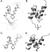
cUBA and UBAb adopt the same fold of highly compact three-helix bundle. (A) Ensemble of the Cα traces representing a bundle of 15 representative structures of cUBA. (B) Ribbon representation of the cUBA structure. The three helices are labeled as α1, α2, and α3. (C) Ensemble of the Cα traces representing a bundle of 15 representative structures of UBAb. (D) Ribbon representation of UBAb. The figures were prepared with MOLMOL.
Figure 3.
Figure 3.
Both cUBA and UBAb domain structures are stabilized by a hydrophobic core. (A) Hydrophobic core of cUBA. The side chains of the residues in the hydrophobic core are displayed in neon style (orange); these residues include Leu7, Ile11, Leu14, Ile24, Leu28, Ile35, Ala38, and Leu42. (B) Hydrophobic core of UBAb. The core residues are Val7, Ile11, Leu14, Val24, Leu28, Val35, Ala38, and Leu42. (C) Overlay of the secondary structure elements of cUBA (cyan) and UBAb (red).
Figure 4.
Figure 4.
The UBAb domain binds to the hydrophobic surface of ubiquitin. (A) Overlay of a representative region of the [1H, 15N]-HSQC spectra of 15N-labeled ubiquitin alone (red) and at a molar ratio of 1:3 with unlabeled cUBA (blue). (B) Overlay of the [1H, 15N]-HSQC spectra of ubiquitin alone (red) and in the presence of an equimolar amount of UBAb (blue). UBAb titration causes severe line broadening and/or peak shifting of certain amide resonances of ubiquitin. (C) Mapping the perturbed resonances of ubiquitin to its ribbon diagram upon titration of UBAb. The residues with broadened resonances are displayed in green (L8, I13, T14, R42, L43, F45, K48, Q49, H68, V70, L71, and R72), and those with chemical shift change are colored in yellow (Q40, Q41, L50, I61, S65, R74). The opposite surface that does not contain perturbed residues is not shown in the figure.
Figure 5.
Figure 5.
The ubiquitin-interacting surface on UBAb. (A) Overlay of a representative region of [1H, 15N]-HSQC spectra of 15N-labeled cUBA alone (red) and at a molar ratio of 1:2 with unlabeled ubiquitin (blue). (B) Overlay of the [1H, 15N]-HSQC spectra of 15N-labeled UBAb alone (red) and with ubiquitin at a molar ratio of 1:0.5 (blue). (C,D) Ribbon and side chain presentation (C) and surface display (D) of the surface of UBAb domain that interacts with ubiquitin. The broadened resonances are displayed in green (N6, D8, A9, K10, A12, G16, and A31), and the shifted ones are colored in yellow (L14, M15, and E22).
Figure 6.
Figure 6.
Comparison of the electrostatic surfaces of cUBA (A) and UBAb (B). The orientation of both cUBA and UBAb is identical to that of panels C and D in Figure 4. The positive charges are shown in blue, and the negative charges are colored in red. The surface presentation shows that the Ub recognition surface on UBAb is predominately hydrophobic with positively charged edges while the corresponding surface on cUBA is something negatively charged in the center. The Ub recognition surface on UBAb and its counterpart on cUBA are highlighted by green parallelograms and labeled with amino acid residues. The dimeric interfaces (indicated by green circles) of cUBA (C) and UBAb (D) are rotated by ∼180° relative to those in A and B, respectively. Residues I30 and I41 that are involved in homodimerization are labeled. The PDB codes for the structures are 2JUJ for cUBA and 2JNH for UBAb. (E) Glutamic acid; (A) alanine; (I) isoleucine.
Figure 7.
Figure 7.
Affinities of the UBA domains and their mutants for Ub. (A) Titration curves for the binding of wild-type cUBA and UBAb to Ub F45W. The relative fluorescence intensity change (ΔRFI) of the F45W mutant of Ub is plotted against the concentration of the UBA domains. (B) Titration curves for the binding of UBAb mutants to Ub F45W. (C) Titration curves for the binding of I30E/I41E mutant of UBAb and E12A/I30E/I41E of cUBA to Ub F45W. (D) Analytical gel filtration of UBAb and its I30E/I41E mutant. The I30E/I41E mutant of UBAb gives an elution volume larger than that of the wild type.

References

    1. Bartkiewicz, M., Houghton, A., Baron, R. Leucine zipper-mediated homodimerization of the adaptor protein c-Cbl. A role in c-Cbl's tyrosine phosphorylation and its association with epidermal growth factor receptor. J. Biol. Chem. 1999;274:30887–30895. - PubMed
    1. Blake, T.J., Langdon, W.Y. A rearrangement of the c-cbl proto-oncogene in HUT78 T-lymphoma cells results in a truncated protein. Oncogene. 1992;7:757–762. - PubMed
    1. Blake, T.J., Shapiro, M., Morse H.C., III, Langdon, W.Y. The sequences of the human and mouse c-cbl proto-oncogenes show v-cbl was generated by a large truncation encompassing a proline-rich domain and a leucine zipper-like motif. Oncogene. 1991;6:653–657. - PubMed
    1. Brunger, A.T., Adams, P.D., Clore, G.M., DeLano, W.L., Gros, P., Grosse-Kunstleve, R.W., Jiang, J.S., Kuszewski, J., Nilges, M., Pannu, N.S., et al. Crystallography and NMR system: A new software suite for macromolecular structure determination. Acta Crystallogr. D Biol. Crystallogr. 1998;54:905–921. - PubMed
    1. Chang, Y.G., Song, A.X., Gao, Y.G., Shi, Y.H., Lin, X.J., Cao, X.T., Lin, D.H., Hu, H.Y. Solution structure of the ubiquitin-associated domain of human BMSC-UbP and its complex with ubiquitin. Protein Sci. 2006;15:1248–1259. - PMC - PubMed

Publication types

LinkOut - more resources