. 2008 Sep;15(9):1330-6.
doi: 10.1128/CVI.00103-08. Epub 2008 Jul 2.

Toxicity of anthrax toxin is influenced by receptor expression

Affiliations

Toxicity of anthrax toxin is influenced by receptor expression

Sarah C Taft et al. Clin Vaccine Immunol. 2008 Sep.

Abstract

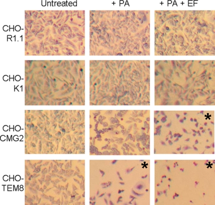
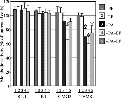
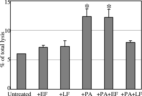
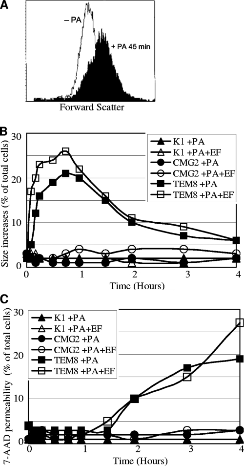
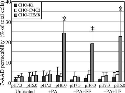
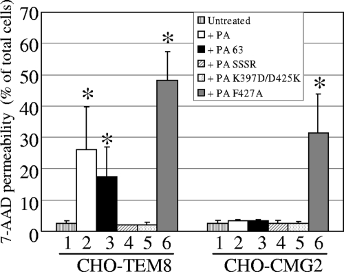
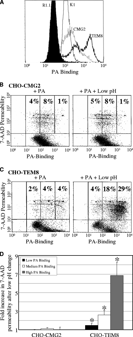
Anthrax toxin protective antigen (PA) binds to its cellular receptor, and seven subunits self-associate to form a heptameric ring that mediates the cytoplasmic entry of lethal factor or edema factor. The influence of receptor type on susceptibility to anthrax toxin components was examined using Chinese hamster ovary (CHO) cells expressing the human form of one of two PA receptors: TEM8 or CMG2. Unexpectedly, PA alone, previously believed to only mediate entry of lethal factor or edema factor, was found to be toxic to CHO-TEM8 cells; cells treated with PA alone displayed reduced cell growth and decreased metabolic activity. PA-treated cells swelled and became permeable to membrane-excluded dye, suggesting that PA formed cell surface pores on CHO-TEM8 cells. While CHO-CMG2 cells were not killed by wild-type PA, they were susceptible to the PA variant, F427A. Receptor expression also conferred differences in susceptibility to edema factor.


Figures

FIG. 1.
FIG. 1.
PA-mediated toxicity to CHO cells is receptor dependent. Mutant CHO cells lacking anthrax toxin receptor (CHO-R1.1), wild-type CHO cells (CHO-K1), and CHO-R1.1 cells expressing the human CMG2 receptor (CHO-CMG2) or TEM8 receptor (CHO-TEM8) were incubated overnight with PA or PA+EF. Cells were fixed, stained with Giemsa, and viewed at ×63 magnification. The asterisk (*) denotes conditions that resulted in a statistically significant loss of cellular metabolic activity (Fig. 2).
FIG. 2.
FIG. 2.
PA alone reduces metabolic activity in CHO-TEM8 cells. CHO-R1.1, CHO-K1, CHO-CMG2, and CHO-TEM8 cells were incubated overnight with: 1, EF; 2, LF; 3, PA; 4, PA+EF; or 5, PA+LF. The mitochondrial conversion of alamarBlue was measured to determine metabolic activity. Values are shown as percentages of untreated control cells. The data are represented as means ± the standard error of the mean (SEM). An asterisk (*) signifies statistical significance compared to untreated cells (P < 0.01).
FIG. 3.
FIG. 3.
PA promotes cellular lysis of CHO-TEM8 cells. CHO-TEM8 cells were incubated overnight with EF, LF, PA, PA+EF, or PA+LF. Cellular LDH release was measured and is presented as the percentage of total lysis. The data are represented as mean ± the SEM. An asterisk (*) signifies statistical significance from untreated cells (P < 0.05).
FIG. 4.
FIG. 4.
PA induces size changes and 7-AAD permeability in CHO-TEM8 cells. CHO-K1, CHO-CMG2, or CHO-TEM8 cells were incubated with PA or PA+EF. (A) Forward scatter (FSC-H) as a measure of cell size for CHO-TEM8 cells untreated (open) or incubated with PA for 45 min (filled). Cell size (B) and permeability to 7-AAD (C) were analyzed by flow cytometry. Values are shown as the percentage of total cells exceeding two standard deviations of the mean of control cells.
FIG. 5.
FIG. 5.
Influence of low pH on PA-mediated permeability. CHO-K1, CHO-CMG2, and CHO-TEM8 cells were incubated with PA for 5 min, and the pH was adjusted to 6.0. The cells were stained and analyzed by flow cytometry. Values are shown as the percentage of total cells exceeding two standard deviations of the mean of control cells. The data are represented as means ± the SEM. An asterisk (*) signifies statistical significance from untreated cells (P < 0.05).
FIG. 6.
FIG. 6.
Analysis of PA mutants. CHO-CMG2 and CHO-TEM8 cells were: 1, untreated; 2, incubated overnight with wild-type PA; 3, incubated overnight with proteolytically activated PA 63; 4, incubated overnight with furin cleavage resistant mutant (PA SSSR); 5, incubated overnight with pore formation resistant mutant (PA K397D/D425K); or 6, incubated overnight with domain 2 mutant (PA F427A). Increases in 7-AAD permeability were analyzed by flow cytometry. Values are shown as the percentage of total cells exceeding two standard deviations of the mean of control cells. The data are represented as means ± the SEM. An asterisk (*) signifies statistical significance from untreated cells (P < 0.02).
FIG. 7.
FIG. 7.
Susceptibility to PA-mediated toxicity is influenced by receptor expression. (A) PA-receptor binding on CHO cells; CHO-R1.1 (filled histogram), CHO-K1 (thin line), CHO-CMG2 (dotted line), and CHO-TEM8 (heavy line) cells were incubated with PA for 5 min, washed, incubated with anti-PA serum for 10 min, washed, incubated with anti-IgG Alexa Fluor 647 antibody for 20 min, and analyzed by flow cytometry. CHO-CMG2 cells (B) and CHO-TEM8 cells (C) were incubated with PA for 5 min and left untreated or treated with buffer to decrease the pH for 5 min. The cells were double labeled for PA-receptor binding and cell permeability with 7-AAD and then analyzed by flow cytometry. (D) Fold increase in 7-AAD permeability after low pH change of PA treated CHO-CMG2 and CHO-TEM8 cells as a function of PA binding (high, >1,000; medium, 100 to 1,000; low, <100). The data are represented as means ± the SEM. An asterisk (*) signifies statistical significance from cells only treated with PA (P < 0.05).

Similar articles

Cited by

References

    1. Abrami, L., S. Liu, P. Cosson, S. H. Leppla, and F. G. van der Goot. 2003. Anthrax toxin triggers endocytosis of its receptor via a lipid raft-mediated clathrin-dependent process. J. Cell Biol. 160:321-328. - PMC - PubMed
    1. Ballard, J. D., A. M. Doling, K. Beauregard, R. J. Collier, and M. N. Starnbach. 1998. Anthrax toxin-mediated delivery in vivo and in vitro of a cytotoxic T-lymphocyte epitope from ovalbumin. Infect. Immun. 66:615-619. - PMC - PubMed
    1. Beauregard, K. E., R. J. Collier, and J. A. Swanson. 2000. Proteolytic activation of receptor-bound anthrax protective antigen on macrophages promotes its internalization. Cell. Microbiol. 2:251-258. - PubMed
    1. Bell, S. E., A. Mavila, R. Salazar, K. J. Bayless, S. Kanagala, S. A. Maxwell, and G. E. Davis. 2001. Differential gene expression during capillary morphogenesis in 3D collagen matrices: regulated expression of genes involved in basement membrane matrix assembly, cell cycle progression, cellular differentiation and G-protein signaling. J. Cell Sci. 114:2755-2773. - PubMed
    1. Blaustein, R. O., T. M. Koehler, R. J. Collier, and A. Finkelstein. 1989. Anthrax toxin: channel-forming activity of protective antigen in planar phospholipid bilayers. Proc. Natl. Acad. Sci. USA 86:2209-2213. - PMC - PubMed

Publication types

LinkOut - more resources