. 2008 Sep;295(3):C752-60.
doi: 10.1152/ajpcell.00228.2008. Epub 2008 Jul 2.

P2X7 receptor-Pannexin1 complex: pharmacology and signaling

Affiliations

P2X7 receptor-Pannexin1 complex: pharmacology and signaling

R Iglesias et al. Am J Physiol Cell Physiol. 2008 Sep.

Erratum in

  • Am J Physiol Cell Physiol. 2009 Mar;296(3):C639

Abstract

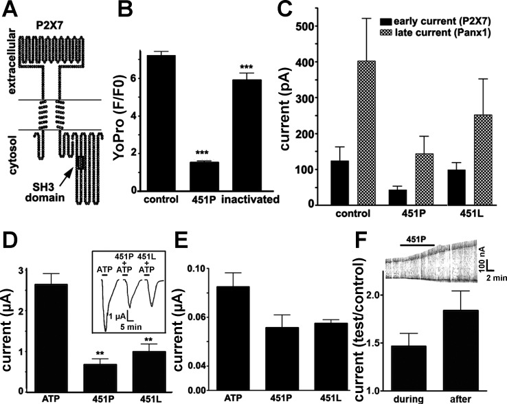
Pannexin 1 (Panx1), an ortholog to invertebrate innexin gap junctions, has recently been proposed to be the pore induced by P2X(7) receptor (P2X(7)R) activation. We explored the pharmacological action of compounds known to block gap junctions on Panx1 channels activated by the P2X(7)R and the mechanisms involved in the interaction between these two proteins. Whole cell recordings revealed distinct P2X(7)R and Panx1 currents in response to agonists. Activation of Panx1 currents following P2X(7)R stimulation or by membrane depolarization was blocked by Panx1 small-interfering RNA (siRNA) and with mefloquine > carbenoxolone > flufenamic acid. Incubation of cells with KN-62, a P2X(7)R antagonist, prevented current activation by 2'(3')-O-(4-benzoylbenzoyl)adenosine 5'-triphosphate (BzATP). Membrane permeabilization to dye induced by BzATP was also prevented by Panx1 siRNA and by carbenoxolone and mefloquine. Membrane permeant (TAT-P2X(7)) peptides, provided evidence that the Src homology 3 death domain of the COOH-terminus of the P2X(7)R is involved in the initial steps of the signal transduction events leading to Panx1 activation and that a Src tyrosine kinase is likely involved in this process. Competition assays indicated that 20 microM TAT-P2X(7) peptide caused 50% reduction in Src binding to the P2X(7)R complex. Src tyrosine phosphorylation following BzATP stimulation was reduced by KN-62, TAT-P2X(7) peptide, and by the Src tyrosine inhibitor PP2 and these compounds prevented both large-conductance Panx1 currents and membrane permeabilization. These results together with the lack Panx1 tyrosine phosphorylation in response to P2X(7)R stimulation indicate the involvement of an additional molecule in the tyrosine kinase signal transduction pathway mediating Panx1 activation through the P2X(7)R.


Figures

Fig. 1.
Fig. 1.
Blockade of P2X7 receptor (P2X7R)-induced currents by gap junction channel blockers and a P2X7R antagonist. A: representative current traces induced by 5-s application of 2′(3′)-O-(4-benzoylbenzoyl)adenosine 5′-triphosphate (BzATP, 50 μM; traces on top) and ATP (1 mM; traces on bottom) are shown to be greatly reduced by carbenoxolone (CBX, 50 μM); after washout of CBX, both agonists induced currents of similar amplitudes as those recorded after the first application. For all electrophysiological experiments, membrane potential was held at −60 mV. Note that after 50 μM BzATP application two distinct currents were recorded (an early and smaller current followed by a larger one). Bar histograms (right) show the mean ± SE values obtained from 6 experiments. B: representative current traces induced by 5-s application of BzATP (50 μM; top and bottom) are shown to be greatly reduced by 0.1 mM (top) and 0.3 mM (bottom) flufenamic acid (FFA); after washout of FFA, currents induced by BzATP were of slightly lower amplitudes than those recorded after the first application. Bar histograms (right) show the mean ± SE values obtained from 4 experiments. C and D: representative current traces induced by 5-s application of BzATP (50 μM) are shown to be greatly reduced by mefloquine (MFQ; 100 nM; C) and totally prevented by KN-62 (1 μM; D); after washout of MFQ and KN-62, BzATP induced currents with similar amplitudes as those recorded after the first application. E: representative current traces induced by 5-s application of BzATP (50 μM) in cells untreated (control) and treated for 48 h with pannexin1 (Panx1) small-interfering RNA (siRNA). Bar histograms (right) show the mean ± SE values obtained from 5 experiments; *P < 0.05, **P < 0.01, and ***P < 0.001. P values were obtained by ANOVA followed by Newman Keuls' test.
Fig. 2.
Fig. 2.
Pharmacology of outward (Panx1) currents and of the permeabilization pore in J774 cells. A: top, examples of outward currents recorded from J774 cells in the absence (control) and presence of MFQ (50 nM), and after washout. Bottom, bar histograms showing the mean ± SE values of the fraction of current amplitudes at the end of the current ramp (+100 mV) relative to control recorded from J774 macrophages bathed in solution containing CBX (10 and 50 μM), FFA (0.1 and 0.3 mM), and MFQ (10, 50, and 100 nM) and the P2X7R antagonist KN-62 (1 μM). Note that all three compounds known to block gap junction channels but not the mouse P2X7R antagonist greatly reduced voltage-activated outward (Panx1) currents. The last two bars on the right show the mean ± SE values of the fraction of current amplitudes recorded from untreated and Panx1 siRNA-treated cells. Data are from 6 independent experiments. B: top, representative curves of the mean value of relative YoPro fluorescence intensity (F/F0) changes recorded from J774 cells (n = 100) untreated (control) and treated with Panx1 siRNA following 300 μM BzATP application. Bottom, bar histogram showing that CBX (50 μM), MFQ (10 nM), and Panx1 siRNA greatly reduce YoPro uptake in BzATP (300 μM)-stimulated J774 cells (n = 300–400 cells/group from 3 independent experiments) bathed in a Ca2+-containing solution. C: representative Western blots of immunoprecipitates using P2X7 antibodies showing that the expression levels of Panx1 present in the P2X7R complex in the J774 cell line are greatly reduced (62%) following 48 h transfection with Panx1 siRNA (left) and that the expression level of P2X7R present in the whole cell lysates used for immunoprecipitation (right) is not altered by Panx1 siRNA treatment compared with untreated cells. Three independent experiments were performed. D: representative fluorescence images of J774 cells showing a reduced number of cells loaded with YoPro following 30 h transfection with Panx1 siRNA compared with control cultures. Arrows illustrate YoPro-loaded cells. Bar: 185 μm. *P < 0.05, **P < 0.01, and ***P < 0.001. P values obtained using ANOVA followed by Newman-Keuls' test.
Fig. 3.
Fig. 3.
The proline-rich region of P2X7R COOH terminus contributes to Panx1 activation. A: membrane topology of the P2X7R showing the region of the SH3 domain that was used to generate the TAT-P2X7 peptide. B: bar histograms showing the mean ± SE values of relative YoPro fluorescence intensity in J774 cells untreated and treated for 15 min with 10 μM active 451P TAT-P2X7 and heat-inactivated peptides following 10 min stimulation with 300 μM BzATP. Dye uptake was measured from cells bathed in a Ca2+-free solution. Note that the active TAT-P2X7 peptide greatly attenuated BzATP-induced dye uptake and that heat-inactivated peptide had only a minor effect. Data are from 3–4 independent experiments. C: bar histograms of the mean ± SE values obtained for current amplitudes induced by ATP (5 mM) in untreated (control n = 9 cells) and 10 μM TAT-P2X7 peptide (n = 14 cells)-treated J774 macrophages. The two TAT-P2X7 peptides (451P and 451L) used have the same amino acid sequence, except at position 451 where in one case a proline (P) was substituted for a lysine (L). About 10 cells/group were used; membrane potential was held at −60 mV, and channels were activated using the ramp protocol. D: bar histograms of the mean ± SE values (n = 3–4 independent experiments) obtained for current amplitudes induced by ATP (500 μM) in untreated and 10 μM TAT-peptide-treated Xenopus oocytes coexpressing P2X7R and Panx1 (see text for details). Inset: example of currents recorded from oocytes. E: bar histograms of the mean ± SE values (n = 3–4 independent experiments) obtained for current amplitudes induced by ATP (500 μM) in untreated and 10 μM TAT-peptides-treated Xenopus oocytes expressing only the P2X7R. F: bar histograms showing the mean ± SE relative current values (test/control; n = 4 independent experiments) obtained from Xenopus oocytes expressing only Panx1. Current amplitudes induced by voltage steps (−30 to +60 mV) during and after addition of 10 μM TAT peptides were normalized to the values obtained before TAT peptide addition. Inset: representative recording of voltage-activated currents in oocytes expressing Panx1 showing current changes during and after exposure to TAT peptides. The same TAT-P2X7 peptides described in B were used. **P < 0.01 and ***P < 0.001. P values were obtained by ANOVA followed by Newman-Keuls' test.
Fig. 4.
Fig. 4.
Src tyrosine kinase mediates activation of Panx1 through P2X7R in J774 macrophages. A: dose-dependence curve showing the inhibition of src[Y418] binding to the P2X7R by the TAT-P2X7 peptide (451P). The IC50 value (20 μM) was obtained after fitting a single exponential decay curve to the densitometric values obtained for src[Y418] bands obtained in Western blots following pull-downs using P2X7R antibodies of J774 whole cell lysates treated with different concentration of TAT-P2X7 peptide (n = 4 independent experiments). An example of a Western blot obtained from competition assays is shown at top. B: representative current traces induced by 5-s application of BzATP (50 μM) are shown to be reduced by the Src tyrosine kinase inhibitor PP2 (20 μM; 5 min preincubation); after washout of PP2, agonist induced currents of similar amplitudes as those recorded after the first application. Bar histograms show the mean ± SE values obtained from 5 experiments. C: bar histograms of the mean ± SE values of dye uptake induced by 150 μM BzATP, showing that the PP2 (10 μM; 5 min preincubation) significantly decreased agonist-induced YoPro uptake. Dye uptake was measured from cells bathed in a Ca2+-free solution. D: bar histograms of the mean ± SE ratio values of the active (src[Y418]) to total src obtained from Western blot analysis of J774 whole cell lysates (n = 4 independent experiments). Before cell lysis, cultures were treated for 15 min with BzATP (300 μM) in the absence and presence of 1 μM KN-62, 10 μM TAT-P2X7 peptide (451P), and 10 μM PP2; the ratio levels of active src to total src obtained from treated cells was compared with those obtained from BzATP-untreated, control (ctrl) whole cell lysates. Inset: example of Western blot obtained from J774 cells showing that increased amount of tyrosine-phosphorylated src (srcY) induced by agonist stimulation was prevented by KN-62, TAT-P2X7 peptide, and PP2. **P < 0.01. P values obtained using ANOVA followed by Newman-Keuls' test.

Similar articles

Cited by

References

    1. Adriouch S, Dox C, Welge V, Seman M, Koch-Nolte F, Haag F. Cutting edge: a natural P451L mutation in the cytoplasmic domain impairs the function of the mouse P2X7 receptor. J Immunol 169: 4108–4112, 2002. - PubMed
    1. Alves LA, Coutinho-Silva R, Persechini PM, Spray DC, Savino W, Campos de Carvalho AC. Are there functional gap junctions or junctional hemichannels in macrophages? Blood 88: 328–334, 1996. - PubMed
    1. Bao L, Locovei S, Dahl G. Pannexin membrane channels are mechanosensitive conduits for ATP. FEBS Lett 572: 65–68, 2004. - PubMed
    1. Baraldi PG, Makaeva R, Pavani MG, Nunez MC, Spalluto G, Moro S, Falzoni D, Di Virgilio F, Romagnoli R. Synthesis, biological activity and molecular modeling studies of 1,2,3,4-tetrahydroisoquinoline derivatives as conformationally constrained analogues of KN62, a potent antagonist of the P2X7-receptor containing a tyrosine moiety. Arzeimittelforschung 52: 273–285, 2002. - PubMed
    1. Baranova A, Ivanov D, Petrash N, Pestova A, Skoblov M, Kelmanson I, Shagin D, Nazarenko S, Geraymovych E, Litvin O, Tiunova A, Born TL, Usman N, Staroverov D, Lukyanov S, Panchin Y. The mammalian pannexin family is homologous to the invertebrate innexin gap junction proteins. Genomics 83: 706–716, 2004. - PubMed

Publication types

MeSH terms

LinkOut - more resources