. 2008 Aug;14(8):1617-31.
doi: 10.1261/rna.1045408. Epub 2008 Jul 2.

Spliceosomal snRNAs in the unicellular eukaryote Trichomonas vaginalis are structurally conserved but lack a 5'-cap structure

Affiliations

Spliceosomal snRNAs in the unicellular eukaryote Trichomonas vaginalis are structurally conserved but lack a 5'-cap structure

Augusto Simoes-Barbosa et al. RNA. 2008 Aug.

Abstract

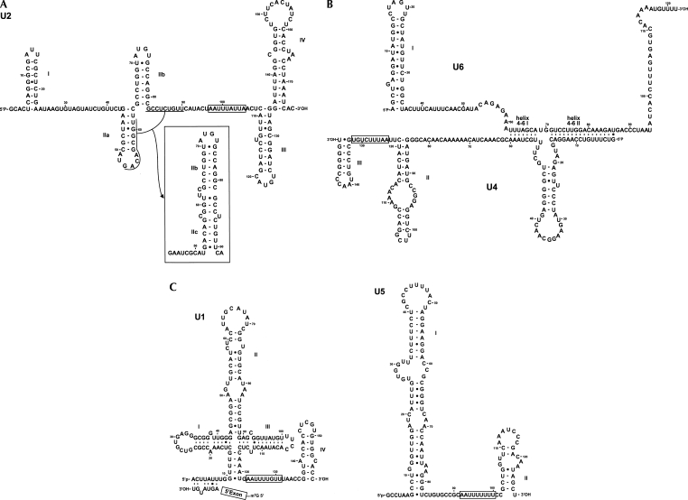
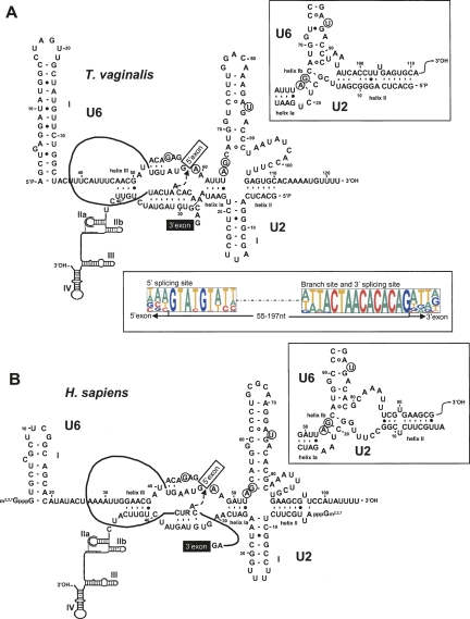
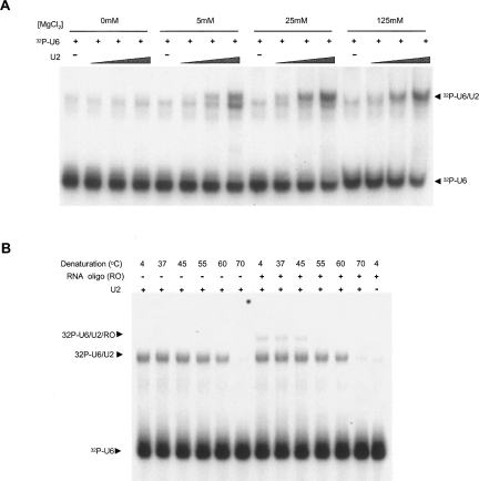
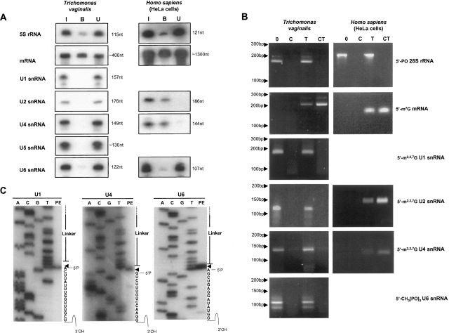
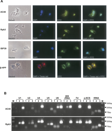
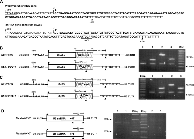
Few genes in the divergent eukaryote Trichomonas vaginalis have introns, despite the unusually large gene repertoire of this human-infective parasite. These introns are characterized by extended conserved regulatory motifs at the 5' and 3' boundaries, a feature shared with another divergent eukaryote, Giardia lamblia, but not with metazoan introns. This unusual characteristic of T. vaginalis introns led us to examine spliceosomal small nuclear RNAs (snRNAs) predicted to mediate splicing reactions via interaction with intron motifs. Here we identify T. vaginalis U1, U2, U4, U5, and U6 snRNAs, present predictions of their secondary structures, and provide evidence for interaction between the U2/U6 snRNA complex and a T. vaginalis intron. Structural models predict that T. vaginalis snRNAs contain conserved sequences and motifs similar to those found in other examined eukaryotes. These data indicate that mechanisms of intron recognition as well as coordination of the two catalytic steps of splicing have been conserved throughout eukaryotic evolution. Unexpectedly, we found that T. vaginalis spliceosomal snRNAs lack the 5' trimethylguanosine cap typical of snRNAs and appear to possess unmodified 5' ends. Despite the lack of a cap structure, U1, U2, U4, and U5 genes are transcribed by RNA polymerase II, whereas the U6 gene is transcribed by RNA polymerase III.


Figures

FIGURE 1.
FIGURE 1.
Sequence and predicted secondary structures of T. vaginalis U1, U2, U4, U5, and U6 spliceosomal snRNAs. Nucleotides are numbered from 5′ to 3′ and putative Sm binding sites are boxed. Helices are indicated in roman numerals. (A) Predicted secondary structure of U2 snRNA; conserved loops are indicated in roman numerals. Sequence underlined shows potential base-pair interactions for formation of helix IIc as indicated in the inset. (B) Predicted secondary structures of U4 and U6 snRNAs, as indicated. U4/U6 intermolecular helices are marked as 4–6 I and 4–6 II. (C) Predicted secondary structure of U1 (left) and U5 (right) snRNAs. Interaction of 5′SS intron sequence from poly(A) polymerase pre-mRNA (Vanacova et al. 2005) with U1 snRNA is shown.
FIGURE 2.
FIGURE 2.
Comparison of U2/U6 snRNA interaction models for T. vaginalis (A) and Homo sapiens (B). Spliceosomal T. vaginalis U2–U6 interactions are modeled after those proposed for yeast and mammalian homologs. Nucleotides proposed to be involved in coordination of metal ions required for catalysis (Fabrizio and Abelson 1992; Yu et al. 1995) are circled. The numbering of nucleotides and helices are indicated. The intron sequence from poly(A) polymerase pre-mRNA (Vanacova et al. 2005) and generic splice site and branch site sequences are incorporated in the T. vaginalis and the H. sapiens models, respectively. The U2/U6 snRNA conformation that allows for an extended U6 intramolecular stem–loop (ISL) proposed to form during the first step of catalysis is shown on the left; the inset on the right shows the proposed second step conformation that allows for formation of helix Ia and Ib. The nucleophilic attack by the branch-site adenosine during the first step of splicing is marked by an arrow. A consensus of the 5′SS, branch site and the 3′SS in identified T. vaginalis introns (Carlton et al. 2007) are shown below the T. vaginalis U2/U6 structure.
FIGURE 3.
FIGURE 3.
In vitro interaction of T. vaginalis U2/U6 catalytic domains. (A) 32P-labeled U6 RNA (32P-U6) was annealed with increasing concentrations of unlabeled U2 RNA (U2) and Mg2+ as indicated. Lower and higher arrows correspond to free 32P-U6 and a U2–U6 complex, respectively. (B) Interaction of U2–U6 catalytic domains with a RNA oligonucleotide that mimics the intron branch site and evaluation of thermostability of the complexes. 32P-labeled U6 RNA (32P-U6) was annealed with unlabeled U2 RNA (U2) and 20 nM of the RNA oligo (RO), as indicated, in the presence of 20 mM of MgCl2. Annealed complexes were then denatured at the indicated temperatures, and complex formation and stability was assessed by nondenaturing PAGE. Lower, middle, and higher arrows correspond to free 32P-U6, the U2–U6 complex, and the U2–U6-RO complex, respectively.
FIGURE 4.
FIGURE 4.
Comparative analysis of snRNA 5′-termini. (A) Anti-TMG immunoprecipitation of total RNA isolated from T. vaginalis (left) and human (right). Collected fractions: (I) input RNA, (B) bound RNA, and (U) unbound RNA were subjected to gel fractionation, blotting, and hybridization to specific probes. Probes (right) and RNA size (left) are indicated. Replica blots from the same immunoprecipitation assay were used. (B) Evaluation of snRNA 5′-ends by RNA Ligase-Mediated Rapid Amplification of cDNA 5′-Ends (RLM-5′RACE). T. vaginalis RNA and human RNA (control) analyses are shown at left and right, respectively. RNAs examined are listed (right) and size markers are indicated (left). Treatments are as follows: (0) untreated; (C) calf intestinal alkaline phosphatase (CIP) treatment only; (T) tobacco acidic pyrophosphatase (TAP) treatment only; (CT) CIP treatment followed by TAP treatment. Controls were a noncapped RNA species (28S rRNA) and a m7G capped mRNA (T. vaginalis ferredoxin and human GAPDH mRNAs). Targets (right) and DNA size (left) are indicated. (C) Primer extension analysis of T. vaginalis U4, U5, and U6 snRNAs subsequent to RLM-5′RACE analyses. Products of primer extension analyses (PE), flanked by sequence reactions (A, C, G, T) for identification of 5′ ends, are shown. Arrowheads mark the single 5′ end nucleotide of each snRNA and correspond to 5′ ends detected using untreated samples.
FIGURE 5.
FIGURE 5.
Chromatin Immunoprecipitation (ChIP) analysis of RNAP components in T. vaginalis. (A) Localization of HA-tagged RNAP core components AC40 and Rpb3 compared with nuclear HA-tagged IBP-39 (positive control) and hydrogenosomal HA-tagged β-HPP (negative control) proteins. The protein detected by FITC staining is indicated to the left. DAPI was used to stain nuclei; merged FITC and DAPI images are shown at the far right. All images are at a 630X magnification. (B) ChIP analysis of AC40 and Rpb3 T. vaginalis transfectants. Target genes are indicated at the top, Molecular weight markers (M) flank the samples, with molecular weight markers listed in base pairs at the far right. Products of independent ChIP assays to detect AC40 and Rpb3 (top and bottom, respectively) are shown. The following DNA samples were tested by PCR: (1) DNA precipitated from ChIP assay done with wild-type nontransfected T. vaginalis cells (negative control); (2) DNA precipitated from ChIP assay done with transfected HA-tagged AC40 (top) or Rpb3 (bottom); (3) total sheared DNA from wild-type nontransfected cells (PCR positive control).
FIGURE 6.
FIGURE 6.
Evaluation of snRNA transcription termination by 3′ RACE. (A) Sequence of endogenous and mutated U6 snRNA genes. A putative TATA-box at −28 nt is underlined, and mapped transcription units are in bold. Both transcription units terminate at a cluster of T residues. (B) 3′ RACE analysis of U6 snRNA from wild-type nontransfected T. vaginalis cells (0) and from T. vaginalis cells transfected with the plasmid constructs: U6ΔT5 (1), U6ΔT5/U2 (2), U6ΔT5/U2+T (3). (C) 3′RACE analysis of U6 snRNA from wild-type nontransfected T. vaginalis cells (0) and T. vaginalis cells transfected with plasmid constructs: U6ΔT5 (1), U6ΔT5/U4 (2), U6ΔT5/U4+T (3). (D) 3′ RACE analysis of U2 snRNA (0 and 1) and U4 snRNA (2 and 3) from wild-type nontransfected T. vaginalis cells (0 and 2) and from T. vaginalis cells transfected with plasmid constructs: MasterU2+T transfectant (1) and MasterU4+T transfectant (3).

Similar articles

Cited by

References

    1. Abrahamsen M.S., Templeton T.J., Enomoto S., Abrahante J.E., Zhu G., Lancto C.A., Deng M., Liu C., Widmer G., Tzipori S., et al. Complete genome sequence of the apicomplexan, Cryptosporidium parvum . Science. 2004;304:441–445. - PubMed
    1. Ares M., Jr, Weiser B. Rearrangement of snRNA structure during assembly and function of the spliceosome. Prog. Nucleic Acid Res. Mol. Biol. 1995;50:131–159. - PubMed
    1. Baldauf S.L. The deep roots of eukaryotes. Science. 2003;300:1703–1706. - PubMed
    1. Braglia P., Percudani R., Dieci G. Sequence context effects on oligo(dT) termination signal recognition by Saccharomyces cerevisiae RNA polymerase III. J. Biol. Chem. 2005;280:19551–19562. - PubMed
    1. Brow D.A., Guthrie C. Spliceosomal RNA U6 is remarkably conserved from yeast to mammals. Nature. 1988;334:213–218. - PubMed

Publication types

LinkOut - more resources